Ultrasound guided cervical interventions for pain management
•Download as PPTX, PDF•
3 likes•829 views

using ultrasound for interventional techniques for cervical pain management, cervical facet pain ,cervical radicular pain,evidence based interventions

Recommended
More Related Content
What's hot
What's hot (20)
Similar to Ultrasound guided cervical interventions for pain management
Similar to Ultrasound guided cervical interventions for pain management (20)
More from mohamed abuelnaga
More from mohamed abuelnaga (9)
Recently uploaded
Recently uploaded (20)
Ultrasound guided cervical interventions for pain management
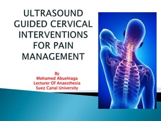
- 1. By Mohamed Abuelnaga Lecturer Of Anaesthesia Suez Canal University
- 2. Why ultrasound ? Ultrasound limitations ? Evidence Based cervical Interventions ? What will we see by ultrasound ?
- 3. 1. Radiation-free imaging. 2. Short procedure time compared to fluoroscopy or CT and the need to insert only 1 needle. 3. Ability to identify and avoid vessels in the trajectory of the needle. 4. Dynamic imaging.
- 4. Not seeing a small blood vessel does not necessarily mean it does not exist. It could be either the limitation of the sonographic resolution or the limitation of the operator’s experience. Also, visualizing such small nerves (cervical medial branches and third occipital nerve) can be very challenging and requires special training and experience. Fluoroscopically guided C7 medial branch blocks are challenges, and the sonographically guided technique is no exception.
- 12. Cervical Facet Joint Injections Cervical Medial Branch Block Identifying Cervical Spine Level &cervical nerve roots
- 15. Posterior Approach. Why? 1. Multilevel injections can be performed with the same view and may even use a single needle entry point. 2. Bilateral injections can be performed without the need to change position. 3. The needle is inserted in plane from a caudal to cranial direction
- 16. Sagittal (longitudinal) sonogram at the level of the articular pillars showing the hyperechoic articular processes of the facet joints as the saw sign and the anechoic facet joint space in between.
- 19. Short-axis (transverse) sonogram at the C6 level
- 20. Short-axis (transverse) sonogram at the C7 level
- 21. Short-axis (transverse) sonogram at the C5 level
- 22. 1-Ultrasound guided pain management interventions is good alternative to both fluoroscopy and CT guidance 2-Transforaminal Cervical nerve root injection may be fatal so it is not recommended for management of cervical radicular pain 3- Non-particulate steroid injection has fewer complications when compared to particulate steroids