
Recommended
More Related Content
What's hot
What's hot (20)
Similar to Insect Wing Venation, Wing coupling apparatus: its structure, function, modification in different order of insect
Similar to Insect Wing Venation, Wing coupling apparatus: its structure, function, modification in different order of insect (20)
More from N.m.c.a
More from N.m.c.a (11)
Recently uploaded
Recently uploaded (20)
Insect Wing Venation, Wing coupling apparatus: its structure, function, modification in different order of insect
- 1. ASSIGNMENT ON Insect Wing Venation, Wing coupling apparatus: its structure, function, modification in different order of insect
- 2. Introduction Among invertebrates, insects are the only winged organism which makes them dominant in animal kingdom. Insects are the only invertebrates that can fly. They have two pairs of wings located on the meso and meta-thoracic segments (jointly known as pterothorax). They always possess three pairs of legs, but do not always possess two pairs of wings e.g., silverfish and springtail have no wings; while, male mealybug have wings and hind pair of wings is reduced or modified. Based on presence of wings, insects are divided into two main categories viz., Apterygota and Pterygota. In Apterygota, wings are absent. e.g., four primitive orders namely Thysanura, Protura, Diplura and Collembola. All winged insects come under Pterygota. In some insects, the wings are highly modified and the number of wings varies from two pairs to none. The veins are the most characteristic structures of the wing, appearing as longitudinal stripes of cells that differentiate darkly pigmented cuticle and are more packed than intervein cells.
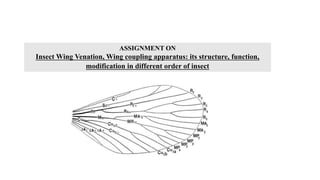
- 3. Wing Venation Some lines run from base of the wing toward the apex these are called veins. The complete system of veins of wings is known as "venation or neuration". Venation is the name given to the arrangement (number and position) of veins within an insect's wing. Most insect groups have lost, or dramatically reduced the number of, these cross veins. Fig 1: Insect wing venation patterns in different insect orders
- 4. Structure of Wing vein Each of the wings consists of a thin membrane supported by a system of veins. The membrane is formed by two layers of integument closely apposed, while the veins are formed where the two layers remain separate; sometimes the lower cuticle is thicker and more heavily sclerotized under a vein. Within each of the major veins there is a nerve and a trachea, and, since the cavities of the veins are connected with the hemocoel, hemolymph can flow into the wings.
- 5. Types of wing veins There are two major types of veins found in insect wings. 1. Longitudinal veins: Runs lengthwise 2. Cross veins: Runs across 1. Longitudinal wing veins/Hypothetical primitive type Costa (C): The vein along the leading edge of the wing, unbranched and convex (+). Sub-costa (SC): The second longitudinal vein, rarely branched and concave (-). Radius (R): The third longitudinal vein, 5- branched, main stem convex (+), divided into 2 main branches viz., R1, which passes directly to wing margin (+) and Rs which is concave and divide into four veins (R2 To R5). Media (M): The fourth longitudinal vein, divided into 2 main branches viz., anterior (MA) and posterior media (MP). MA is convex (+), divided into two branches viz., MA1, and MA2 . MP is concave (-), divided into four branches (MP1, MP2, MP3, and MP4). Cubitus (Cu): The fifth longitudinal vein, divided into 2 main branches viz., first cubitus, Cu1 (+) and second cubitus, Cu₂ (-). Cu1 is subdivided into anterior (Cu1a) and posterior (Cu1b) cubitus. Anal (A): The remaining veins between the cubitus and the trailing edge of the wing are termed as anal veins. There are mainly three anal veins viz. A1, (+), A2(-) and A3(+).
- 6. 2. Cross wing veins The cross veins connect the longitudinal veins and are typically named for the two veins, which they connect: c-sc: Cross vein connecting the costa and subcostal. Radial (r): Cross vein connecting adjacent branches of the radius. r-s: Cross vein connecting the subcosta and radius. Radio-medial (r-m): Cross vein connecting the radius and media. Medial (m): Cross vein connecting adjacent branches of the media. Medio- cubital (m-cu): Cross vein connecting the media and cubitus. Cubito-anal (cu-a): Cross vein connecting the cubitus and the first anal vein. Certain additional cross veins have special names: Humeral (h): Cross vein connects the costa and subcosta near the base of the wing. Sectorial (s): Cross vein that either connects the radius (R1) to the branch R4 to R5 or connects R3 TO R4.
- 7. 1. Odonata (Dragonflies) The two pairs of wings are almost identical in the Zygoptera, but in Anisoptera the hind wings are broader basally and there are minor venational differences. The pterostigma, a thickening of the wing-membrane between C and R, is a very characteristic feature. Costa (C) – at the leading edge of the wing, strong and marginal, extends to the apex of the wing. Subcosta (Sc) – second longitudinal vein, it is unbranched, joins C at nodus. Radius and Media (R+M) – third and fourth longitudinal vein, the strongest vein on the wing, with branches, R1-R4, reach the wing margin, the media anterior (MA) are also reach the wing margin. IR2 and IR3 are intercalary veins behind R2 and R3 respectively. Cubitus (Cu) – fifth longitudinal vein, cubitus posterior (CuP) is unbranched and reach the wing margin. Anal veins (A1) – unbranched veins behind the cubitus. The main veins and the cross veins form the wing venation pattern. The venation patterns are different in different species. There may be very numerous cross veins or rather few. Fig.2: Wing venation of Dragonfly Wing venation of different insect orders
- 8. 2. Orthoptera (Grasshoppers, Locusts, Crickets) The degree of development of the wings varies considerably; in some species both sexes may include normal and brachypterous forms while in others the short-winged condition is characteristic of the female. Costa (C) – at the leading marginal of the forewing and hindwing, unbranched. Subcosta (Sc) – second longitudinal vein, unbranched. Radius (R) – third longitudinal vein, branched to Rs in forewing and hindwing. Media anterior (MA) – fourth longitudinal vein, branched in basal part as Media posterior (MP). Cubitus (Cu) – fifth longitudinal vein, on forewing and hindwing dividing near the wing base into branched CuA, and unbranched CuP. Anal veins (A) – veins behind the cubitus, unbranched, two in forewing, many in hindwing. Fig.3: Wing-venation of Locusta migratoria
- 9. 3. Ephemeroptera (Mayflies) The fluting, which strengthens the wing in flight, is associated with the presence of characteristic intercalary veins, which are not often absent in the Ephemeroptera. The intercalaries appear to be branches which have lost their basal connections with the remaining veins but are united with the latter by a greatly developed system of cross-veins. Its shows the primitive feature of a media retaining its anterior (MA) and posterior (M) divisions in both pairs of wings. Rs is attached basally to MA. Fig.4: Fore wing of Chironeles albimanicalus
- 10. 4. Dictyoptera (Cockroaches and Mantids) The two pairs of wings differ markedly. The front pair, known as tegmina, are rather strongly sclerotized and serve mainly to protect the membranous hind wings. Costa (C) - at the leading edge of the wing. Subcosta (Sc) - second longitudinal vein, it is relatively short. Radius (R) - third longitudinal vein, with many pectinate branches. Media (M) - fourth longitudinal vein, reach the wing margin. Cubitus anterior (CuA) - fifth longitudinal vein, with dichotomous branches occupy large part of tegmen. Cubitus posterior (CuP) - is unbranched, curved and reach the wing margin. Anal veins (A) - veins- behind the cubitus. The veins of hind wing are about the same as front wing but with large anal lobe folded at rest between CuP and 1A. The anal lobe usually folded in a fan-like manner. Fig.5: Wing-venation of Blattella
- 11. 5. Hemiptera (Plant bugs & Cicada ) A. Among Heteroptera there is a marked difference in the consistency of the two pairs of wings. The fore wings are termed hemelytra (hemi-elytra). The hemelytra exhibit much diversity of structure and have been used taxonomically. Costa (C) – at the leading wing marginal, in forewing extends to the node and lies close to Sc+R. Subcosta + Radius (Sc+R) – in forewing Sc and R fused together to the node. Radial sector (Rs) arises near the node and unbranched. Radius anterior (RA) Radius posterior (RP) Media (M) – branches to M1 to M4. Cubitus anterior (CuA) – branches to CuA1 and CuA2. Cubitus posterior (CuP) – unbranched. Anal veins (A) – veins behind the cubitus, 1A and 2A fused in the forewing, CuP and 2A are folded. Fig.7: Wing-venation of Cicada Fig.6: Wing-venation of Plant Bug B. Among the Homoptera the fore wings are of uniform texture. Although there is great diversity of venation which is dealt with under the different families, the occurrence of fossil forms and the nymphal tracheation have made it possible to determine the homologies of the wing-veins.
- 12. 6. Neuroptera (Lace wing, Ant lions, Alder flies) Suborder I. Planipennia (Lace wing, Ant lions) The venation is characterized by Rs arising from the main stem separately from RI which does not fuse distally with Sc and by the formation of a so-called pseudomedia and pseudocubitus. These are highly complex veins, the first being formed by the fusion of M1+2, M3 +4 and portions of the four proximal branches of Rs. The pseudocubitus is formed by Cu, by the distal portion of M1+2 and M3+4 and by part of the three proximal branches of Rs. Suborder II. Megaloptera (Alder Flies, Snake Flies) They differ from other Neuroptera in the hind-wings being broad at their bases with the anal area folded fanwise when at rest. Fig.8: Wing-venation of Chrysopa quadripunctata Fig.9: Wing-venation of Sialis lutaria
- 13. 7. Lepidoptera (Butterflies and Moths) Costa (C) – not found in Butterflies. Subcosta (Sc) + Radius 1 (Sc+R1) – at the leading wing marginal, fused or very close for most of the length, in hindwing fused and well developed in the humeral area, subcosta never branches in butterfly. Radius (R2-R5) – radius divides into branches beyond the middle of the wing up to five branches in Papilionidae. On forewing, the last R is stalked in all butterflies except Hesperiidae is separated. Radius sector (Rs) –Present in hindwing. Media (M1-M3) – the basal section has been lost. Cubitus anterior (CuA1-CuA2) – CuP section has been lost. Anal veins (A, 1A+2A, 3A) – either one vein A, or two veins 1A+2A, 3A. Humeral vein – The hindwing of most butterflies has the humeral vein, except Lycaenidae There is the enlargement of the humeral area of the hindwing. Fig.10: Wing-venation of Ditrysia.
- 14. 8. Hymenoptera (Ants, Bees, Wasps, Sawflies) Costa (C) – not found in Hymenoptera. Subcosta (Sc) – unbranched. Radius (R) – branched to R1-R5. Media (M) – M is unbranched, in forewing M is fused with Rs for part of its length. Cubitus (CuA) – unbranched, CuP is absent in Hymenoptera. Anal veins (A) – only two anal veins 1A and 2A are present, 2A is not distinctive in some species. Line of wing folding – Some species, including Vespidae, the forewing are longitudinally folded along the 'line of wing folding' at rest. Pterostigma – is present for some species. Fig.11: Wing-venation of Honey bee
- 15. 8. Coleoptera (Beetles) The elytra are the highly modified mesothoracic wings and arise simultaneously with the hind wings: they develop in an exactly similar manner during the greater part of the larval life. Costa (C), Subcosta posterior (ScP) - at the leading wing marginal, fused for most of the length. Radius anterior (RA) - divided into two branches beyond the middle of the wing. Radius posterior (RP) - basal connection is lost. Media posterior (MP) - branches, long and strong vein. Cubitus anterior (CuA) Anal veins (AA, AP) - veins behind the cubitus, separated by anal fold. Three general types are recognizable: A. The Adephagid type: All the principal veins remain more or less completely developed and are usually joined by a greater number of cross-veins than occur in other Coleoptera. M1 is connected with M2 by means of one or two transverse veins: when two are present an oblong cell, the oblongum, is formed which is very characteristic of the type. B. The Staphylinid type: Here the chief characteristic is exhibited in the disappearance of all the cross-veins, and the atrophy of the proximal portion of M, the remainder of that vein being isolated in the apical portion of the wing. C. The Cantharid Type. A B Fig.12: Wing-venation of Beetles
- 16. 9. Diptera (Two-Winged or True Flies) Costa (C) – not found in Diptera. Subcosta (Sc) – became the leading wing vein, unbranched. Radius (R) – branched to R1-R5. Media (M) – branched to M1-M4. Cubitus anterior (CuA)- unbranched, CuP is reduced in Diptera. Some species CuA and 1A are separated, some species meets when reaching the wing margin, some species fused. Anal veins (A) – only two anal veins 1A and 2A are present, 2A is not distinctive in some species. Fig.13: Wing-venation of Brachycera. A.Geosargus Fig.14: Wing-venation of Calliphora erythrocephala 10. Isoptera (Termites or White Ants) There is a striking absence of regular cross-veins though in more primitive genera the membrane is stiffened by an irregular sclerotized network between the main veins. There is no true costal vein: Sc is 2-branched, and R1- 5 are recognizable as separate branches. Both M and Cu are well developed, Cu2 forms a curved veins dividens and there are no distinct anal veins. In the hind wing Sc is unbranched and M arises from the stem of R4+5. Three anal veins are present and support a well-developed anal lobe which recalls the Blattarian hind wing Fig.15: Wing-venation of Termite
- 17. Function Of Wing Venation The venation patterns in wings are important for the functioning of the wings, as well as for the classification of insects. The veins are extensions of the body’s circulatory system. Veins give structural support to the wing membrane. Veins not only provide mechanical integrity but also act as conduits, containing tracheal tubes for gas exchange, nerves that provide sensory information in flight, and perhaps most importantly, hemolymph that serves multiple functions. Hemolymph actively circulates through wing veins and is critical for maintaining sensory structures that populate the wing. In membranous wings, the veins provide strength and reinforcement during flight. Wing shape, texture, and venation are quite distinctive among the insect taxa and therefore highly useful as aides for identification.
- 18. Wing Coupling For taking flight, insect need to keep both the fore and hindwings together as a single unit. The structures in the form of lobes, bristles, hairs or spines that help the wings to be together are known as wing coupling organs. The fore and hind-wings are coupled together (2 wings of each side) which brings more synchronized action and improves the aerodynamic efficiency of flight. In many insects, special structures of wing coupling mechanism are found. These structures overlap during flight and simple at repose. These structures are: 1. Jugal lobe (Retinaculum): Present on posterior margin of fore wing which ensure fly simply before wing overlapping. 2. Humeral lobe (Frenulum): Present of anterior margin of hind wings. In Primitive pterygota - fore wings and hind wings Moved independently of each other like Isoptera and Odonata. After course of evaluation Specialization set in and two wings began to work together after development of some structures.
- 19. Wing Coupling of different insect orders 1. Mecoptera Types of coupling: Primitive/ Mecopteran type wing coupling Both jugal and humeral lobes possess bristle. During flight, jugal bristle lies on top of hind wings while humeral bristle forms frenulum which presses the underside of forewing. As a result, greater part of inner margin of forewing overlaps the hind wings e.g., Mecoptera (Scorpionfly)
- 20. 2. Hymenoptera Type of coupling: Hamulate type wing coupling A row of small hooks is present on the coastal margin of the hind wing which is known as “hamuli”. Which lock onto the forewing, keeping them held together e.g., Hymenoptera (honey bees). 3.Hemiptera Type of coupling: Fold and hook type wing coupling Wings are held together in flight by various small hooks or folds along the wing margin e.g., Aphid 4. Lepidoptera I. Type of coupling: Amplexiform type wing coupling It is the simplest form of wing coupling a linking structure is absent. Wings are coupled simply by overlapping basally e.g., butterflies (Papilionidae), Silk moths (Bombycidae).
- 21. II. Type of coupling: Frenate type wing coupling (Male) Found in male butterfly where frenulam bristles are fused together and formed single stout structure. This structure is held by curved process from sub costal vein of forewing Retinaculum works as cuticular clasp. e.g., fruit sucking moth (Erebidae). III. Type of coupling: Frenate type wing coupling (Female) This type of wing coupling is found in female butterfly. consists of a group of stout bristles (2 - 20 bristles) lying beneath the extended forewing where it engages in a retinaculam formed from a path of hairs near cubitus (cubital vein). e.g., Cabbage butterfly (Pieridae) 5. Trichoptera and some Lepidopteran Type of coupling: Jugate type wing coupling Jugal area is produced into lobe like fibula or more elongated jugam, which lies on top of hind wings during flight and folded beneath forewings at rest. Frenular bristles wanting or small e.g., Trichoptera and some lepidoptera (Hepialid moth).