04 Bone non neoplastic part-2
•Download as PPT, PDF•
0 likes•136 views

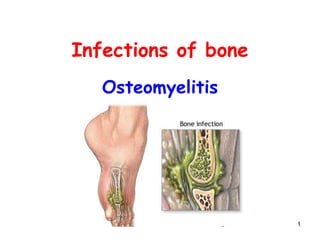
Refers to inflammation of bone and bone marrow. Most commonly occurs due to infections by: Pyogenic bacteria Pyogenic Osteomyelitis

Recommended
More Related Content
What's hot
What's hot (20)
Similar to 04 Bone non neoplastic part-2
Similar to 04 Bone non neoplastic part-2 (20)
More from med_students0
More from med_students0 (20)
Recently uploaded
Recently uploaded (20)
04 Bone non neoplastic part-2
- 2. 2 Osteomyelitis • Refers to inflammation of bone and bone marrow. • Most commonly occurs due to infections by: – Pyogenic bacteria • Pyogenic Osteomyelitis
- 3. 3 Pyogenic Osteomyelitis • Usually occurs in children and young adults • Due to pyogenic infection of bone by: – Staphylococcus aureus* (most common) – Escherichia coli & Streptococci - neonates – Salmonella (common in sickle cell disease) – Pseudomonas : common in intravenous drug abusers, diabetics and puncture of foot through rubber footwear.
- 4. 4 How do these organisms reach the bone? • Three routes 1. Hematogenous spread (most common) • Seeding of bone after bacteremia 2. Direct inoculation 3. Spread from an adjacent site of infection. • Bones affected: – Infant and children :(long bones)-tibia, femur, humerus – Adults: (small bones) - vertebrae, pelvic bones • Region of the bone affected – metaphysis (most vascular part of bone)
- 6. 6
- 7. 7
- 8. 8 Blood vessel Organism from Septic focus = boil Metaphysis of long bone Acute inflammation Formation of PUS Subperiosteal abscess Soft tissue Surface Sinus tract Periosteal elevation + Septic thrombosis
- 9. 9 Periosteal elevation + Septic thrombosis Stoppage of blood supply + Enzymatic destruction Involucrum New bone formation Sequestrum Death of part of bone
- 10. 10 Morphology • Depends upon the stage of Osteomyelitis • Acute Osteomyelitis: – Acute inflammation: cell death and pus formation. – Pus may reach the Periosteum subperiosteal abscess. – Rupture of Periosteum abscess in surrounding soft tissue drain to surface via a draining sinus tract. – Neutrophils enzymatically destroy the bone. • Devitalized bone is called sequestrum.
- 11. 11
- 12. 12 Morphology • Chronic disease: – Is characterized by replacement of acute inflammatory cells by chronic inflammatory cells. – A sleeve of new bone formation may surround the infected necrotic area • This reactive new bone is k/a involucrum.
- 14. 14 Clinical findings • Rapid onset with C/O feeling ill. • Most frequent manifestation: – Fever and severe pain over the affected area. – reluctance to use affected extremities. • On examination: – Localized area of tenderness – Erythema and – Swelling.
- 15. 15 • Investigations: – Leukocytosis – Raised ESR • X ray – Early stage (10 days) - may be normal. – Slow periosteal elevation – Lytic focus of bone destruction with surrounding sclerosis. • Radionuclide bone scan: – Best for detection ( even in early cases) – Localized increased uptake of traces • Needle aspiration • Blood cultures • Bone biopsy and culture
- 16. 16
- 17. 17 Sequestration of bone In area of destruction
- 19. 19 Draining sinus Squamous cell carcinoma
- 20. 20 Complications • Most important – Draining Sinus tract to the skin surface • Danger of Squamous cell carcinoma developing at orifice of sinus tract. – Extension of infection to adjacent joint pyogenic arthritis. – Septicemia and infective endocarditis – Chronic Osteomyelitis • Others – Fractures – Retardation of growth from damage to epiphyseal cartilage. – Amyloidosis – Osteogenic sarcoma (rare).
- 21. 21 Tuberculous Osteomyelitis • Occurs secondary to tuberculous infection located elsewhere. – Active tuberculosis of lung. – TB of GIT and lymphnodes • Characteristically occurs in: – Vertebrae (Pott’s spine or disease)
- 22. 22 Pott’s disease • Infection begins at the anterior margin of vertebral body near inter-vertebral disc. • Complete destruction of inter-vertebral disc with partial destruction of two adjacent vertebrae. • Collapse of vertebral bodies anteriorly.
- 23. 23
- 24. 24 • Clinical findings: – Back pain – Stiffness, – Deformity (kyphosis) , – Neurological abnormalities (Pott’s paraplegia) – Fever, night sweats and weight loss. • Pathology: – Granulomas with caseous necrosis on histology.
- 25. 25 Spinal TB - Potts Disease
- 27. 27 Langhans Giant cell Caseous necrosis
- 28. 28 Osteomyelitis: Note • MC organism : Staphylococcus aureus • In patient’s with: – Sickle cell anemia Salmonella paratyphi* – Genitourinary tract infections • E coli, Pseudomonas, Klebsiella – IVDA pseudomonas. – Dog and cat bites Pasteurella multocida (a gram negative rod) – Nail puncture through rubber footwear pseudomonas*. – AIDS: ??**
- 29. 29 Fracture (#) of bones
- 30. 30 Fracture (#) • Is a complete break in the continuity of bone or – it may be an incomplete break or crack. • CLASSIFICATION: – Closed and open fractures • Closed or simple fracture: no communication between site of fracture and exterior of body. • Open or compound fracture: direct communication between the skin surface and the fracture site through the skin wound.
- 31. 31 Closed # Open #
- 32. 32 Open or compound fracture
- 33. 33 Classification: According to etiology • Fractures caused solely by sudden injury – Most common ; Caused by : – Direct violence, Indirect violence • Fatigue or stress fracture: – Due to oft-repeated stress – Risk group: • Athletes, New military recruits • Bone : metatarsal • Pathological fracture – Fracture through a bone already weakened by disease. – Cause of # :Trivial trauma
- 34. 34 Patterns of fracture 1. Transverse fracture 2. Oblique fracture 3. Spiral fracture 4. Comminuted fracture (more than two fragments) – The bone is broken into several pieces 5. Greenstick fracture (incomplete break) – The bone is cracked, but not broken into two pieces. "Incomplete" fracture. 6. Compression or crush fracture
- 35. 35
- 36. 36 Healing of fractures • Process divided into 4 stages 1. Hematoma formation and Inflammation 2. Formation of soft callus or procallus 3. Formation of Hard callus or bony callus 4. Bone Remodeling
- 37. 37 Stage 1 – Hematoma & Inflammation • Fracture causes hemorrhage and tissue destruction ; blood clot (hematoma) forms (hours). • Swelling and inflammation around the fracture site.
- 38. 38 Stage 2- Soft Callus • Fibrin mesh of hematoma: Acts as a framework. – inflammatory cells (neutrophils and macrophages) move in ; phagocytize debris. – Fibroblasts and capillary grow into blood clot forming granulation tissue. • Cartilage is formed ( from primitive mesenchymal cells) • Granulation tissue + cartilage = soft tissue callus or procallus (provisional callus) • Soft callus bridges the fractured bone.
- 39. 39 Stage 3 - Hard Callus • Osteoblasts proliferate • Form new bone • New bone is mineralized • Soft callus converted into bony callus (or osseus callus.) • This spans across the fracture and fills in the space between broken bone ends. • The fracture is now stable.
- 40. 40 • Portions of callus that are not under physical stress are resorbed. • continual reduction in the size of callus • Restoration of marrow cavity. • Strengthening of bone along the lines of stress. Stage 4 - Bone Remodeling
- 41. 41
- 42. 42 Factors which affect # healing • Age • Type of fracture (Simple Vs. Compound/ comminuted/spiral etc.) • Favorable factors: – Good blood supply – Immobility • Factors that hinder union: – Impaired blood supply – Movement between the fragments – Infection – Interposition of soft tissue – Systemic disorders : DM , vitamin deficiency
- 43. 43 Fracture healing: Complications – delayed union – nonunion – pseudoarthrosis (false joint)
- 44. 44 Important fractures • Femoral neck #: – Bleeds into capsule – Compromises Medial femoral circumflex artery avascular necrosis of femoral head. • Scaphoid bone #: – MC # of carpal bones – Susceptible to avascular necrosis and nonunion • Colles’ #: – Person falls on outstretched hand – # distal end of radius dinner fork deformity • Supracondylar #: – Distal # of humerus – Compromises brachial artery with danger of Volkmann’s ischemic contracture of forearm muscles
- 46. 46 Miscellaneous bone disorders Myositis ossificans
- 47. 47 Myositis ossificans • Is characterized by development of bony tissue in: – Skeletal muscle or soft tissue – Following a blunt trauma. • Favored Locations: – Quadriceps or brachialis muscle • Pathogenesis: – Soft tissue trauma hematoma formation bone formation. • Differential diagnosis: – Bone sarcomas ( osteosarcoma)