Cerebral angiography - medical information
•Download as PPTX, PDF•
2 likes•2,360 views

Cerebral angiography is an X-ray examination method with the help of which the state of blood vessels, their functional abilities, roundabout blood circulation, the presence of deviations and the size of pathological zones are determined. i hope this will be useful among the medical professionals. please comment thank u

Recommended
More Related Content
What's hot
What's hot (20)
Similar to Cerebral angiography - medical information
Similar to Cerebral angiography - medical information (20)
More from martinshaji
More from martinshaji (20)
Recently uploaded
Recently uploaded (20)
Cerebral angiography - medical information
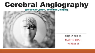
- 1. Cerebral Angiography(procedure ,price , definition ,images) PRESENTED BY MARTIN SHAJI PHARM D
- 2. Cerebral angiography is an X-ray examination method with the help of which the state of blood vessels, their functional abilities, roundabout blood circulation, the presence of deviations and the size of pathological zones are determined. Introduction
- 3. Angiography: Basic information Modern angiography permits detecting vascular diseases at an early stage of their development, figuring out the state of the lymphatic system, and also identifying pathologies of inner organs. Often, diagnostics are carried out to detect formed atherosclerotic deposits on the walls of blood vessels. Angiography may be very indicative when inspecting the heart muscle, detecting neoplasms, cysts, assessing the functional characteristics of the kidneys.
- 4. Brain angiography is used to detect arterio venous shunts, irregular dilatations of the vascular walls, and the presence of blood clots in the brain. The method can be used to detect diseases of the retina. It can be indispensable earlier than open-heart or brain surgical procedure to find out the affected person's situation.
- 5. Where to do a cerebral angiography? Angiography of the vessels of the brain or other areas could be performed in clinics and diagnostic centers with X-ray equipment. Sterility is maintained in the angiography rooms and modern devices are used. Among them: 1) high-speed fluorographic camera;
- 6. 2) angiograph - special X-ray equipment for diagnosing the vascular system; 3) equipment for video recording and X-ray multi shooting. In modern medical centers with a good supply, there are CT angiography machines, that are distinguished by increased visualization of high quality and higher picture detail.
- 8. Find out the Angiography / - procedure. The process is carried out by intravenous injection of a contrast agent into the body earlier than starting the diagnosis. The place where the contrast is entered is decided according to the area to be rendered. To make sure the accuracy of the examination, you need to stop eating 6 hours earlier than the examination. If the puncture will be performed in the area of the groin fold or axillary region, hygiene of those areas should be performed and the hair should be shaved off.
- 9. Angiography is performed utilizing local anesthesia in combination with common sedative and antihistamines. This allows you to prevent the development of an allergic response and reduce the affected person's experience. The place for the injection of the distinction agent is treated with an antiseptic. After that, the physician makes a small incision in the skin, opens access to the artery, so that the needle is inserted as easily as possible. Then a short, hollow tube called an introducer is inserted. To prevent vascular spasm and reduce the irritating impact of the contrast agent on the walls, a solution of novocaine is injected into the vessel.
- 11. A thin flexible tube 1–2 mm in diameter (catheter) is passed into the introducer. The physician slowly advances the catheter to the start of the visualized vessel, controlling the process due to the monitor of the X-ray machine. Contrast is introduced and a collection of pictures is taken (sometimes this process is carried out several times). For higher visibility, the affected person could also be requested to change position.
- 12. The action of the contrast agent is sometimes accompanied by short-term complications: dizziness, facial flushing, headache, heart palpitations, a sense of heat, painful sensations within the chest, metallic taste within the mouth, nausea. Cerebrovascular angiography should be performed with specific care and accuracy. After examination, the catheter is removed and the bleeding is stopped with a sterile pressure bandage. It is left for a day. To prevent blood clot formation, it is strongly recommended to stay in bed for 6-10 hours.
- 13. Circle of Willis
- 14. Angiography of cerebral vessels Brain angiography is performed by inserting a catheter into the ulnar, right femoral, brachial, carotid, or subclavian arteries. The catheter is advanced to the branch of the desired vessel and contrast is injected into its lumen. After the initial dose of contrast agent is injected, the head pictures are taken in numerous projections (side and front). Re-introduction of contrast and re-shooting enable assessing the situation of distant parts of the vascular system.
- 15. Angiography of cerebral vessels doesn't cause pain and damage to the vascular walls. Brain angiography is prescribed when the clinical image indicates critical pathologies. These include narrowing or aneurysms, hematomas or neoplasms in the brain, vital impairment of blood circulation, and the presence of a blood clot. Angiography of the vessels of the brain allows you to establish an accurate diagnosis and determine the need for an operation.
- 16. Angiography: price For angiography of blood vessels, the price primarily is dependent upon the standing of the medical institution and the country in which you decide to be examined. To find out how much angiography costs, you can use information from the web sites of clinics or the assistance of operators.
- 17. The cost of angiography is determined by the size and location of the area of interest. Simultaneous examination of the situation of arteries and veins is more expensive. And if angiography of vessels is complemented by magnetic resonance imaging, then the price also will increase.
- 18. Thank you…