AN OUTLINE ON HERPESVIRAL DISEASES IN MARINE TURTLES

Abstract: Marine turtles are calm, long-living reptiles with complicated lifestyle. They have their attendance all over the globe. The survival of turtles became so pathetic by various means of human development exercises. Now, on the other hand, emerging diseases came to the platform. Disease indications are alarming high in the coastal belts where human agitation is heavier. Herpesviridae family members are the top most pathogenic agents that are disturbing the survival of these long-living animals. They are a total of five herpesviruses that are associated with diseases in turtles; in them, Fibropapillomatosis is the catastrophic disease, which is characterized by tumors over smooth surfaces of the body. Disease had turned to be an upcoming invader for the maritime turtle’s community. The establishment and potential of this disease is under the hands of environmental factors. The longevity of marine turtles, coupled with their close association with inshore habitats and seagrass meadows and coral reefs in these habitats, has led to the proposal that they may act as sentinel indicators of marine ecosystem health.

Recommended
More Related Content
What's hot
What's hot (20)
Similar to AN OUTLINE ON HERPESVIRAL DISEASES IN MARINE TURTLES
Similar to AN OUTLINE ON HERPESVIRAL DISEASES IN MARINE TURTLES (20)
Recently uploaded
Recently uploaded (20)
AN OUTLINE ON HERPESVIRAL DISEASES IN MARINE TURTLES
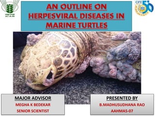
- 1. MAJOR ADVISOR MEGHA K BEDEKAR SENIOR SCIENTIST PRESENTED BY B.MADHUSUDHANA RAO AAHMA5-07
- 2. INTRODUCTION • Order : Testudines • Family : Cheloniidae • Poikilotherms • 110 million years, since the time of the dinosaurs.
- 3. Turtles are exposed to a no. of threats • Degradation, urbanisation & pollution of nesting habitats & foraging areas. • Traditional hunting and egg harvest • The impacts of climate change on the marine and terrestrial environment.
- 4. “Cayman Turtle Farm” (CTF) • Grand Cayman island, West Indies in 1968 with 23 acres. • Opens the door for – exchange of views on the pros and cons of sea turtle farming. High demand for food & medicinal purposes.
- 5. HERPESVIRUSES Generally characteristic features of HV: Variable host range, Short replication cycle, and • Linear, ds DNA genome with Icosahedral symmetry • Replication is within the nucleus.
- 7. HERPESVIRUSES • Greek word herpein , referring to the LATENT. • Establish latent infection. During which viral gene expression is minimized • Expression of these latency- associated genes may function to keep the viral genome from being digested by cellular ribozymes or being found out by the immune system.
- 8. HERPESVIRUSES ChHV 1 (Grey Patch Disease) ChHV 2 ChHV 3 ChHV 4 ChHV 5 (Fibropapillomatosis) ChHV 6 (Lung-Eye- and Tracheal disease) HV affecting F.W Turtles
- 9. Strains of Chelonid Herpesvirus 5 FIBROPAPILLO MATOSIS A B D C Genome -----23 kb Atlantic ( Florida & Barbados) Middle Pacific (Hawaii) Eastern Pacific. Western Pacific (Australia)
- 10. FIBROPAPILLOMATOSIS (FP) • FP - 1st reported in 1938 at Key West, Florida. • V. limited focus is done on diseases of sea turtles. But greatest concern - Green turtles as it has only reached a panzootic status in this species (Williams et al., 1994). FP is a neoplastic disease characterized by cutaneous, ocular tumours that has been documented to infect all sea turtle species.
- 11. AETIOLOGY • Research to date suggests that FP is associated with a Herpesvirus infection (Herbst et al., 1995; Quackenbush et al., 1998, 2001; Lackovich et al., 1999). This virus cannot be cultured in vitro and therefore Koch's postulates have not been fulfilled.
- 12. AETIOLOGY • Agent : Chelonid Herpesvirus 5 (ChHV5) or "Green turtle fibropapillomatosis” (GTFP) because it was first recorded in Green turtles. Family : Herpesviridae Subfamily : Alphaherpesvirinae Genus : Scutavirus (ICTV, 2011).
- 13. 1938 (1.5%) Disease prevalence and impact FP emerge in the Eastern Pacific, Hawaiian Islands, Indonesia and Australia (40%). PANZOOTIC Over 140 countries (Groombridge and Luxmoore, 1989). 50% of the population of green turtles in the world are affected by the disease (Balazs 1991, Herbst 1994). 1980s (20-60%) 1990s
- 14. Epidemiology • FP typically occurs in marine turtles inhabiting neritic areas – most frequently observed in juvenile turtles. • Horizontal. • The higher incidence in females may be – behavioral ecology in life cycle. • FP rates are correl- ated with degraded habitat quality.
- 15. Epidemiology • HV have the potential to be maintained on various surfaces in marine environment (sediments, rubbing rocks, foods sources, etc.) that could serve as vectors for transmission. Chemical pollution can activate latent viruses or indirectly increase its virulence (Aguirre et al., 1994). Most prominent in warmer climates, affecting 50%- 70% of popn.
- 16. Epidemiology • FPHV DNA was detected on the gills of wrasse cleaner fish (Thalassoma duperrey) – It groom sea turtles - possible vector for transmission (Lu et al. 2000b).
- 17. • Epidemiological links are seen between FP rates, nitrogen footprints, and invasive macroalgae – ARGININE. • Water temperature - lesions of a debilitating size by autumn. • Susceptible stages : Juveniles 40 - 70 cms length/ (10– 30 kg) most effected. • FP is rare (0-12%) among nesting adult females and lesions tend to be focal and mild. Epidemiology
- 18. Epidemiology PROTEIN KINASE C (PKC) ACTIVATORS OKADAIC ACID LYNGBYATOXIN A DEBROMOAPLY SIATOXIN (DAT) Prorocentrum lima Lyngbya majuscula Epidemiology
- 19. Note the Fibropapilloma with highly rugose structure (black arrow) and presence of leeches - Ozobranchus (white arrow). Photo credit: Tim Collins.
- 20. MULTIFACTORIAL AETIOLOGY U.V LIGHT (Aguirre, 1991), CONTAMINATS (Aguirre et al. 1994a) METALS (Monserrat et al., 2007) TEMP (Herbst et al., 1995) BIOTOXINS (ARGININE) (Landsburg et al. 1999). PARASITES (Dailey & Morris, 1995)
- 21. CLINICAL SIGNS Lesions that are comprised primarily of proliferating epidermis with little or no underlying dermal involvement are properly called PAPILLOMAS. Those masses in which both tissues are hyperplastic are termed FIBROPAPILLOMAS. While those lesions predominantly comprised of proliferating dermal components with relatively normal epidermis are called FIBROMAS.
- 22. Severe fibropapillomas in the cervical, periocular, and right front limb regions of one green turtle
- 23. The conjunctiva (especially bulbar conjunctiva) is a common site for GTFP, and masses may grow over the cornea.
- 24. STAGES OF LESION DEVELOPMENT (Herbst, 1994; Kang et al., 2008). • Papilloma lesions EARLY PHASE • Fibropapill -omas lesions INTERMEDIATE PHASE • Fibromas lesions CHRONIC PHASE Internal - mouth, heart, lungs, kidneys, liver and gastrointestinal tract (Mascarenhas and Iverson, 2008). Occasional visceral Fibromas, Myxofibromas and Fibrosarcomas
- 25. HISTOLOGICAL STUDIES • Histological studies on FP lesions have observed orthokeratotic hyperkeratosis (a)and varying degrees of epidermal hyperplasia (b) (upto 30 cells thick), and papillary projections (c)
- 26. Key features in FP is proliferative epidermis presenting ballooning cells with intracellular inclusion bodies (IIBs) suggestive of viral infection, shown with arrows. HISTOLOGICAL STUDIES
- 27. VIRULENT FACTORS • Four members of the C-type lectin-like domain superfamily (F-lec1, F-lec2, F-sial and F-M04) were found to be present in the ChHV5 genome. Two of these has been suggested that these genes may play a role in FP pathogenesis & lesions formation (Ackermann et al., 2012).
- 29. GRAY PATCH DISEASE • Tissues affected – SKIN, SHELL. • Occurs in two forms Non spreading Papules/ pustular like lesions resolve spontaneously. Benign, a necrotizing dermatitis of post-hatching green turtles 1st Spreading Gray patches to large areas of the epidermal surface. Malignent, often these animals die 2nd
- 30. Clinical symptoms • Necrotizing dermatitis of post - hatching. • Epizootics of small, circular papular skin lesions that coalesced into patches were described in young sea turtles. • Stressful environmental conditions such as over crowding and higher temp. seemed to enhance the onset and severity of lesions.
- 31. Proliferative dermatitis lesions on subadult loggerhead turtle (A) Early skin lesions with typical raised white appearance. (C) Resolving lesions on scales of front flipper (B) Advanced skin lesions showing area of discoloration during healing.
- 32. • The first critical time - 2-6 weeks of age. • At this age – no typical skin lesions of gray-patch disease; however, there is a consistent level of mortality (20%). Affects 90-100% hatchlings Clinical symptoms The only obvious clinical sign is impacted fecal material in the lower intestinal tract.
- 33. • The second critical - 8 to 15 weeks of age. • Mortality is less compare to the first stroke. Clinical symptoms • Turtles over I year of age do not have the natural lesions of gray-patch disease. • Infection is in higher degree in summer. • Inactivated vaccine – skin scraping. • Transmission - horizontal This is the period of time that we are able to obtain large amounts of skin scrapings containing the herpesvirus.
- 35. LUNG-EYE-TRACHEAL DISEASE(LETD) • Agent : (LETV) or (Chelonid herpesvirus 6) • Name implies, the virus affects the eyes and respiratory tract. • And has a clinical course of 2-3 weeks. Characterized by conjunctivitis, pharyngitis, tracheitis, periglottal necrosis and pneumonia (Jacobson et al. 1986).
- 36. EPIZOOTOLOGY • Transmission is thought to be vertical or water- borne. Affects turtles worldwide, and mortality in post- hatchling and juvenile - can reach 70%.
- 37. MODEL CHELONID HERPES VIRUS • The virus was cultured in green turtle kidney cells that helped in further research. • LETV - gathers information about the general characteristics of this largely unexplored group of viruses. • In addition, the prevalence of LETV infections in wild populations.
- 38. LOGGERHEAD GENITAL- RESPIRATORY HERPESVIRUS” (LGRV) Ulcers in the trachea, around the cloaca and on the base of the phallus. Venereal transmission. LOGGERHEAD OROCUTANEOUS HERPESVIRUS” (LOCV) Ulcers in the oral cavity & cutaneous plaques that can be covered with exudate & have an erythematous border. ------ Both viruses were thought to be opportunistic in debilitated turtles.
- 39. CONCLUSION • The longevity of marine turtles, coupled with their close association with inshore habitats and seagrass meadows and coral reefs in these habitats, has led to the proposal that they may act as sentinel indicators of marine ecosystem health. • Further investigations has to be carried to rule out several confusions about remaining viruses which will in turn inform the management and conservation of a vulnerable species.
- 40. REFERENCE • Orós, J., Torrent, A., Calabuig, P. and Déniz, S., 2005. Diseases and causes of mortality among sea turtles stranded in the Canary Islands, Spain (1998– 2001). Diseases of aquatic organisms, 63(1), pp.13-24. • EAZWV Transmissible Disease Fact Sheet Sheet No. 23 1 FIBROPAPILLOMATOSIS (FP) OF SEA TURTLES Rachel E. Marschang 2009 • EAZWV Transmissible Disease Fact Sheet Sheet No. 26 GPD • Select conditions in sea turtle pathology ;Judy St. Leger, DVM, DACVP • Haines, H.G., Rywlin, A. and Rebell, G., 1974, March. A herpesvirus disease of farmed green turtles (Chelonia mydas). In Proceedings of the annual meeting‐World Mariculture Society (Vol. 5, No. 1‐4, pp. 183-195). Blackwell Publishing Ltd. • Alfaro-Núñez, A., Bertelsen, M.F., Bojesen, A.M., Rasmussen, I., Zepeda-Mendoza, L., Olsen, M.T. and Gilbert, M.T.P., 2014. Global distribution of Chelonid fibropapilloma-associated herpesvirus among clinically healthy sea turtles. BMC evolutionary biology, 14(1), p.1. • Jones, A.G., 2004. Sea turtles: old viruses and new tricks. Current biology, 14(19), pp.R842-R843. • Stacy, B.A., Wellehan, J.F., Foley, A.M., Coberley, S.S., Herbst, L.H., Manire, C.A., Garner, M.M., Brookins, M.D., Childress, A.L. and Jacobson, E.R., 2008. Two herpesviruses associated with disease in wild Atlantic loggerhead sea turtles (Caretta caretta). Veterinary microbiology, 126(1), pp.63-73.
- 41. • Jacobson, E.R., 1996. Guest editorial: marine turtle farming and health issues. Marine Turtle Newsletter, 72, pp.13-15. • Herbst, L., Ene, A., Su, M., Desalle, R. and Lenz, J., 2004. Tumor outbreaks in marine turtles are not due to recent herpesvirus mutations. Current Biology, 14(17), pp.R697-R699. • Greenblatt, R.J., Work, T.M., Balazs, G.H., Sutton, C.A., Casey, R.N. and Casey, J.W., 2004. The Ozobranchus leech is a candidate mechanical vector for the fibropapilloma-associated turtle herpesvirus found latently infecting skin tumors on Hawaiian green turtles (Chelonia mydas). Virology, 321(1), pp.101-110. • Arthur, K., Limpus, C., Balazs, G., Capper, A., Udy, J., Shaw, G., Keuper-Bennett, U. and Bennett, P., 2008. The exposure of green turtles (Chelonia mydas) to tumour promoting compounds produced by the cyanobacterium Lyngbya majuscula and their potential role in the aetiology of fibropapillomatosis. Harmful Algae, 7(1), pp.114-125. • Sounguet, A.G., Villarubia, A., Balazs, G.H. and Spraker, T.R., 2007. Fibropapillomatosis confirmed in Chelonia mydas in the Gulf of Guinea, West Africa. Marine Turtle Newsletter, (116), p.20. REFERENCE
- 42. • Manire, C.A., Stacy, B.A., Kinsel, M.J., Daniel, H.T., Anderson, E.T. and Wellehan, J.F., 2008. Proliferative dermatitis in a loggerhead turtle, Caretta caretta, and a green turtle, Chelonia mydas, associated with novel papillomaviruses. Veterinary microbiology, 130(3), pp.227-237. • Herbst, L.H., Jacobson, E.R., Klein, P.A., Balazs, G.H., Moretti, R., Brown, T. and Sundberg, J.P., 1999. Comparative pathology and pathogenesis of spontaneous and experimentally induced fibropapillomas of green turtles (Chelonia mydas). Veterinary Pathology Online, 36(6), pp.551-564. • Coberley, S.S., Herbst, L.H., Brown, D.R., Ehrhart, L.M., Bagley, D.A., Schaf, S.A., Moretti, R.H., Jacobson, E.R. and Klein, P.A., 2001. Detection of antibodies to a disease-associated herpesvirus of the green turtle, Chelonia mydas. Journal of clinical microbiology, 39(10), pp.3572-3577. • Curry, S.S., Brown, D.R., Gaskin, J.M., Jacobson, E.R., Ehrhart, L.M., Blahak, S., Herbst, L.H. and Klein, P.A., 2000. Persistent infectivity of a disease-associated herpesvirus in green turtles after exposure to seawater. Journal of Wildlife Diseases, 36(4), pp.792-797. REFERENCE
- 43. • Duarte, A., Faísca, P., Loureiro, N.S., Rosado, R., Gil, S., Pereira, N. and Tavares, L., 2012. First histological and virological report of fibropapilloma associated with herpesvirus in Chelonia mydas at Príncipe Island, West Africa. Archives of virology, 157(6), pp.1155-1159. • Reséndiz, E., Flores-Ramírez, S., Koch, V. and Cordero-Tapia, A., 2016. First Record of Fibropapillomatosis in a Green Turtle Chelonia mydas from the Baja California Peninsula. Journal of Aquatic Animal Health, 28(4), pp.252-257. • Warwick, C., Arena, P.C. and Steedman, C., 2013. Health implications associated with exposure to farmed and wild sea turtles. JRSM short reports, 4(1), p.8. • EAZWV transmissible disease fact sheet sheet no. 37 Loggerhead genital- respiratory herpesvirus (lgrv) • Monezi, T.A., Mehnert, D.U., de Moura, E.M., Müller, N.M., Garrafa, P., Matushima, E.R., Werneck, M.R. and Borella, M.I., 2016. Chelonid herpesvirus 5 in secretions and tumor tissues from green turtles (Chelonia mydas) from Southeastern Brazil: A ten-year study. Veterinary microbiology, 186, pp.150-156. • Reckseit, S.M., 2016. Volunteer Programs in the Conservation of Sea Turtles in Costa Rica (Doctoral dissertation, Texas A&M University). REFERENCE
- 44. • Jones, K., Ariel, E., Burgess, G. and Read, M., 2016. A review of fibropapillomatosis in green turtles (Chelonia mydas). The Veterinary Journal, 212, pp.48-57. • da Silva, C.C., Klein, R.D., Barcarolli, I.F. and Bianchini, A., 2016. Metal contamination as a possible etiology of fibropapillomatosis in juvenile female green sea turtles Chelonia mydas from the southern Atlantic Ocean. Aquatic Toxicology, 170, pp.42- 51. • Bunkley-Williams, L., Williams Jr, E.H., Horrocks, J.A., Horta, H.C., Mignucci-Giannoni, A.A. and Poponi, A.C., 2008. New leeches and diseases for the hawksbill sea turtle and the West Indies. Comparative Parasitology, 75(2), pp.263-270. • Jacobson, E.R., BUERGETT, C., Williams, B. and Harris, R.K., 1991. Herpesvirus in cutaneous fibropapillomas of the green turtle Chelonia mydas. Diseases of Aquatic Organisms, 12(1), pp.1-6. • Haines, H. and Kleese, W.C., 1977. Effect of water temperature on a herpesvirus infection of sea turtles. Infection and immunity, 15(3), pp.756-759. • Rodenbusch, C.R., Almeida, L.L., Marks, F.S., Ataíde, M.W., Alievi, M.M., Tavares, M., Pereira, R.A. and Canal, C.W., 2012. Detection and characterization of fibropapilloma associated herpesvirus of marine turtles in Rio Grande do Sul, Brazil. Pesquisa Veterinária Brasileira, 32(11), pp.1179-1183. REFERENCE
Editor's Notes
- found today have been around for
- Ingestion of marine debris. Nest and hatchling depredation by wild, feral and domestic animals, boat strike, and entanglement in fishing nets and lines.
- Severely debilitating disease
- disease has a worldwide, circumtropical distribution in florida, mexico, costa rica, australia. Perhaps due to limited funding or human resources, FP has gone unnoticed in other parts of the world until current times.
- These new recruits may be exposed to several stressors associated with migration, adaptation to a new environment, and changes in population density, diet and pathogen exposure, which may all combine to reduce the efficacy of the immune system and make these immature and juveniles more susceptible to infection.
- There appears to be a link between the development of FP and the immune system. Immunosuppression is strongly correlated with FP in captive green turtles
- Arginine is a regulator of immune activity & promote herpesviruses and contribute to tumour formation
- Okadaic acid is a known tumor promotor and is produced by the Prorocentrum dinoflagellate that grows on sea grasses. Similarly, lyngbyatoxin A & debromoaplysiatoxin (DAT) are produced by Lyngbya majuscula (filamentous cyanobacterium found in marine and estuarine environments). act as protein kinase C (PKC) activators and are potent tumour promoters
- Persistent chlorinated compounds, such as polychlorinated biphenyls(PCBs) and chlorinated pesticides,
- interfere with the hydrodynamics and locomotion.
- These three proliferative lesions are the hallmarks of the disease referred to as green turtle fibropapillomatosis (GTFP).
- hypertrophy of the stratum corneum of the skin No nucleus is seen in the cells.
- Conjunctivitis, also known as pink eye, is inflammation of the outermost layer of the white part of the eye and the inner surface of the eyelid.
- In addition, this research has provided the means to assess the prevalence of LETV infections in wild populations and the threat such infections may present for marine turtle health and conservation via serodiagnosis.