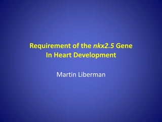
2. Introduction
Background
• Genetic disorders are caused by abnormalities in an
organism’s genome.
• Over 20% of infant mortality is caused by genetic and
birth defects (Hoffman et al.).
• Affecting over 40,000 newborns per year in the
United States, congenital heart defects are the most
common genetic defects (CHD; 2014).
• Congenital heart defects can present in many forms
including, atrial and ventricular septal defect (ASD
and VSD), aortic and pulmonary valve stenosis (AVS
and PVS), single ventricle defects, and many others.
3. Introduction (cont.)
Literature Review
• When the formation of the heart in a developing fetus is disrupted
it results in congenital heart disease.
• Mutations in NKX2.5 result in CHD in humans. (Benson et al., 1999).
• In order to fully understand how mutations in nkx2.5 lead to cardiac
malformations, it was essential to discover the roles this
transcription factor plays during cardiogenesis (Targoff et al., 2008).
• nkx2.5 is essential in limiting atrial cell number, promoting
ventricular cell number, and preserving chamber-specific identity in
zebrafish (Targoff et al., 2013).
• Loss of nkx2.5 leads to substantial impairment in proliferation of
cardiomyocyte precursors in zebrafish (Prall et al., 2007).
• To analyze the early and late functions of nkx2.5, I evaluated the
influence of timing of its expression on cardiac chamber formation,
through the use of an nkx2.5 transgene.
4. Methodology
• Transgene, Tg(hsp70l:nkx2.5-EGFP) expressed nkx2.5 tagged to Green
Fluorescent Protein (GFP)
• All embryos were taken out of their protective shell before each
experiment.
• The in situ hybridization method allowed for the visualization of
transcribed genes through the use of probes specific to their particular
mRNA molecules
• The immunofluorescent staining method enabled the study of translated
genes by using antibodies for specific protein molecules
• After the experimentation was complete, the fish samples were imaged
• Genotyping was performed immediately after each method and embryos
were classified based on their phenotype and genotype.
5. In situ and Immunofluorescent images of transgenic
and non-transgenic zebrafish embryos.
Results
6. Results (cont.)
Non-transgenic phenotypes post in situ and immunofluorescence.
The images above illustrate the imaged embryos after in situ and
immunofluorescence procedures. The nkx2.5 deficient embryos (D-F) show a bulbous
atrium and a shrunken ventricle.
7. Results (cont.)
Transgenic phenotypes post in situ and immunofluorescence.
The phenotypes of nkx2.5 deficient embryos with activated transgene
(M-R) show the embryos rescued by the transgene. These embryos’ hearts
are similar to that of wild type embryos (G-L).
8. Results (cont.)
Transgenic and non-transgenic
phenotypes.
This image shows the comparison
of the phenotypes of transgenic
(B, D, G, H), non-transgenic (A, C,
E, F), wild type (A, B, E, G) and
nkx2.5 deficient (C, D, F, H)
embryos. The graphs show the
percent of embryos with wild type
and nkx2.5 phenotypes per heat
shock group. During 7 somites
(11hpf) to 26hpf, the percent of
visible mutant phenotype was
diminished.
9. Discussion
• The discoveries demonstrate that nkx2.5 expression is
only essential for about 15 hours during embryogenesis
for normal heart development.
• The transgene is only effective for a period of eight
hours and would have to be activated at a specific
moment in development for the heart to express nkx2.5,
and be able to rescue an nkx2.5 deficient embryo.
• The results show that the time period in which the
transgene is most effective for rescuing nkx2.5 deficient
embryos is within 7 somites (11 hpf) and 26hpf.
• The mutant fish did not express the gene during that
time were not able to survive to adulthood.
10. Discussion (cont.)
Limitations:
• The results demonstrated that transgenic nkx2.5
mutant embryos were able to survive to
adulthood, however, the morphology of the
rescued nkx2.5 mutants has yet to be fully
analyzed. It is unknown if the transgenic mutant
hearts are impaired during the adult stages of
development.
• The research was limited to analysis of nkx2.5
gene function only during embryonic heart
development.
11. Conclusion
• The results illustrate the specific time interval during
which nkx2.5 is essential and most effective.
• These findings can help to further advancements in
treating congenital heart defects brought about
through mutations in NKX2.5 for humans.
• The discovery of the effectiveness of the transgene
can contribute to future research and provide a more
precise way of analyzing the effects of nkx2.5 at
more specific time periods in development.
13. References
1) Benson, D., et al., 1999. Mutations In The Cardiac Transcription Factor NKX2.5 Affect Diverse Cardiac Developmental Pathways.
Journal of Clinical Investigation. 1567-573.
2) Boston Children’s Hospital. 2014. Congenital Heart Defects in Children. http://www.childrenshospital.org/health-
topics/conditions/congenital-heart-defects
3) Buckingham, M., et al., 2005. Building the mammalian heart from two sources of myocardial cells. Nat. Rev. Genet. 6, 826–835.
4) Centers for Disease Control and Prevention. 2014. Congenital Heart Defects.
http://www.cdc.gov/ncbddd/heartdefects/index.html
5) Hoffman JL, Kaplan S. The incidence of congenital heart disease. J Am Coll Cardiol. 2002;39(12):1890-1900.
6) Inoue, D., & Wittbrodt, J. 2011, May 13. One for All-A Highly Efficient and Versatile Method for Fluorescent Immunostaining in
Fish Embryos. PLoS ONE, 6(5), 1-7. doi:10.1371/journal.pone.0019713
7) Prall, O.W., et al., 2007. An Nkx2-5/Bmp2/Smad1 negative feedback loop controls heart progenitor specification and
proliferation. Cell 128, 947–959.
8) Targoff, K. L., Colombo, S., George, V., Schell, T., Kim, S. H., Solnica-Krezel, L. and Yelon, D. (2013). Nkx genes are essential for
maintenance of ventricular identity. Development 140, 4203–4213.
9) Targoff, K.L., et al. (2008). Nkx genes regulate heart tube extension and exert differential effects on ventricular and atrial cell
number, Dev. Biol. doi:10.1016/j.ydbio.2008.07.037
10) Thisse, C., & Thisse, B. (2007, December 20). High-resolution in situ hybridization to whole-mount zebrafish embryos. Nature
Protocols, 3(1), 59-69. doi:10.1038/nprot.2007.514
11) Tu, Shu, and Neil C. Chi. (2012) "Zebrafish Models in Cardiac Development and Congenital Heart Birth Defects."
Differentiation: 4-16.
We used zebrafish emryos because their heart characteristics and development are similar to humans and easy to visualize because of their transparency.
Find out RNA and protein used????
The color embryos under UV light determined if it expressed the gene.
Figure 1: In situ and Immunofluorescent images of transgenic and non-transgenic zebrafish embryos.
In-situ (B-E) and Imuunoflourescent (F-I) images of zebrafish embryos with and with out the heat shock nkx2.5 transgene (A) at 24 somites, 30 somites, and 28hpf. Embryos were heat shocked at 22 somites. The transgenic embryos show the presence of GFP within eight hours of heat shock.