Cell cycle presentation by Salman Ul Islam.
•Download as PPTX, PDF•
6 likes•2,707 views


Recommended
More Related Content
What's hot
What's hot (20)
Viewers also liked
Viewers also liked (18)
Similar to Cell cycle presentation by Salman Ul Islam.
Similar to Cell cycle presentation by Salman Ul Islam. (20)
More from Salman Ul Islam
Recently uploaded
Recently uploaded (20)
Cell cycle presentation by Salman Ul Islam.
- 1. REGULATION OF CELL CYCLE SALMAN UL ISLAM, (MS), CELLULAR BIOCHEMISTRY LAB. 2013298039
- 2. CONTENTS Introduction Overview of the cell cycle Cell-cycle control system Summary
- 3. INTRODUCTION “Where a cell arises, there must be a previous cell, just as animals can only arise from animals and plants from plants. A cell reproduces by carrying out an orderly sequence of events in which it duplicates its contents and then divides in two. This cycle of duplication and division, known as the cell cycle, is the essential mechanism by which all living things reproduce.
- 4. How is cell division and growth regulated? Growth factors -- stimulate cell growth Mitogens -- trigger cell division Survival signals -- disable apoptotic mechanisms Cell Cycle Regulation
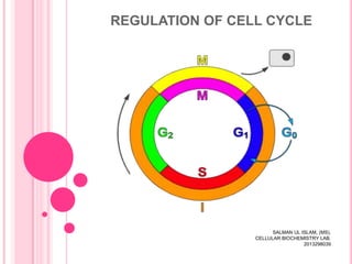
- 5. OVERVIEW OF THE CELL CYCLE The eucaryotic cell cycle is traditionally divided into four phases: M phase: constituted by mitosis (nucleus division) and cytokinesis (cell splits in two). The period between one M phase and the next is called interphase. The interphase encompasses the remaining three phases of the cell cycle. S phase ( S=synthesis): the cell replicates its nuclear DNA. S phase is flanked by two phases in which the cell continues to grow. G 1 phase (G= gap): it is the interval between the completion of M phase and the beginning of S phase (DNA synthesis). G2 phase: interval between the end of S phase and the beginning of M phase.
- 6. Essential Cell Biology (© Garland Science 2010)
- 7. CHECKPOINTS IN CELL-CYCLE REGULATION Two important checkpoints occur in G1 and G2. The G1 checkpoint allows the cell to confirm that the environment is favorable for cell proliferation and its DNA is intact before committing to S phase. G2 checkpoint ensures that cells do not enter mitosis until damaged DNA is repaired and DNA replication is complete.
- 8. Essential Cell Biology (© Garland Science 2010)
- 9. CELL-CYCLE CONTROL SYSTEM DEPENDS ON CYCLICALLY ACTIVATED PROTEIN KINASES Key proteins are activated and then inactivated that regulate DNA replication, mitosis, and cytokinesis. Phosphorylation followed by dephosphorylation is one of the most common ways to switch the activity of proteins on and then off. Protein kinases are activated at appropriate times in the cycle, after which they quickly become deactivated again. Switching these kinases on and off is done by cyclins, so kinases are therefore known as cyclindependent protein kinases or Cdks.
- 10. Essential Cell Biology (© Garland Science 2010)
- 11. CDKS ACTIVITY IS ALSO REGULATED BY PHOSPHORYLATION AND DEPHOSPHORYLATION For M-Cdk to be maximally active, it has to phosphorylated at one or more sites by a specific protein kinase, and dephophorylated at other sites by specific protein phosphatase. The removal of the inhibitory phosphate groups by the phosphatase is the final step that activates the M-Cdk at the end of interphase. Then M-Cdk complex can activate more of the same complexes. This positive feedback produces the sudden, explosive increase in M-Cdk activity that drives the cell abruptly into M phase.
- 12. Essential Cell Biology (© Garland Science 2010)
- 13. Essential Cell Biology (© Garland Science 2010)
- 14. CDKS ARE REGULATED BY ACCUMULATION AND DESTRUCTION OF CYCLINS Cyclin concentration plays an important part in timing the events of cell cycle. M cyclin= cyclin that helps drive cells into M phase. M-cyclin synthesis starts immediately after cell division and continues steadily throughout interphase. The cyclin concentrates, so that its concentration rises gradually and helps time the onset of mitosis; its rapid elimination then helps initiate the exit from mitosis.
- 15. Essential Cell Biology (© Garland Science 2010)
- 16. Essential Cell Biology (© Garland Science 2010)
- 17. CDKS ARE REGULATED BY ACCUMULATION AND DESTRUCTION OF CYCLINS (CONT…) As mitosis nears completion, multiple molecules of the protein ubiquitin are covalently attached to the M-cyclin by anaphase promoting complex (APC). This ubiquitination marks the cyclin for degradation in proteosomes, large proteolytic machines found in all eucaryotic cells. Destruction of the cyclin inactivates the Cdk.
- 19. DIFFERENT CYCLIN-CDK COMPLEXES Essential Cell Biology (© Garland Science 2010)
- 20. Distinct Cdks associate with different cyclins to trigger the different events of the cell cycle.
- 21. S-PHASE CYCLIN-CDK COMPLEXES INITIATE DNA REPLICATION ONCE PER CELL CYCLE Initiates DNA replication and helps block Rereplication. DNA replication begins at origins of replication. Origin recognition complex (ORC) remains bound to the origin of replication; serves as a sort of landing for other regulatory proteins. Cdc6 binds to ORC in G1, promotes additional proteins binding to form pre-replicative complex, making the replication origin ready to “fire”. S-Cdk then pulls the “trigger” initiating DNA replication. S-Cdk helps phophorylate Cdc6, causing it and the other proteins in the pre-replicative complex to dissociate from the ORC after an origin has fired.
- 22. Essential Cell Biology (© Garland Science 2010)
- 23. CELL CYCLE ARREST The cell-cycle control system can arrest the cycle at specific checkpoints. The molecular mechanisms of these are poorly understood. In some cases, however, Cdk inhibitor proteins come into play. For example DNA damage causes increased concentration and activity of p53 (gene regulatory protein) which activates the transcription of a gene encoding a Cdk inhibitor protein called p21. The p21 protein binds to G1/S-Cdk, preventing them from driving the cell into S phase.
- 24. Essential Cell Biology (© Garland Science 2010)
- 25. CELLS CAN DISMANTLE THEIR CONTROL SYSTEM AND WITHDRAW FROM THE CELL CYCLE This is a different matter from pausing in the middle of a cycle. In human body, e.g, nerve cells and skeletal muscle cells persist for a lifetime without dividing, they enter G0. G0 is modified G1 state in which the cell-cycle control system is largely dismantled, in that many of the Cdks and cyclins disappear. The G1 checkpoint is therefore sometimes called Start, passing it represents a commitment to complete a full dividion cycle.
- 26. Essential Cell Biology (© Garland Science 2010)
- 27. Essential Cell Biology (© Garland Science 2010)
- 28. Eucaryotic Cell-Cycle Times • Cell type • Early frog embryo cells • Yeast cells • Intestinal epithelial cells • cultured fibroblasts • Human liver cells Cell cycle time ~30 minutes 1.5–3 hours ~12 hours ~20 hours ~1 year
- 29. SUMMARY The control system depends on a set of proteins kinases, each composed of Cyclin and Cdk. The control system also depends on protein complexes such as APC. The Cdks are cyclically activated by both cyclin binding and the phosphorylation of some amino acids and the dephosphorylation of others; when activated, Cdks phophorylate key proteins in the cell. The cell cycle control system can halt the cycle at specific check points to ensure that the next step in the cycle does not begin before the previous one has finished, and intracellular and extracellular conditions are favorable.