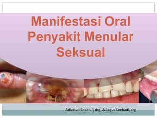
2. Latar Belakang, Tujuan ,
Manfaat
Lesi ulser oral sering dijumpai
Penyebab ulser : faktor lokal, infeksi, penyakit sistemik
Gejala & tanda klinis dari suatu kelainan hampir sama
Meningkatkan ketelitian dalam pemeriksaan subyektif,
obyektif lesi ulser oral melalui pemeriksaan visual
Meningkatkan dalam membedakan lesi ulser oral berdasar
gejala & tanda klinis
Identifikasi ulser oral dapat menentukan pemriksaan
pendukung
Holistic Approach : pendekatan terhadap suatu
masalah/gejala dengan memandang bahwa masalah/gejala
yang ditemukan sebagai suatu kesatuan yang utuh
5. 5
ETIOLOGY AND
PREDISPOSING FACTORS
Caused by Treponema pallidum
through contact with primer
lesion
Transmitted by sexual activity
with manifestation on glans penis,
vulva, vagina, and cervix
Transmitted by intimate contact
with manifestation on lips,
tongue, or finger
SYPHILIS
7. PRIMARY SYPHILIS
(STAGE I OF SYPHILIS)
Ulseration and
erithematous lession on
buccal mucossa
(Neville, 2016)
7
8. PRIMARY SYPHILIS
(STAGE I OF SYPHILIS)
Lesions are seen on the lips, oral mucosa, lateral of the tongue,
soft palate, tonsils, pharynx and gingiva
Intraoral lesions are coated by a grayish-white coating and
cause pain. Tonsillar involvement with edema, redness,
accompanied by ulceration and erosional lesions
(Ghom, 2014)
8
10. SECONDARY SYPHILIS
(STAGE II OF SYPHILIS)
Sign of mucus patch
Lesions are usually found on the tongue, buccal mucosa,
tonsillar, pharyngeal, and lips
Lesions are covered by a grayish-white membrane. The
presence of trauma results in bleeding in the lesion.
Usually accompanied by pain in the lesion.
(Ghom, 2014)
10
12. TERTIER SYPHILIS
(STAGE III OF SYPHILIS)
Gumma can occur in all parts of the oral cavity, but most often
on the palate, and tongue
Lesions of the gingiva can manifest as deep solitary lesions,
accompanied by ulceration of the mucos
characterized by the perforation of the palate.
13. DIAGNOSIS
SUBJECTIVE EXAMINATION
The history of syphilis begins with the chronology of
the patient's complaints. Syphilis develops in one third
of untreated patients. Patients are usually infected by
other people through social (rare) and sexual contact,
especially in the first year (primary and secondary
syphilis). Exploring the social life history of the patient
is very important.
(Janier, 2014)
14. DIAGNOSIS
OBJECTIVE EXAMINATION
The clinical presentation of syphilis is very diverse and
occurs within a few decades after the initial infection.
Syphilis, if left untreated, can go through four stages:
primary, secondary, latent and late. (Kinghorn, 2016)
15. CONGENITALSYPHILIS
Hutchinson’s Triad (Interstitial keratitis, malformed teeth
Hutchinson incisors and mulberry molars, and eighth nerve
deafness).
There may also be a deformity on the nose known as saddle
nose deformity
destruction of the palate, and development of the nasal septum
(Ghom, 2014)
16. CONGENITAL SYPHILIS
Chronic fluid, and
lesions in the corner of
the mouth
Hutchinson’s incisors
Mulberry Molar
17. SUPPORTIVE
EXAMINATION
Dark Field Examination Microscopy
used to identify Spirochete in stage I and II syphilis. Cannot be used for oral lesions.
Biopsy
useful when the lesion contains various microorganisms, in the case of stage III syphilis.
Treponemal Antigen Test
to evaluate the success of syphilis therapy.
Non Treponema Antigen Test
used on Veneral Disease Research Laboratory (VDRL), and Rapid Plasma Reagin (RPR).
(Ghom,2014)
20. DIFFERENTIAL DIAGNOSIS
Primary Syphilis
Herpes simplex is easily distinguished from syphilis because in
herpes simplex there are multiple lesions, and vesicles (Bill and
Dirk, 2016)
Infeksius Noninfeksius
Herpes simplex
Chancroid
Granuloma inguinal
Vaccinia
Limfogranuloma venereum
Ulkus aphthous
Vulvitis atau balanitis candida erosif
Erosi akibat trauma atau ulkus
Penyakit Behcet
Karsinoma sel skuamosa
Karsinoma sel basal
Fixed drug eruption
21. DIFFERENTIAL DIAGNOSIS
Secondary Syphilis
In stage II syphilis the differential diagnosis is candidiasis, leukoplakia, hairy
leukoplakia, lichen planus, herpetic gingivostomatitis, erythema multiforme,
tuberculosis. (Greenberg, et al. 2014)
22. DIFFERENTIAL DIGNOSIS
Tertiary Syphilis
Palatal perforation due to syphytic gumma may be
confused by clinical features of granulomatosis,
midline lethal granuloma, malignant reticulosis,
mucormycosis, and antral carcinoma. (Silverman,2011)
23. DIFFERENTIAL DIGNOSIS
Congenital Syphilis
Some conditions that may be diagnosed differentially
with congenital syphilis lesions such as traumatic ulcer,
apthous ulcer, herpetic ulcer, candidiasis, and
mononuclear infection. (Delong, L., 2008)
24. COMPREHENSIVE
TREATMENT
benzathine penicillin IM, tetracycline hydrochloride 500 mg orally 4 times a
day for 15 days. Patients who are allergic to the drug penicillin, can be given
erythromycin 500 mg orally 4 times a day for 15 days. Treponema pallidum can
disappear 24 hours after treatment begins.
Follow-up on the patient is a physical examination, and repetition of the VDRL
test at month 1, 3, 6, 9, 12, 18, and 24. At the end of the 24th month if the
VDRL test results are negative , the patient is cured from syphilis.
sexual education of patients, not changing sexual partners, and not having
sexual relations with people who have been infected. Local antibiotic use in
pregnant women suspected of being infected. Prevention of congenital syphilis
can be done by examining pregnant women for antenatal and postnatal
examinations.
(Ghom, 2014)
25. PROGNOSIS
the discovery of penicillin, the prognosis for syphilis
becomes better. Microbiologic healing (all T.
pallidum dies) is not possible. Cured syphilis means
clinically cured for life, not transmitted to others
(Bill and Dirk, 2016)
27. Definition
Gonorrhoea is sexually transmitted infection caused
by Neiserria gonorrhoea
N gonorrhoea is a gram negative aerob bacteria
Gonorrhoea in Mouth is oropharyngeal gonorrhoea
29. ETHIOLOGY AND PREDISPOSING FACTOR
Ethiology of Oropharyngeal gonorrhoeae was
Neisseria gonorrhoea
Predisposing factor :
Oral sex with person infected of gonorrhoea
Men sex Men
31. DIAGNOSIS AND SUPPORTIVE
EXAMINATION
Oropharyngeal gonorrhoea is rare in symtom
Symptom could be shown in 7 – 21 days after contact
Intra oral lesion show redness on throat and tongue,
inflammation.
Supportive Examination :
Kultur
(nucleic acid amplification testing (NAAT)
38. APA ITU?
APA BEDANYA?
BAGAIMANA PENULARANNYA?
BAGAIMANA PENGOBATANNYA?
BAGAIMANA PENCEGAHANNYA?
APA KAITANNYA DENGAN KITA,
PARA DOKTER GIGI?
Infeksi HIV dan AIDS
39. Why so destructive ?
Sel target untuk inangnya = CD4+
Komponen sistem imun
CD4+ ↘ ↘ ↘
Sistem imun ↘ ↘ ↘
Infeksi oportunistik ↗↗↗
†
46. Dapatkah disembuhkan ?
Dapat diobati
Highly Active Anti Retroviral Therapy
(HAART)
Menggunakan 1 macam ARV atau kombinasi
beberapa jenis mampu menekan HIV sehingga
tidak bereplikasi
Orang dengan HIV dan AIDS (ODHA) harus
didukung untuk tertib minum obat dan kontrol
Kualitas hidup ↗ usia harapan hidup ↗
50. WHO ( 2003 )
Salah satu Program Kesehatan Mulut :
Mengutamakan Pencegahan efektif
dari Manifestasi Rongga Mulut
HIV/AIDS
melalui IDENTIFIKASI lesi Mulut
51. Radithia, D., Soebadi, B., Hendarti, HT., Triyono, EA. 2009
Common Dental-
Related Complaints
Adult
Childre
n
Dental caries 80% 100%
Dental & root decay 97% -
M3 impaction 26% -
Marginal gingivitis 68% 80%
52. Bagaimana cara mengenali
ODHA di antara pasien yang
datang
ke praktek kita?
Manifestasi HIV/AIDS di rongga mulut
Kelainan khas terkait infeksi HIV dan AIDS
Penampilan fisik
Keadaan umum
Penampilan & gaya hidup berisiko
Anamnesis
Screening
58. Oral Hairy
Leukoplakia
•Lesi putih
berombak atau
berambut pada
lateral lidah
•Tidak dapat
dikerok ,
asimtomatik.
•Terasosiasi
infeksi virus
Epstein-Barr
•sering
ditumpangi
candida
59. Linear Gingival Erythema
•pita merah
•OH tidak selalu buruk
•tanpa ulserasi, tanpa poket
•asimtomatik, kadang berdarah
•Kemungkinan subgingival candida infection