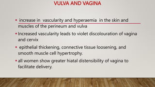
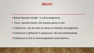
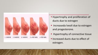
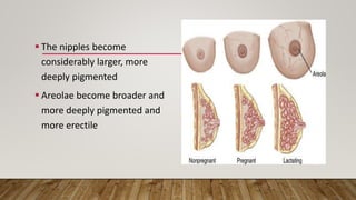
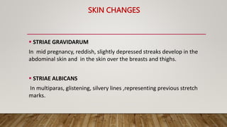
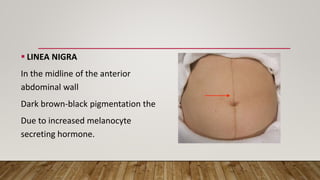
2. INTRODUCTION
Physiological changes begin soon after fertilisation and continues throughout pregnancy.
Following changes will be discussed:
Reproductive tract
Breast
Skin
Metabolic changes
3. VULVA AND VAGINA
increase in vascularity and hyperaemia in the skin and
muscles of the perineum and vulva
Increased vascularity leads to violet discolouration of vagina
and cervix
epithelial thickening, connective tissue loosening, and
smooth muscle cell hypertrophy.
all women show greater hiatal distensibility of vagina to
facilitate delivery.
4. • Vaginal ph decreases during pregnancy (~3.5)
• due to increase doderlline bacteria more glycogen is
converted into lactic acid .
5. CERVIX
About 1 month after
conception, the cervix begins to
soften and had bluish tone.
increase vascularity edema and
collagen change
hypertrophy and hyperplasia of
the cervical glands
6. Rearrangement of collagen-rich tissue aids the cervix in retention
of the pregnancy and dilatation.
Endocervical mucosal cells produce copious amounts of tenacious
mucus
It obstruct the cervical canal to protect the uterine contents
against infection
At labour onset, , this mucus plug is expelled, resulting in a bloody
show
CERVIX
7. SIGNS IN PREGNANCY
Chadwick’s Sign
A dark blue to purple –red congested
appearance of vaginal and cervical
mucosa
Hegar’s Sign
Occurs in first trimester
Softening of uterus at isthmus
Goodell’s sign
Softening of uterus after 6 weeks
8.
9. CERVICAL RIPENING
connective tissue remodelling
lowers collagen and proteoglycan concentrations
raises water content
result of progesterone
10. UTERUS
UTERINE ENLARGEMENT
stretching ,hypertrophy and hyperplasia of muscle cells
stimulated by estrogen and progesterone
it occurs maximum in fundus
Fibrous tissue accumulates
• MYOMETRIUM
muscle layer of uterus thicken and strengthen during first half of pregnancy
Gradually becomes thin in second half.
By term, the myometrium is only 1 to 2cm thin.
12. • By the end of 12 weeks, the enlarged
uterus extends out of the pelvis.
• It gradually increases in size reaching
xiphi-sternum at 38 weeks
• The longitudinal axis of the uterus
corresponds to the pelvic inlet axis.
13. BRAXTON HICKS CONTRACTIONS
In second trimester, can be
detected by bimanual examination
unpredictable and sporadic and
usually nonrhythmic
may cause some discomfort and
account for false labour.
Intrauterine pressure : 5-25mm of
Hg.
14.
15. UTEROPLACENTAL BLOOD FLOW
• Placental perfusion depends on total uterine
blood flow (uterine, ovarian and collateral)
• Uteroplacental blood flow increase
progressively
• Approx. 450 mL/min in the mid trimester
• Nearly 500 to 750 mL/min at 36 weeks.
• Fall in vascular resistance accelerates the
flow velocity
• Shear stress in vessels lead to
circumferential vessel growth.
• Nitric oxide—a potent vasodilator has role
16. OVARIES AND FALLOPIAN TUBE
Ovulation ceases during pregnancy
maturation of new follicles is suspended.
corpus luteum functions during the first 6 to 7 weeks of pregnancy
diameter of the ovarian vascular pedicle increased from 0.9 cm to
approx. 2.6 cm at term..
The fallopian tube musculature, myosalpinx, undergoes little
hypertrophy
17. BREAST
Breast become tender in early pregnancy.
From second month, the breasts grow in size.
Colostrum- can be seen as early as 12weeks of pregnancy
Colostrum is deficient in potassium, fat and carbohydrate.
Colostrum is rich in immunoglobulin and vitamins.
18. Hypertrophy and proliferation of
ducts due to estrogen
increaseda lveoli due to estrogen
and progesterone.
Hypertrophy of connective tissue
Increased ducts due to effect of
estrogen.
19. The nipples become
considerably larger, more
deeply pigmented
Areolae become broader and
more deeply pigmented and
more erectile
20. Scattered through areola, the
glands of Montgomery
Hypertrophic sebaceous glands.
Enlargement of these called
Montgomery tubercle
21. SKIN CHANGES
STRIAE GRAVIDARUM
In mid pregnancy, reddish, slightly depressed streaks develop in the
abdominal skin and in the skin over the breasts and thighs.
STRIAE ALBICANS
In multiparas, glistening, silvery lines ,representing previous stretch
marks.
23. LINEA NIGRA
In the midline of the anterior
abdominal wall
Dark brown-black pigmentation the
Due to increased melanocyte
secreting hormone.
24. Brownish patches of varying size appear on the face and neck,
giving rise to chloasma or melasma gravidarum—the mask of
pregnancy.
Angiomas / vascular spiders, common on the face, neck, upper
chest, and arms.
Minute, red skin papules with radicles branching out from a central
lesion.
All the skin changes are due to increased estrogen except increase
basal body temperature which is due to progesterone.
25.
26. METABOLIC CHANGES
Basal Metabolic Rate
Increases by 20% by third trimester
30% in twins
Total energy demand associated in pregnancy is 77000 kcal
85, 285, and 475 kcal/d during the first, second, and third trimester,
respectively
27. • Pregnancy is an anabolic state.
• Total serum calcium decreases
• Foetus is dependent on mother for: glucose, thyroxine,
calcium
• Vit. D requirement during pregnancy: 10mcg(400IU)/day
• Calcium requirement in pregnancy: 1200mcg/day
28.
29. WEIGHT GAIN
weight gain of approx. 12.5 kg
mainly by -
uterus and its contents
breasts
expanded blood and extravascular extracellular fluid
volumes.
smaller fraction from accumulation of cellular water, fat, and
protein.
30.
31. PROTEIN METABOLISM
• The products of conception, uterus and maternal blood are relatively
rich in protein
• placenta concentrates amino acids into the fetal circulation by facilitated
transport
• placenta also involved in protein synthesis, oxidation, and
transamination of some nonessential amino acids
• average requirements
• 1.22 g/kg/d of protein for early pregnancy
• 1.52 g/kg/d for late pregnancy.
32. WATER RETENTION
Due to reset in osmotic threshold for thirst
Vasopressin secretion
Drop in plasma osmolarity of 10 mosm/kg.
The amount of extra water that gets accumulated during
pregnancy (approx. 6.5 L)
33. CARBOHYDRATE
mild fasting hypoglycaemia, postprandial hyperglycaemia, and hyperinsulinemia .
peripheral insulin resistance, for supply of glucose to the fetus.
insulin sensitivity in late normal pregnancy is 30 to 70 percent lower
Insulin resistance is maximum between 24 to 28 weeks
hormones such as progesterone, placentally derived growth hormone, prolactin,
human placental lactogen (HPL) and cortisol has role in this.
Hepatic gluconeogenesis is augmented.
Plasma concentrations of free fatty acids, triglycerides, and cholesterol are also
higher in the fasting state.
34. LIPID
there is increase levels of lipid , lipoproteins and apolipoprotein in plasma
increase insulin resistance due to estrogen.
Triacylglycerol and cholesterol levels in very low-density lipoproteins (VLDLs),
low-density lipoproteins (LDLs), and high-density lipoproteins (HDLs) are
increased during the third trimester
35. ELECTROLYTES
During normal pregnancy
1000 mEq of sodium
300 mEq of potassium
(nearly retained)
serum concentrations are diminished slightly ( expanded plasma volume)
Magnesium
both total and ionized magnesium concentrations are significantly lower
during normal pregnancy.
36. CALCIUM
Total serum calcium levels, decrease during pregnancy.
Serum ionized calcium level remains unchanged
the fetal skeleton needs approximately 30 g of calcium (80% is
deposited in third trimester)
demand is largely met by a doubling of maternal intestinal calcium
absorption mediated partly by 1,25-dihydroxyvitamin D3 and
adequate dietary intake.
37. IODINE
Requirements increase during normal pregnancy
1.Greater thyroid hormone production for maternal euthyroidism
2.Transfer thyroid hormone to the fetus
3.Fetal iodine requirements , fetal thyroid hormone production
augmented renal clearance of iodine
4.Glomerular filtration rate increases by 30 to 50 percent.