Multimodal imaging in MacTel-2
•
7 likes•1,604 views

A brief description of retinal findings in MacTel-2 patients using different imaging modalities

Recommended
More Related Content
What's hot
What's hot (20)
Similar to Multimodal imaging in MacTel-2
Similar to Multimodal imaging in MacTel-2 (20)
Recently uploaded
Recently uploaded (20)
Multimodal imaging in MacTel-2
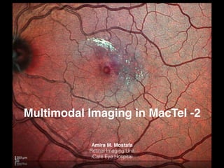
- 1. Multimodal Imaging in MacTel -2 Amira M. Mostafa Retinal Imaging Unit iCare Eye Hospital
- 2. • “Multimodal Retinal Imaging” is a trending concept of combining different imaging modalities to improve preclinical assessment, diagnostics, and therapeutic monitoring.1,2 • Hybrid imging platforms • Including: 1. Digital color fundus photography 2. Multicolor scanning laser fundus photography 3. cSLO 4. Dye-based angiography 5. FAF 6. SD-OCT 1. Keane PA, Sadda SR. Retinal imaging in the twenty-first century: state of the art and future directions. Ophthalmology. 2014;121(12):2489-2500. 2. Yannuzzi LA, Ober MD, Slakter JS, et al. Ophthalmic fundus imaging: today and beyond. Am J Ophthalmol. 2004;137(3):511-524.
- 3. • Concurrent assessment of different retinal structures:3-5 1. Better understanding and evaluation of retinal diseases 2. Adding up new entities to the spectrum of retinal diseases 3. Improving treatment modalities 4. Improving interpretation of newer modalities e.g. OCT-A 3. Kim YG, Baek SH, Moon SW, Lee HK, Kim US. Analysis of spectral domain optical coherence tomography findings in occult macular dystrophy. Acta Ophthalmol. 2011;89(1):e52-56. 4. Park SJ, Woo SJ, Park KH, Hwang JM, Chung H. Morphologic photoreceptor abnormality in occult macular dystrophy on spectral-domain optical coherence tomography. Invest Ophthalmol Vis Sci. 2010;51(7):3673-3679. 5. Christenbury JG, Klufas MA, Sauer TC, Sarraf D. OCT angiography of paracentral acute middle maculopathy associated with central retinal artery occlusion and deep capillary ischemia. Ophthalmic Surg Lasers Imaging Retina. 2015;46(5):579-581.
- 5. • The term of “retinal telangiectasia” has been used to describe dilated retinal vessels on all three components of the circulation; arterioles, capillaries, and venules, and ass. abnormalities such as aneurysmal dilatations, vascular leakage and exudation.6,7 • Classifications:7-9 6. Aung KZ, Wickremasinghe SS, Makeyeva G, et al.: The prevalence estimates of macular telangiectasia type 2. Retina. 30:473-478 2010. 7. Gillies MC, Zhu M, Chew EY, et al.: Familial asymptomatic macular telangiectasia type 2. Ophthalmology. 116:2422-2429 2009. 8. Gass JD, Blodi BA: Idiopathic juxtafoveolar retinal telangiectasis. Update of classification and follow-up study. Ophthalmology. 100:1536-1546 1993. 9. Yannuzzi LA, Bardal AM, Freund KB, et al.: Idiopathic macular telangiectasia. Arch Ophthalmol. 124:450-460 2006
- 6. • MacTel Research Group classification:8-10 • MacTel-1……”congenital or developmental” • MacTel-2 …… “idiopathic” 10.Clemons TE, Gillies MC, Chew EY, et al.: Baseline characteristics of participants in the natural history study of macular telangiectasia (MacTel) MacTel Project Report No. 2.Ophthalmic Epidemiol. 17:66-73 2010
- 7. • Bilateral • Asymmetric • Females • 6th decade • Systemic HTN “52%”, DM “28%” MacTel-2
- 8. MacTel-2 • Degeneration of parafoveal retinal Müller cells with subsequent vascular changes: 1. Pericyte degeneration and lipid accumulation in capillary walls. 2. Multilaminated basement membrane. 3. Dilation and proliferation of retinal capillaries into the outer retinal, subretinal, and preretinal spaces.
- 9. MacTel-2 • Diagnostic challenge ???????????? 1. Bilateral, Vision threatening 2. Progressive 3. Usually asymptomatic , Easily over-looked 4. Overlapping retinal vascular diseases such as DR 5. Complications as a presenting signs such as SRNVM, MH
- 10. • Appropriate combination of imaging modalities • Including: 1. BAF 2. Multicolor scanning laser fundus photography 3. SD-OCT 4. cSLO Dye-based angiography
- 11. • Appropriate combination of imaging modalities • Including: 1. BAF 2. Multicolor scanning laser fundus photography 3. SD-OCT 4. cSLO Dye-based angiography
- 12. BAF • Identification of early disease process • Before evidence of vascular changes • Pathognomonic loss of normal foveal hypo-AF caused by luteal pigment depletion Normal MacTel2
- 13. BAF • Features of progressing disease: • Telangiectatic vascular changes TEMPORAL to the fovea
- 14. BAF • Features of progressing disease: • Pigmentary migration
- 15. • Appropriate combination of imaging modalities • Including: 1. BAF 2. Multicolor scanning laser fundus photography 3. SD-OCT 4. cSLO dye-based angiography
- 16. • Earliest fundoscopic feature: • Subtle loss of TEMPORAL perifoveal retinal transparency Multicolor cSLO Fundus Photography
- 17. • Parafoveal TEMPORAL capillary dilatation Multicolor cSLO Fundus Photography
- 18. • Crystalline deposits at vitreoretinal interface Multicolor cSLO Fundus Photography
- 19. • Blunted diluted “diving” venule Multicolor cSLO Fundus Photography
- 20. • Pigment migration along dilated capillaries Multicolor cSLO Fundus Photography
- 21. • Complications: 1. SRNVM & disciform scarring Multicolor cSLO Fundus Photography
- 22. • Complications: 2. Macular hole formation Multicolor cSLO Fundus Photography
- 23. • Appropriate combination of imaging modalities • Including: 1. BAF 2. Multicolor scanning laser fundus photography 3. SD-OCT 4. cSLO dye-based angiography
- 24. • Diagnosis & Monitoring • Outer nuclear and/or photoreceptor layer loss • Disruption of the photoreceptor IS/OS junction SD-OCT
- 25. • Hyporeflective inner retinal cavities • Hyporeflective outer retinal cavities SD-OCT
- 26. • Pseudolamellar MH • Outer retinal atrophy SD-OCT
- 27. • SRNVM SD-OCT
- 28. • Appropriate combination of imaging modalities • Including: 1. BAF 2. Multicolor scanning laser fundus photography 3. SD-OCT 4. cSLO dye-based angiography
- 29. • Traditional • Camera vs. cSLO • Hallmark…… characteristic telangiectactic ? leaking capillaries TEMPORAL to the fovea. cSLO Fluorescein Angiography
- 30. • Progression …… entire perifoveal involvement cSLO Fluorescein Angiography
- 31. • Overlapping conditions cSLO Fluorescein Angiography
- 32. • Overlapping conditions cSLO Fluorescein Angiography
- 33. • Bilateral assessment cSLO Fluorescein Angiography
- 34. Take Home Message • MacTel-2 is a primary neurodegenerative macular disease with secondary vascular involvement. • One of the earliest changes reported in MacTel-2 is the pathognomonic loss of normal foveal hypo-AF in BAF. • Clinico-pathological correlation demonstrates that loss of perifoveal Müller cells correlate with areas of luteal pigment loss • Data provided by multimodal imaging approach may help understanding finding provided by newer technologies such as OCT-A. • Future development of the therapeutic agents that rescue Müller cells