1. 140 Sri Lanka Journal of Obstetrics and Gynaecology December 2013
CLINICAL GUIDELINE www.slcog.lk/sljog
Guideline on fetal monitoring in labour
Fetal monitoring in labour could be done by:
• Intermittent auscultation (preferably by a hand
held Doppler device)
• Intermittent or continuous electronic monitoring
Intermittent auscultation is recommended for low-risk
women in spontaneous labour.
Electronic monitoring is recommended when:
• The baby is growth restricted
• There is significant meconium staining of
amniotic fluid
• Abnormal fetal heart rate detected by intermittent
auscultation
• Fresh vaginal bleeding
• Maternal pyrexia
• Use of oxytocin for augmentation or induction of
labour
• Women with a scarred uterus
• Women on epidural analgesia
Intermittent auscultation
This could be done by using either a Pinnard's
stethoscope or preferably a hand-held Doppler device.
Auscultation should be carried out immediately after a
contraction for one full minute.
The maternal pulse should be palpated if there is
suspected fetal bradycardia or any other FHR anomaly
to differentiate the two heart rates.
The normal rate is between 110 - 160 beats per minute in
a term fetus.
The frequency of auscultation should be as specified in
the partogram.
Electronic fetal monitoring (EFM)
EFM is carried out by external cardiotocography (CTG).
The following are recommended at the commencement
of a CTG.
1. The paper speed must be set at 1 cm per minute.
2. The date and time settings on the machine must
be validated.
3. The CTG must be labeled with the mother's
name, BHT number and date and time.
4. Maternal heart rate should be noted on the CTG.
5. The presence and the point at which the fetal
heart rate is best heard must be delineated by
auscultation and the probe placed at that point.
6. Ensure that the contraction probe is functioning
properly and used for the recording.
7. The woman should be positioned in such a way
that aortocaval compression is avoided.
8. It should be interpreted without delay and the
categorization recorded as either normal or
suspicious or pathological, as per Table 1, and
signed by the responsible officer. The entry on
the BHT must include a plan for management.
9. If the CTG is categorized as suspicious or
abnormal, the Consultant must be informed.
10. For the management plan the overall clinical
picture must be taken into account. e.g. the rate
of progress of labour, presence or absence of fetal
growth restriction, meconium staining of
amniotic fluid and the evolution of the CTG
abnormalities.
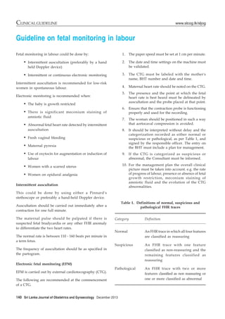
Table 1. Definitions of normal, suspicious and
pathological FHR traces
Category Definition
Normal An FHR trace in which all four features
are classified as reassuring
Suspicious An FHR trace with one feature
classified as non-reassuring and the
remaining features classified as
reassuring
Pathological An FHR trace with two or more
features classified as non reassuring or
one or more classified as abnormal
2. December 2013 Sri Lanka Journal of Obstetrics and Gynaecology 141
CLINICAL GUIDELINEwww.slcog.lk/sljog
Further useful information on FHR patterns
• If repeated accelerations are present with reduced
variability, the FHR trace should be regarded as
reassuring.
• True early uniform decelerations are rare and
benign, and therefore they are not significant.
• Most decelerations that occur during labour are
variable.
• If a bradycardia occurs in the baby for more than
3 minutes, urgent medical aid should be sought
and preparations should be made to urgently
expedite the birth of the baby, i.e. immediate
commencement of cesarean section. This could
include moving the woman to theatre if the fetal
heart has not recovered by 9 minutes. If the fetal
heart recovers within 9 minutes the decision to
deliver should be reconsidered in conjunction with
Feature Baseline (bpm) Variability (bpm) Decelerations Accelerations
Reassuring 110-160 ≥ 5 None Present
Non-reassuring 100-109 <5 for 40-90 Typical variable The absence of
161-180 minutes decelerations with accelerations with
over 50% of contractions, otherwise normal
occurring for over 90 trace is of uncertain
minutes significance
Abnormal < 100 < 5 for 90 minutes Either atypical variable
> 180 decelerations with over
Sinusoidal 50% of contractions or late
pattern ≥ 10 decelerations, both
minutes for over 30 minutes
Table 2. Classification of fetal heart rate patterns
the woman if the post-recovery tracing is
reassuring.
• A tachycardia in the baby of 160-180 bpm, where
accelerations are present and no other adverse
features appear, should not be regarded as
suspicious. However, an increase in the baseline
heart rate, even within the normal range, with
other non-reassuring or abnormal features should
increase concern. In such cases inquiry must be
made to ascertain if the fetus was active during
the recording.
When women are having continuous EFM, systematic
assessment of above definitions and classification should
be undertaken with every review.
During episodes of abnormal FHR patterns, if the woman
is lying supine, advise her to adopt the left lateral
position.