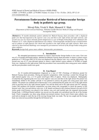
Percutaneous Endovascular Retrieval of Intravascular foreign body in pediatric age group
•
1 like•148 views

The document describes two cases of percutaneous retrieval of intravascular foreign bodies in pediatric patients. In the first case, a guidewire displaced during a central line procedure in an 18-month old and migrated to the heart. In the second case, a catheter in a 4-year old broke off and migrated to the heart. Both foreign bodies were successfully retrieved using a gooseneck snare catheter without complications. The technique of percutaneous retrieval using snares is an effective non-surgical approach for removing intravascular objects and avoiding potential complications of leaving them in place.

Recommended
Recommended
More Related Content
What's hot
What's hot (20)
Viewers also liked
Viewers also liked (20)
Similar to Percutaneous Endovascular Retrieval of Intravascular foreign body in pediatric age group
Similar to Percutaneous Endovascular Retrieval of Intravascular foreign body in pediatric age group (18)
More from iosrjce
More from iosrjce (20)
Recently uploaded
Recently uploaded (20)
Percutaneous Endovascular Retrieval of Intravascular foreign body in pediatric age group
- 1. IOSR Journal of Dental and Medical Sciences (IOSR-JDMS) e-ISSN: 2279-0853, p-ISSN: 2279-0861.Volume 14, Issue 11 Ver. VI (Nov. 2015), PP 12-14 www.iosrjournals.org DOI: 10.9790/0853-141161214 www.iosrjournals.org 12 | Page Percutaneous Endovascular Retrieval of Intravascular foreign body in pediatric age group. Shivaji Pole, Vivek S. Shah, Monarch V. Shah (Department of Interventional Radiology, Mahatma Gandhi Missions Medical College and Hospital, Aurangabad, India) Abstract:An 18 months-old-female patient admitted for Blalock-Taussig shunt procedure with a displaced guide wire that had migrated to the superior vena cava and then to the right atrium and right ventricle; and second case was of a 4years-old-male patient diagnosed as a case of Dengue hemorrhagic fever admitted in the Pediatric I.C.U. with a displaced right femoral vein catheter. Ultrasonography and fluoroscopy revealed broken end of catheter of right femoral vein which had travelled to the right atrium and right ventricle. Both cases referred to Interventional Radiology were managed by percutaneous retrieval of the foreign bodies using goose snare catheters. Keywords:foreign body, goose snare catheter, intravascular, percutaneous. I. Introduction We attempted percutaneous treatment to retrieve intravascular foreign bodies in two cases. First case was of a 18 months-old-female patient admitted for Blalock-Taussig shunt procedure in whom a central venous guidewire of 5.5F(Vygon 20G) of 10.3cms was displaced into the superior vena cava and the right atrium. The second case was of a 5 year-old-male patient in whom a right femoral venous catheter of 3F(20G) of length 9cms was displaced in the right atrium. Both the procedures were successfully done using goose snare catheter and the post procedure results were good. II. Case Report An 18 months-old-femalepatient diagnosed as a case of TOF (Tetralogy of fallot)was posted for Blalock-Taussig shunt procedure. During pre-operative preparation, while inserting a central line through the right internal jugular vein, a 2.5 Fr guide wire(Vygon 20G) of approx. 10cms was displaced. Radiography revealed that the migrated guide wire was in the superior vena cava and then in the right atrium.The patient was immediately shifted to the cardiac cath-lab. It was planned to remove the guide wire using goose neck snare catheter. The patient was sedated and on fluoroscopy it was confirmed that the guide wire was in the right atrium with its proximal end in the right internal jugular vein and the distal end in the right atrium and IVC opening. Under USG guidance left femoral venipuncture was done along with venography to check the patency of the femoral veins, iliac vein and inferior vena cava. A 4F sheath was placed in the common femoral vein and a 0.014 inches guide wire(Terumo) was placed in the inferior vena cava and reached upto the migrated intravascular foreign body. A 4F(Head Huntar) catheter was moved over the guide wire and placed near the tip of the foreign body. Check venography was done using diluted non-ionised contrast. Goose neck (3mm) snare catheter was used and an attempt to catch the tip of the moving foreign body was made. The tip was caught and the foreign body was removed successfully through the left femoral vein.
- 2. Percutaneous Endovascular Retrieval of Intravascularforeign bodyin pediatric age group. DOI: 10.9790/0853-141161214 www.iosrjournals.org 13 | Page AB C Case 1- A: Fluoroscopic view showing part of the guide wire in the superior vena cava and in the right atrium. B:Goose snare (3mm) catheter catching the tip of the distal part of the guide wire. C: Guide wire being successfully removed through the left femoral access. Second case was of a 4 years-old-male patient diagnosed as a case of Dengue hemorrhagic fever was admitted in the Pediatric I.C.U. A right femoral vein catheter of 3F (Vygon 20G) of length approx. 20cms was secured for medications and transfusions. Coming morning on general examination it was noticed that the femoral catheter was fractured and the broken end was displaced. The child was immediately referred to the Interventional Radiology Department for further management. USG and Fluoroscopy revealed broken end of catheter of approximately 8-10cms in the right femoral vein. During the preoperative preparation it was evident on fluoroscopy that the foreign body had travelled to the right atrium and right ventricle and the patient was having ectopic beats on ECG due to constant moving of the foreign body in the right ventricle. Aim was to remove the foreign body urgently by percutaneous retrieval with help of Goose snare catheter (3mm). Central femoral vein access was secured and a 0.035 inches guide wire (Terumo) was placed in the inferior vena cava and reached to the migrated intravascular foreign body. A 4F (Head Huntar) catheter was moved over the guide wire and placed near the tip of the foreign body. Check venography was done using diluted non-ionized contrast. Goose neck (3mm) snare catheter was used and an attempt to catch the tip of the moving foreign body was made. The foreign body measuring 9 cms and 3F (20G)diameter was successfully removed with the help of a snare catheter by pulling it out from distal to proximal end from the right atrium. A B C Case 2- A: Fluoroscopic image of foreign body (catheter) in the right atrium. B: Goose neck (3mm) snare catheter catching the distal tip of the catheter. C: The catheter being successfully removed through the right femoral access.
- 3. Percutaneous Endovascular Retrieval of Intravascularforeign bodyin pediatric age group. DOI: 10.9790/0853-141161214 www.iosrjournals.org 14 | Page Both the procedures were uneventful with no noted complications and good post-operative recovery of the patients. III. Discussion: Percutaneous retrieval of intravascular foreign bodies was first introduced in 1964 by Thomas et al, presenting non-surgical retrieval of a broken segment of steel spring guide from right atrium and inferior vena cava[1]. Commonly encountered intravascular foreign bodies include fragments of central venous catheters (most common), knotted pulmonary artery (Swan Ganz) catheters, lost guidewires or guidewire fragments, misplaced embolization coils and metallic stents. Fracture of central venous access catheter is although rare, yet can be associated with serious complications like thromboembolism, sepsis, cardiac arrhythmias, pericardial effusion, myocardial lesion and bacterial endocarditis, unretrieved intravascular foreign bodies may cause severe or life threatening complications in upto 71%patients[2] with mortality ranging from 24% and 60%[3]. Retrieval of foreign objects with theuse of baskets or forceps was no longer the method of choice after introduction of the nitinol goose neck micro snare[4,5]. The loop snare is being recognized as the most effective and preferred devicefor atraumatic removal of the foreign bodies [6]. In our patients this technique was successful and was without any pre-operative or post-procedure complications. IV. Conclusion: The high success rate of percutaneous endovascular techniques makes it superior to the other techniques in retrieving intravascular foreign bodies. This case report highlights the importance of goose snare catheter and the complications if the intravascular foreign body is left untreated. References: [1]. Thomas J, Sinclair-Smith B, Bloomfield D, Davachi A. Non-Suurgi-calretrievaof a brokensegment of steel springguide from the right atrium and inferiorvenacava. Circulation. 1964; 30:106-8.[14197828]. [2]. SmouseHB, Fox PF, Brady TM,SwischukJL, Castaneda F, Pham MT, Intravascular foreign bodyremoval.SeminInterventradiol 2000; 17:201-12. [3]. Gabelmann A, Kramer S,GorichJ.Percutaneous retrieval of lost or misplaced intravascular objects. AJR Am J Roentgenol 2001; 176:1509-13. [4]. Yedlicka JW Jr, Carlson JE, Hunter DW, Castañeda-Zúñiga WR, Amplatz K. Nitinol gooseneck snare for removal of foreign bodies: experimental study and clinical evaluation. Radiology 1991; 178:691-3.[1994404]. [5]. Cekirge S, Weiss JP, Foster RG, Neiman HL, McLean GK. Percuta- neous retrieval of foreign bodies: experience with the nitinol Goose Neck snare:JVascIntervRadiol 1993;4:805-10. [8281004]. [6]. Andrews RE, TullohRM, Rigby ML. Percutaneous retrieval of central venous catheter fragments. Arch Dis Child 2002; 87:149-50.