

Recommended
More Related Content
What's hot
What's hot (20)
Similar to Microtubules
Similar to Microtubules (20)
More from Intesar Aba-Conding
Recently uploaded
Recently uploaded (20)
Microtubules
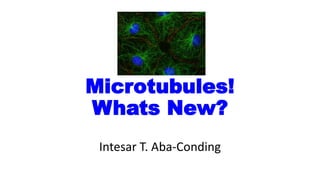
- 1. Microtubules! Whats New? Intesar T. Aba-Conding
- 2. What bones are to bodies, the cytoskeleton is to cells. The cytoskeleton maintains cellular structure, builds appendages like flagella and, together with motor proteins, powers cellular movement, transport, and division. Microtubules are a critical component of the cytoskeleton, vital for cell division and, because of that, an excellent target for chemotherapy drugs.
- 3. History of Microtubules • Microtubules were first discovered by De Roberties and Franchi (1953) • Term “Microtubules” was given by Slautterback (1963)
- 4. Chemistry of Microtubules • The chemistry of microtubules has been studied in some detail with the help of colchicines and its derivative colcemide other chemicals like vincristine, vinblastini and podophyllotoxin, which inhabit assembly of microtubules. • These chemicals bind to the globular subunit and prevent polymerization.
- 7. Structures and Functions of Microtubules • Microtubules are filamentous intracellular structures that are responsible for various kinds of movements in all eukaryotic cells. • Microtubules are involved in nucleic and cell division, organization of intracellular structure, and intracellular transport, as well as ciliary and flagellar motility. • Because the functions of microtubules are so critical to the existence of eukaryotic cells (including our own), it is important that we understand their composition, how they are assembled and disassembled, and how their assembly/disassembly and functions are regulated by cells.
- 8. • All eukaryotic cells produce the protein tubulin, in the usual way. • The usual way, of course, is by transcription of genes coding for tubulin to produce messenger RNA, followed by the translation of mRNA by the ribosomes in order to produce protein. • Cells maintain at least two types of tubulin, which we call alpha tubulin and beta tubulin. • However, it is doubtful that the two types can found in cells as individual proteins.
- 9. Microtubule Structure • Alpha and beta tubulin spontaneously bind one another to form a functional subunit that we call a heterodimer. • A heterodimer is a protein that consists of two different gene products. • The term is entirely descriptive - the prefix hetero-means "different," the prefix di- means "two," and the suffix - mer refers to a unit, in this case a single polypeptide. • Obviously, cells do not continue to make tubulin (or any other protein) until they run out of resources. Some process must regulate the synthesis of tubulin. A common regulatory mechanism is feedback inhibition.
- 10. Microtubule Structure • The alpha-tubulin and beta-tubulin molecules are 4-5 nm in diamater and 55 kDa, have almost identical shapes but only share 40% amino acid sequence identity. • A microtubule is a hollow cylinder of 13 protofilaments around a lumen with an outer diameter (25 nm), inner diameter (15 nm) and dimer width (8 nm).
- 11. Assembly of Microtubules • When intracellular conditions favor assembly, tubulin heterodimers assemble into linear protofilaments. Protofilaments in turn assemble into microtubules. All such assembly is subject to regulation by the cell. • Microtubules form a framework for structures such as the spindle apparatus that appears during cell division, or the whip like organelles known as cilia and flagella. Cilia and flagella are the most well-studied models for microtubule structure and assembly, and are often used by textbooks to introduce microtubules.
- 12. Dynamic instability of microtubules • Under steady state conditions a microtubule may appear to be completely stable, however there is action taking place constantly. • Populations of microtubules usually consist of some that are shrinking and some that are growing. A single microtubule can oscillate between growth and shortening phases. • During growth, heterodimers are added on to the end of a microtubule, and during shrinkage they come off as intact subunits. The same heterodimer can come off and go back on. • Since even apparently stable microtubular structures have an intrinsic instability, they are considered to be in a dynamic equilibrium, or steady state.
- 13. A research was recently published in the journal eLife https://www.sciencedaily.com/releases/2016/01/160128122350.htm • Microtubules can spontaneously self-organize, transforming from many singular components into one large cellular structure capable of performing specific tasks. • A researchers at the Harvard John A. Paulson School of Engineering and Applied Sciences (SEAS) have observed how microtubules and motor proteins assemble into macroscopic networks. • Their observation provides a better understanding of cytoskeletal self- organization in general, which may in turn lead to better drug design and new materials that can mimic cellular behaviors. • Spindles are cellular structures that play an important role in cell division, separating chromosomes and pulling the duplicated DNA from the mother cell into the daughter cell. They are made up of microtubules and many other proteins, including the motor protein dynein.
- 14. A research was recently published in the journal eLife https://www.sciencedaily.com/releases/2016/01/160128122350.htm • "What we are really looking for is a grand unified theory of spindle assembly," said Peter Foster, a graduate student at SEAS and the paper's first author. "We know how motor proteins interact with microtubules but how do you go from individual microtubules and motor proteins to large networked structures?" • To gain insight into how spindles assemble, Foster and his team, under the leadership of Dan Needleman, associate professor of applied physics and of molecular and cellular biology, built a simple experiment. • They extracted cytoplasm from frog eggs, which contains dynein and all of the components needed to make spindles, added fluorescent protein and the chemotherapy drug Taxol to create and stabilize microtubules, and loaded the mixture into "the world's simplest microfluidic chamber."
- 15. Two type of motor proteins 1) Kinesins its responsible for moving vesicles and organelles from the cell body toward the synaptic terminals. This motor protein, which was named, Kinesin, it constructed from two identical heavy chains and two light chains. 2) Cytoplasmic Dyneins its responsible for the movements of cilia and flagella. Cytoplasmic dyneins is huge protein composed of two identical heavy chain and a variety of intermediate and light chains.
- 16. A research was recently published in the journal eLife https://www.sciencedaily.com/releases/2016/01/160128122350.htm • "Very quickly, we saw that these microtubules organize into networks that spontaneously contract," Foster said. "The question is why?" • The answer lay not in the microtubules but in the behavior of the motor protein. • Microtubules have plus and minus ends and researchers have observed dynein moving from the plus end to the minus. • As a result, the motor protein draws the minus ends of microtubules together, creating star-like clusters called asters. • The dynein drives these small clusters together, fusing them to create larger and larger networks. As the motor protein continues to jam the microtubules together, the network contracts, until it can't get any smaller.
- 17. Another Recent Findings Developmentally Regulated GTP binding protein 1 (DRG1) controls microtubule dynamics Anna Katharina Schellhaus, Daniel Moreno-Andrés, Mayank Chugh, Hideki Yokoyama, Athina Moschopoulou, Suman De, Fulvia Bono, Katharina Hipp, Erik Schäffer & Wolfram Antonin Published online: August 30, 2017 www.nature.com/scientificreports
- 18. • Several types of microtubules are found in the mitotic spindle. • The microtubules of the kinetochore, a protein complex assembled on centromeric chromatin, connect the centrosome with the kinetochore. • Usually 20–30 kinetochore microtubules are bundled into stable k-fibers, which mediate chromosomal movement. • The non-kinetochore microtubules are part of the spindle body, without being attached to the kinetochore. They are important for separating the poles and mitotic spindle stability. • Lastly, astral microtubules radiate from the centrosomes toward the cell cortex and position the • Several classes of microtubule-associated proteins are known. These include microtubule polymerases and de- polymerases, nucleation factors, severing enzymes, microtubule bundling/crosslinking proteins, motor proteins that are essential to establish the bipolar array e.g. by sliding microtubules, microtubule capping/ end- binding/tracking factors and many more.
- 19. • Here, they identify Developmentally regulated GTP-binding protein 1 (DRG1) as a microtubule polymerase that also bundles and stabilizes microtubules. • Developmentally regulated GTP-binding proteins (DRGs) are a deeply conserved group of proteins belonging to the subfamily of Obg GTPases. • DRGs are conserved from archaebacterial having one DRG to eukaryotes from yeast to human, containing DRG1 and DRG2. • Plants even have three DRGs. Beside the canonical G-domain they do not share similarities with other known GTPases and their function is still largely unclear.
- 20. • The function of DRG1 has been long debated. Considering its high evolutionary conservation, it was suggested that DRG1 has an important function in a fundamental cell biological process. • They identify here that DRG1 is involved in spindle assembly. DRG1 binds microtubules and can diffuse on the microtubule lattice in vitro. DRG1 promotes microtubule polymerization and bundling and stabilizes them. • To perform these latter activities, DRG1 does not require GTP hydrolysis, but does require each of its domains as only the full-length protein is functional in these assays. • Consistent with these observations DRG1 is also involved in spindle dynamics in HeLa cells: microtubules regrow faster after a cold shock induced disassembly if DRG1 is present; early mitotic progression is extended if DRG1 is down regulated and a high number of asymmetric monoasters forms upon monastrol treatment in cells lacking DRG1.
- 21. • The polymerization, bundling and stabilization activities of DRG1 could be completely independent functions or connected to each other: the bundling of microtubules could also stabilize them • The polymerization activity could increase the amount of microtubules in a population that is in the growth phase and thereby stabilize them; the bundling could increase polymerization by increasing the microtubule density close to DRG1. • In this respect, it is surprising that the GTPases α- and β-tubulin are directly regulated by another GTPase, DRG1, although not using its GTP hydrolysis activity in this context.
- 22. • DRG1 has been suggested to possess a function connected to ribosomes and translation as it co-fractionates with ribosomes. Its function in this context is still not fully understood. • The likely independent functions of DRG1 concerning microtubules and translation could be spatially or temporally regulated e.g. DRG1 might have different functions during different cell cycle stages or one of the functions could be induced upon stress situations as previously suggested. • Likewise, its function could be regulated by its binding partners. Analysis shows that DRG1 is a microtubule binding, bundling, polymerization and stabilization factor. It does not need its GTPase activity to perform these functions. • Truncated versions bind microtubules but have highly reduced or none of the other activities. Down regulation of DRG1 in HeLa cells indicated that the protein is involved in mitotic spindle assembly. • Deregulation of DRG1 was suggested to be involved in cancer formation and it is conceivable that the function of DRG1 in mitotic spindle assembly is connected to this. It is also possible that the microtubule function of DRG1 is not limited to mitosis. • How DRG1 potentially affects interphase microtubule function is an interesting question awaiting detailed investigation.
- 23. To summarize: 1. DRG1 directly interacts with microtubules 2. DRG1 diffuses on microtubules 3. DRG1 binds microtubules via multiple regions 4. DRG1 binds to microtubules lacking the negatively charged C-terminus of tubulin 5. DRG1 bundles microtubules 6. DRG1 promotes microtubule polymerization 7. DRG1 stabilizes microtubules 8. The GTPase activity of DRG1 is not necessary for its microtubule functions 9. Full-length DRG1 is necessary to bundle, polymerize and stabilize microtubules 10. DRG1 impacts spindle dynamics in cells
- 24. Figure 1. DRG1 and DFRP1 bind microtubules. (a) 4 μM taxol-stabilized microtubules (MTs) were incubated with Xenopus cytostatic factor arrested (CSF) extract. Microtubules were co-sedimented together with Mtbinding proteins and eluted by 500 mM NaCl in CSF-XB buffer. The pellet and the elution were analysed by western blotting. (b) Recombinant Xenopus laevis DRG1 and DRFP1 as well as human DFRP2 were incubated with 12 μM taxol-stabilized microtubules to test if the observed binding is direct. RanQ69L served as a negative control (neg. ctrl.). S: supernatant, P: pellet. (c) Coomassie stainings of recombinant proteins in binding experiments as in were quantified using ImageJ. The columns represent the averages of the protein fractions found in the pellet from at least three different experiments with the individual data points indicated. (d) Recombinant Xenopus laevis DRG1 was incubated with different concentrations of taxol-stabilized microtubules. (e) Coomassie staining of recombinant DRG1 from (d) was quantified using ImageJ and blotted in dependence to the microtubule concentrations. The binding curve was fitted to the data points calculating a KD of 0.47 (+/−0.05) μM.
- 25. Figure 2. DRG1 interacts with the microtubule lattice in distinct binding modes. (a) Kymographs showing two different binding modes (diffusion and immobile) of eGFP-DRG1 over four different concentrations (0.08 nM, 0.4 nM, 4 nM, 40 nM). On top of each kymograph, the respective image of the rhodamine-labelled microtubule is shown. Every kymograph represents a microtubule on its horizontal axis observed over time (vertical). (b) The proportions of the different DRG1 binding populations are shown at the aforementioned concentrations. (c) Residence times of diffusive and immobile DRG1 molecules on microtubule lattice are 12.0 ± 0.8 s (mean ± S.E.M., 0.08 nM), 12.4 ± 0.9 s (0.4 nM), 11.1 ± 0.9 s (4 nM), 5.4 ± 0.6 s (40 nM) for the immobile fraction and 2.2 ± 0.2 s (4 nM) and 2.6 ± 0.5 s (40 nM) for the diffusive population. Color scheme: diffusion (cyan), immobile (green). Exemplary events are pointed out by arrows.
- 26. Figure 3. Different DRG1 domains interact with microtubules. (a) Scheme of DRG1 indicating the different domains. (b) Full-length and truncated versions of Xenopus laevis DRG1 were incubated and co-sedimented with taxol-stabilized MTs as in Fig. 1b. (c) Full-length DRG1 and its truncated versions lacking both the HTH and TGS domains were incubated and co-sedimented with taxol-stabilized MTs. (d) Coomassie stainings of recombinant proteins in binding experiments as in (b) and (c) were quantified using ImageJ. The column represents the average of the protein fractions found in the pellet from three different experiments with the individual data points indicated (e) Structure prediction of Xenopus DRG1 modeled with Swiss-Model46. Blue color represents the positively charged surface and red the negative charges (±5 kT/e). Lower structure shows a cartoon representing the different domains using the color code from (a). (f) Taxol-stabilized MTs were digested by the protease subtilisin and employed in the co- sedimentation assay with full-length DRG1.
- 27. • Figure 4. DRG1 bundles and polymerizes microtubules in vitro (a,b) 0.3 μM taxol-stabilized, Cy-3 labelled MTs were incubated with 1 μM DRG1, BSA or buffer for 10 min at RT. Samples were analyzed by confocal microscopy (a) or electron microscopy (b). (c) 5 μM tubulin (mixed in a 1:4 Cy3 labeled:unlabeled ratio) were incubated with 1 mM GTP and 1 μM DRG1, BSA or buffer for 30 min at 37 °C, fixed with BRB80 buffer containing 0.25% glutaraldehyde, 10% glycerol and 0.1% Triton X-100, spun down on coverslips and postfixed with cold methanol. Samples were analyzed by confocal microscopy. MT only polymerized if DRG1 was present. (d) Light-scattering experiments were carried out by mixing 2.5 μM tubulin, 1 mM GTP and 1 μM DRG1 in a 96-well plate which was followed by immediately measuring absorbance at 340 nm every 38 seconds for 2:10 hours.
- 28. Figure 5. DRG1 stabilizes microtubules in the cold. (a) 12 μM tubulin was polymerized in the absence or presence of 5 μM DRG1 for 1 h at 37 °C and placed on ice for 30 min. MTs were then pelleted by centrifugation, while free tubulin stays in the supernatant. Pellet and supernatant were analyzed by SDS-PAGE. The quantification shows the tubulin fraction found in the pellet. Columns represent the average of five independent experiments with the individual data points indicated. (b) Western blotting shows that DRG1 was knockeddown in HeLa cells stably expressing histone H2B-mCherry and eGFP-tubulin by siRNA 72 h post-transfection. (c) siRNA treated HeLa cells were placed 72 hrs after transfection on ice for 1 hour to induce MT disassembly. Warm medium was then added to the cells which were fixed with 4% PFA at indicated time points. Maximum intensity projections of Z-stacks from representative prometaphase cells at the given time points are shown. (d) The spindle size in voxels was quantified in 20 random prometaphase cells (from 2 independent experiments) per time point and siRNA (mean and SD are plotted). ***P < 0.001; **P < 0.01; *P < 0.01. Insert shows the spindle size at 0 min enlarged.
- 29. Figure 6. Microtubule binding, bundling, polymerization and stabilization activity of DRG1 does not require GTP hydrolysis. (a) MT co-sedimentation with 12 μM taxol-stabilized microtubules and DRG1 WT, DRG1 S78N and DRG1 P73V. The quantification shows the fraction of the proteins in the pellet. Columns represent the average of three independent experiments, individual data points are indicated. (a) MT-polymerization assay was done as in Fig. 4 using 1 μM DRG1 WT, S78N and P73V. (b) MT bundling assay was done as in Fig. 4 using 1 μM DRG1 WT, S78N and P73V. (c) DRG1 S78N and DRG1 P73V were employed in the microtubule stabilization assay as in Fig. 5. The quantification shows the tubulin fraction found in the pellet. Columns represent the average of four independent experiments, individual data points are indicated.
- 30. Figure 7. DRG1 regulates mitotic progression and spindle assembly in cells. (a) HeLa cells stably expressing histone H2B-mCherry and tubulin-eGFP were transfected with siRNA oligonucleotides against DRG1 or a control. The cells were imaged every 3 min for 48 h starting at 24 h post-transfection. (b) A cumulative histogram of the timing from prophase to anaphase onset based on chromatin morphology (based on H2BmCherry) and (c) of the timing from aster formation to anaphase spindle (based on eGFP- tubulin) are shown. Mean and SD from 3 independent experiments containing more than 150 cell trajectories per siRNA and experiment are plotted.
- 31. Figure 8. DRG1 plays a role in spindle dynamics in vivo. (a) HeLa cells were seeded on glass coverslides and transfected with DRG1 or control siRNA oligonucleotides. 72 h post-transfection cells were incubated with 70 μM monastrol with or without 2 nM taxol for 3 h33, fixed and stained with antibodies against α-tubulin (green) and anti-human centromere (magenta). DAPI in blue. (b,c) Quantitation of cells with asymmetric asters. Z-Stacks from five to eight random positions per condition were acquired and quantified (4 independent experiments for monastrol treatment (b) and two independent experiments (represented by the two dots/ squares) for monastrol treatment with rescue by taxol (c)). Between 15 and 98 cells with monopolar spindles were evaluated per siRNA knockdown per experiment. Mean and SD are plotted. ***P < 0.001; **P < 0.01; *P < 0.01.
- 32. Another Interesting Findings • Cells need microtubules for intracellular transport and to avoid being crushed, ACETYLATION is a must because this makes microtubules STRONG! Acetylation Keeps Microtubules Strong Stella M. Hurtley Science 21 Apr 2017: Vol. 356, Issue 6335, pp. 280-283 DOI: 10.1126/science.356.6335.280-k http://science.sciencemag.org/content/356/6335/280.11/tab-pdf
- 33. THANK YOU …