MUCOEPIDERMOID CARCINOMA /certified fixed orthodontic courses by Indian dental academy
•
42 likes•6,520 views

The document discusses Mucoepidermoid carcinoma, which is the most common malignant salivary gland tumor. It arises from the ductal system of major and minor salivary glands. The document describes the clinical features, histopathological features, grading, variants, differential diagnosis and management of Mucoepidermoid carcinoma. It is graded as low, intermediate or high grade based on histopathological characteristics like presence of cystic spaces, cellular atypia and proportion of cell types. Low grade tumors have a better prognosis compared to intermediate and high grade tumors.

Recommended
More Related Content
What's hot
What's hot (20)
Viewers also liked
Viewers also liked (20)
Similar to MUCOEPIDERMOID CARCINOMA /certified fixed orthodontic courses by Indian dental academy
Similar to MUCOEPIDERMOID CARCINOMA /certified fixed orthodontic courses by Indian dental academy (20)
More from Indian dental academy
More from Indian dental academy (20)
Recently uploaded
Recently uploaded (20)
MUCOEPIDERMOID CARCINOMA /certified fixed orthodontic courses by Indian dental academy
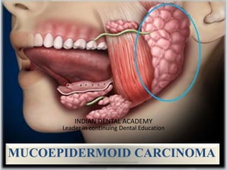
- 1. MUCOEPIDERMOID CARCINOMA INDIAN DENTAL ACADEMY Leader in continuing Dental Education www.indiandentalacademy.com
- 3. At the end of the presentation learner should be able to describe clinical, radiological , histopathological features, differential diagnosis and plan management of patient with gingival swelling www.indiandentalacademy.com
- 4. S.No Learning objectives Domain level criteria condition 1 Initiate examination of gingival swelling Cognitive & psychomotor Must know All 2 Explain clinical and radiological features of gingival swelling Cognitive & psychomotor Must know All 3 Explain histopathological features of gingival swellings Cognitive & psychomotor Must know All 3 Explain and plan the management of gingival swellings Cognitive & psychomotor Must know All www.indiandentalacademy.com
- 5. At the end of the seminar the learner should be able to know about Mucoepidermoid carcinoma:- Introduction Clinical features Histological features Variants of tumor Treatment and prognosis www.indiandentalacademy.com
- 6. •Tumors of salivary glands constitute a heterogeneous group of lesions of great morphologic variation. •Neoplasms of the major salivary glands constitute minor portion of head and neck neoplasms •Less than 2% are malignant. www.indiandentalacademy.com
- 7. Most neoplasms in parotid 75%, 0.8% in sublingual glands. Remainder equally distributed between submandibular gland and minor salivary glands Incidence of malignant neoplasm increases after 4increases 4th and 5th decades and peaks 65and 65-75 years. Benign neoplasm present slightly earlier. Malignant neoplasm occur most often in men Cancers of the salivary glands account for only 6% of H&N cancers Only 0.3% of all cancers www.indiandentalacademy.com
- 8. Saliva transported from central structure (acini) in complex ductal system to the oral cavity. System is a bilayer with internal luminal layer and external reserve layer. Internal layer forms acini and ductal epithelium. External layer forms myoepithelium and reserve cells www.indiandentalacademy.com
- 10. INTERCALATED DUCTS • Pleomorphic adenoma • Warthin’s tumor • Oncocytoma • Acinic cell • Adenoid cystic carcinoma EXCRETORY DUCTS • Squamous cell carcinoma • Mucoepidermoid carcinoma www.indiandentalacademy.com
- 11. Striated duct - Oncocytic Tumors Acinar cells - Acinic Cell Carcinoma Excretory Duct - Squamous cell and Mucoepidermoid carcinoma Intercalated duct and myoepithelial cells – Pleomorphic tumors www.indiandentalacademy.com
- 13. It was first studied and described as a separate entity by Stewart et al. in 1945. Definition (WHO) : Mucoepidermoid carcinoma is a malignant glandular epithelial neoplasm characterized by mucous, intermediate and epidermoid cells, with columnar, clear cell and oncocytoid features. www.indiandentalacademy.com
- 14. Mucoepidermoid carcinoma (MEC) is the most common primary salivary gland malignancy in both adults and children It is the common malignant neoplasm observed in both major and minor salivary glands www.indiandentalacademy.com
- 16. CLINICAL FEATURES: Adulthood tumor Significant female predilection (3:2 ) Peak incidence in 3rd and 5th decade of life Most common in children previously exposed to radiation Slowly enlarging, painless mass www.indiandentalacademy.com
- 17. CLINICAL FEATURES: Facial nerve palsy (high grade) Trismus, drainage for ear and dysphagia. Numbness of adjacent areas. Sometimes, ulceration is seen www.indiandentalacademy.com
- 18. SITE: Extraorally: 50% MEC occur in the parotid gland (arising in superficial lobe) Intraorally : 20% occur on the palate Rest of the lesions arising from the minor salivary glands with the buccal mucosa, lips, tongue and retro molar areas to be the favored sites. www.indiandentalacademy.com
- 19. Clinical manifestation: Slowly-enlarging, Painless mass (low grade), Painful (high grade), Seldom exceeds 5 cm, Not completely encapsulated, Often contains cysts, Maybe filled with a viscoid, mucoid material www.indiandentalacademy.com
- 20. Clinical manifestation: High grade: trismus, Drainage from the ear, Dysphagia, Numbness of the adjacent areas and ulceration. www.indiandentalacademy.com
- 21. Well circumscribed. Partially encapsulated. Firm , pinkish, or yellowish tan. Extreme induration is occasionally present. www.indiandentalacademy.com
- 22. Majority of cut surface is cystic. If cyst space present it contain viscid, translucent mucoid material that may be blood tinged. www.indiandentalacademy.com
- 23. It is basically composed of mucous secreting cells, epidermoid cells and intermediate cells. Mucous cells:- are of various shapes and sizes with abundant, pale, foamy cytoplasm staining positive for mucin. Epidermoid cells:- having squamatoid features having polygonal shape with intercellular bridges and sometimes keratinization. www.indiandentalacademy.com
- 24. A group of highly prolific, basaloid cells referred to as the intermediate cells is seen being larger than basal cells and smaller than Squamous cells. They are believed to be progenitors of epidermoid and mucous cells. Occasionally, clusters of clear cells can be present which are mucin and glycogen free. www.indiandentalacademy.com
- 25. Epidermoid cells, together with intermediate and mucous cells line cystic spaces or form solid masses or cords. Other types of cell : Basal cells called as maternal cells. www.indiandentalacademy.com
- 26. Mucoepidermoid carcinoma is graded into:- Low grade Intermediate grade High grade www.indiandentalacademy.com
- 27. Low grade:- well formed glandular structures and prominent mucin filled cystic spaces, minimal cellular atypia and a high proportion of mucous. Intermediate grade:- they have solid areas of epidermoid cells or squamous cells with intermediate basaloid cells. Fewer cyst formations as compared to low grade. High grade:- it consists of cells present as solid nests and cords of intermediate basaloid cells and epidermoid cells. Prominent nuclear pleomorphism and mitotic activity. Necrosis and perineural invasion maybe present. www.indiandentalacademy.com
- 28. Histology – Low grade Mucus cell > epidermoid cells Prominent cysts Mature cellular elements www.indiandentalacademy.com
- 29. Histology –Intermediate grade Mucus = epidermoid Fewer and smaller cysts Increasing pleomorphism and mitotic figures www.indiandentalacademy.com
- 30. Histology - High grade Epidermoid > mucus Solid tumor cell proliferation Mistaken for SCCA Mucin stainingMucin staining www.indiandentalacademy.com
- 31. Mucoepidermoid Carcinoma LOW GRADE / HIGH GRADE www.indiandentalacademy.com
- 32. CLINICAL • Minor Salivary Gland Malignancy • Peripheral Odontogenic Tumor • Connective Tissue Malignancy HISTOPATHOLOGICAL • Cystadenoma. • Adenosquamous carcinoma. • Polymorphous low grade adenocarcinoma. • Primary or metastatic squamous cell carcinomas of major salivary gland. • Cystadenocarcinoma www.indiandentalacademy.com
- 35. Sclerosing mucoepidermoid carcinoma:- This variant is extremely rare Tumor infarction and extravasations of mucin resulting in reactive fibrosis is suggested as the cause of this morphologic variant. It is characterized by an intense central sclerosis that occupies the entirety of an otherwise typical tumor frequently with an inflammatory infiltrate of plasma cells,eosinophils or lymphocytes. www.indiandentalacademy.com
- 36. Sheets of cells Cystic Space with basophilic material in the lumen www.indiandentalacademy.com
- 37. Intraosseous mucoepidermoid carcinoma:- It may originate within the jaws. it is thought to form by malignant transformation of epithelial lining of odontogenic cysts. It presents as an asymptomatic radiolucent lesion and histologically of low grade malignancy. The mandible is three times more commonly affected than the maxilla. www.indiandentalacademy.com
- 38. Conservative excision with preservation of facial nerve. Radical neck dissection. Low-grade lesions had a 5 year rate of 92%,intermediate- grade and 49% five-year curerate. www.indiandentalacademy.com
- 39. Shafer’s Oral Pathology(5 th edition) Surgical pathology of salivary gland (Ellis) Neville Textbook of oral pathology www.indiandentalacademy.com