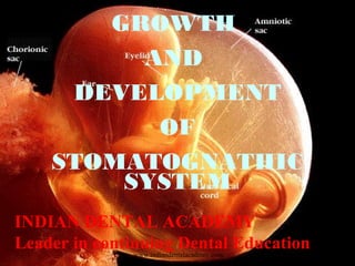
Growth and development of stomatognathic system/ dental crown & bridge courses
•
2 likes•1,034 views

The Indian Dental Academy is the Leader in continuing dental education , training dentists in all aspects of dentistry and offering a wide range of dental certified courses in different formats.for more details please visit www.indiandentalacademy.com

Recommended
More Related Content
What's hot
What's hot (20)
Similar to Growth and development of stomatognathic system/ dental crown & bridge courses
Similar to Growth and development of stomatognathic system/ dental crown & bridge courses (20)
More from Indian dental academy
More from Indian dental academy (20)
Recently uploaded
Recently uploaded (20)
Growth and development of stomatognathic system/ dental crown & bridge courses
- 1. GROWTH AND DEVELOPMENT OF STOMATOGNATHIC SYSTEM INDIAN DENTAL ACADEMY Leader in continuing Dental Educationwww.indiandentalacademy.com
- 2. Contents of this seminar 1. Introduction 2. Definitions 3. Phases of Development 4. Early orofacial development 5. Formation of face 6. Development of maxilla 7. Branchial arches 8. Bone development and growth 9. Growth factors 10. Differential growth 11. The mandible 12. Temporomandibular joint 13. Tongue 14. Palate 15. Anomalies of development www.indiandentalacademy.com
- 3. Over the structure of the cell rises the structure of plants and animals, which exhibit the yet more complicated, elaborate combinations of millions and billions of cells coordinated and differentiated in the most extremely different ways. - Hertwig www.indiandentalacademy.com
- 4. Introduction: Human development is a continuous process that begins when an oocyte(ovum) from a female is fertilized by a sperm(spermatozoon) from a male. Development involves many changes that transform a single cell, the zygote(fertilized ovum), into a multicellular human being. Most development changes occur before birth, but important changes also occur during the later periods of development; infancy childhood, adolescence, and adulthood. www.indiandentalacademy.com
- 5. The mating of male and female gametes in the maternal uterine tube initiates the development of a zygote-the first identification of an individual. The union of the haploid number of chromosomes (23) of each gamete confers the hereditary material of each parent upon the newly established diploid number of chromosomes (46) of the zygote. All the inherited characteristics of an individual and its sex are thereby established at the time of union of the gametes. The single totipotential cell of approximately 140 µ m diameter resulting from the union very soon commences mitotic division to produce a rapidly increasing number of smaller cells, so that the 16-cell stage, known as the morula, is not much larger than the initial zygote. www.indiandentalacademy.com
- 6. These cells of the early zygote reveal no significant outward differences of form. However, the chromosomes of these cells must necessarily contain the potential for differentiation of subsequent cell generations into the variety of cell forms that later constitute the different tissues of the body. The differentiation of the these early pluripotential cells into specialized forms is dependent upon genetic, cytoplasmic and environmental factors that act at critical times during their proliferation and growthwww.indiandentalacademy.com
- 7. A compendium of manifold biochemical reactions leads to cytodifferentiation and histodifferentiation, resulting in the formation of epithelial and mesenchymal tissues that acquire specialized structure and function. Epithelial mesenchymal interaction that provide for reciprocal cell differentiation are essential to organogenesis, i.e. the production of organs and systems. Growth is a fundamental attribute of developing organisms. The dramatic increase in size that characterizes the living embryo is a consequence of : www.indiandentalacademy.com
- 8. 1) Increased number of cells resulting from mitotic divisions (hyperplasia) 2) Increased size of individual cells (hypertrophy) 3) Increased amount of non-cellular material (accretion). Hyperplasia tends to predominate in the early embryo, whereas hypertrophy largely prevails later. Once differentiation of the tissue has been established, further development is predominantly that of growth .The rate of growth of the tissue is inherently determined, but is, of course is also dependent upon environmental conditions. The health, diet, race and sex of an individual influence the rate and extent of growth. www.indiandentalacademy.com
- 9. Growth may be interstitial, where increase in bulk occurs within a tissue or organ, or appositional, where surface deposition of tissue enlarges its size. Interstitial growth characterizes soft tissues, whereas hard tissues (bone, dental tissues) necessarily increase by apposition. The growth and development sequence is genetically determined and operates through the mechanism of inductors, metabolic modulators, neurotrophic and hormonal substances and interacting systems of contact stimulation and inhibition of contiguous tissues. Should these differential, but integrated, rates of development fail to maintain their normal determined ‘pace’, aberrations of overall development will manifest themselves as malformations that may require clinical interception for correction. www.indiandentalacademy.com
- 10. Maturation is a counterpart of growth; it indicates not only the attainment of adult size and proportions but also the full adult constituents of tissues (e.g. mineralization) and the complete capability for performance of each organ’s destined functions. www.indiandentalacademy.com
- 11. Definitions: The British Medical Dictionary defines growth “As the progressive development of a living being or part of an organism from its earliest stage to maturity including the resultant increase in size”. o J.S. Huxley – Growth is defined as the self- multiplication of a living substance. o Krogman – Increase in size, proportion and progressive complexities. o Mevedith – Entire series of sequential anatomic and physiologic changes taking place from the beginning of parental life to senility. o Todd - Growth is an increase in size. www.indiandentalacademy.com
- 12. o Moyers – Quantitative aspect of biologic development per unit of time. o Moss – change in any morphologic parameter that is measurable. Growth can result in an: Increase in size, Decrease in size, change in form, change in proportion, change in complexity, change in texture. Decrease in size, e.g. Thymus Gland. www.indiandentalacademy.com
- 13. Development: - The term connotes an increasing degree of organization. - It is series of changes by which the individual embryo becomes a mature organization. - Development is a progress towards maturity – Todd - It is the sequences of changes from cell fertilization to maturity. It relates to cell division, growth, differentiation and maturation – J.H. Salsamann. - Development refers to all the changes that are natural and unidirectional in life of an individual from its existence as a single cell to its elaboration as a multifunctional unity terminating in death. – Moyers. www.indiandentalacademy.com
- 14. Growth and development though closely related are not synonymous. According to W.R.Proffit, Growth is largely an anatomic phenomenon, whereas development is physiologic and behavioral. Development = Growth and differentiation & Translocation www.indiandentalacademy.com
- 15. Phases of Growth and Development –Pre Natal Life: Period of the ovum – The period from fertilization to the end of the 14th day. Embryonal period – From the 14th day to about 56th day. The fertilized ovum differentiates rapidly into an organism that has most of the gross anatomic features of human. 1st week : Active cell division 2nd week : Tissue differentiation into ectoderm and endoderm 3rd Week : The 3rd layer Mesoderm is added 4th to 8th week : Active & Rapid differentiation. www.indiandentalacademy.com
- 16. Fetal Period – the second trimester of pregnancy ending by about the 28th week is characterized by rapid fetal growth especially primarily. The 3rd trimester shows further increase in size of the new viable fetus involving primarily subcutaneous tissue and muscle mass. www.indiandentalacademy.com
- 18. STAGE TIME RELATED SYNDROMES Formation of organ systems- primary palate Days 28-38 Cleft lip & palate, facial clefts Secondary palate Days 42-55 Cleft of palate Final differentiation of tissues Days 50- birth Achondroplasia, Crouzon’s disease Apert’s syndrome www.indiandentalacademy.com
- 19. Post Natal Life: 1. Infancy: Refers to first year after birth and the first four weeks are designated as the new born or neonatal period. 2. Childhood: this is a period from 15 months to 12-13 years. During early childhood, there is active ossification, but this rate goes down, as the child grows older. 3. Puberty: This is the period between the ages of 12-15 years in girls and 13-16 years in boys, now is the time when secondary sexual characteristics develop and are expressed. www.indiandentalacademy.com
- 20. 4 Adolescence: Period of 3 to 4 years after puberty extending from the earliest signs of sexual maturity until, the attainment of physical, mental and emotional maturity. 5 Adulthood: Ossification and growth are virtually completed during early adulthood, 18-25 years thereafter development changes occur slowly. www.indiandentalacademy.com
- 21. Phases of development: Embryogenesis is divided into three distinct phases during the 280 days of gestation (ten 28-day menstrual cycles). The phases are the preimplantation period (the first 7 days), the embryonic period (the next 7 weeks), and the fetal period (the next 7 calendar months) www.indiandentalacademy.com
- 22. The preimplantation period(1-7 days) During the first 2-3 days post conception the zygote progresses from a single cell to a 16 cell cluster, the morula, no larger than the original ovum . The early totipotential blastomeres can develop into any tissue, but later differentiation creates an approximately 100-cell fluid-filled blastocyst. The outer sphere of cells forms the trophoblast, and the inner cell mass will form the embryo. During this period, the conceptus passes along the uterine tube to enter the uterus, where it implants into the uterine endometrium on the 7th postconception day. The trophoblast converts into the chorion by sprouting villi. Chorionic implantation establishes the placenta, the organ of fetomaternal exchange of nutrition and waste disposal.www.indiandentalacademy.com
- 27. The embryonic period(2-8 weeks) This phase, from the end of the 1st week until the 8th week, can be subdivided into three periods: presomite (8- 21 days postconception), somite (21-31 days) and postsomite (32-56 days postconception). During the presomite period, the primary germ layers of the embryo and the embryonic adnexa (fetal membranes) are formed in the inner cell mass. In the somite period, characterized by the appearance of prominent dorsal metameric segments, the basic patterns of the main body systems and organs are established. The postsomite period is characterized by formation of body’s external features. www.indiandentalacademy.com
- 28. The fetal period The beginning of this long phase, from the 8th week until term, is identified by the first appearance of ossification centers and the earliest movements by the fetus. There is little new tissue differentiation or organogenesis, but there is rapid growth and expansion of the basic structures already formed. Embryonic adnexa form membranes surrounding cavities in which the embryo (and subsequently the fetus) develops. These fluid filled cavities, membranes, and organs protect the fetus physically and save its nutritional and waste disposal requirements, casting off at birth. The main cavities and their membranes are the chorion and amnion, surrounding the fetus. Lesser cavities and their membranes are the yolk sac and a transient diverticulum, the allantois, that become incorporated into the umbilical cord, connected to the placenta, the chief organ of fetal sustenance. www.indiandentalacademy.com
- 29. The chorion arises from the trophoblast as an all encompassing membrane that, with the maternal endometrium, forms the placenta. The amnion and yolk sac, which form and fluid filled sacs in the inner cell mass within the chorion, are separated by a bilaminar plate; this plate forms the embryonic disk that later gives rise to the definitive embryo. The attachment of the inner cell mass to the chorion constitutes the connecting (body) stalk that contains the yolk sac and allantois. The stalk converts into the umbilical cord. www.indiandentalacademy.com
- 30. The presomite period (8-21 days) The primordial embryonic germ disk is composed of two primary germ layers; the ectoderm, which forms the floor or the amniotic cavity, and the endoderm, which forms the roof of the yolk sac. Early embryogenesis: Embryonic period (8-56 days) www.indiandentalacademy.com
- 31. There is early demarcation, at the 14th day, of the anterior pole of the initially oval disk; an endodermal thickening, the prechordal plate appears in the future midcephalic region. The prechordal plate preface the development of the orofacial region, giving rise later to the endodermal layer of the orpharyngeal membrane.www.indiandentalacademy.com
- 32. The third primary germ layer, the mesoderm, makes its appearance at the beginning of the 3rd week as a result of ectodermal cell proliferation and differentiation in the caudal region of the embryonic disk. The resultant bulge in the disk is grooved cranicaudally, by which characteristic it is termed primitive streak. From the primitive streak the rapidly proliferating tissue known as mesenchyme forms the intraembryonic mesoderm which migrates in all direction between the ectoderm and endoderm except the sites of the oropharyngeal membrane anteriorly and the cloacal membrane posteriorly. www.indiandentalacademy.com
- 33. The appearance of the mesoderm coverts the bilaminar disk into a trilaminar structure. The midline axis becomes defined by the formation of the notochord from the proliferation and differentiation of the cranial end of the primitive streak. The notochord terminates anteriorly at the prechordal plate at the future site of the pituitary gland. The notochord serves as an axial skeleton of the embryo, and induces formation of the neural plate in the overlying ectoderm (neural ectoderm), and the lateral mesoderm induces epidermal development (cutaneous ectoderm). www.indiandentalacademy.com
- 34. The three primary germ layers serve as a basis for differentiating the tissues and organs that are largely derived from each of the layers. The cutaneous and neural systems develop from the ectoderm. The cutaneous structures include the skin and its appendages, the oral mucous membrane, and the enamel of teeth. The neural structures include the central and peripheral nervous systems. The mesoderm gives rise to the cardiovascular system (heart and blood vessels), the locomotor system (bones and muscles), connective tissues and dental pulp. The endoderm develops into the lining epithelium of the respiratory system and of the alimentary canal between the pharynx and the anus as well as the secretory cells of the liver and pancreas. www.indiandentalacademy.com
- 35. Development of the ectoderm into its cutaneous and neural portions occurs at 20 days, by infolding of the neural plate ectoderm along the midline forming the neural folds; this creates a neural groove. Neural folds and neural groove give rise to neural crest cells, which are pleuripotential and give rise to following structures. www.indiandentalacademy.com
- 36. Derivatives of neural crest cells Connective tissues:Ectomesenchyme of facial prominences and branchial arches Bones and cartilages of facial and visceral skeleton (basicranial and brachial arch cartilages) - Dermis of face and ventral aspects of neck - Stroma of salivary, thymus, thyroid, parathyroid and pituitary glands - Corneal mesenchyme - Sclera and choroids optic coats -Blood vessel walls (excepting endothelium); aortic arch arteries - Dental papilla (dentine); portion of periodontal ligament; cementum www.indiandentalacademy.com
- 37. Muscle tissues: - Ciliary muscles - Covering connective tissue of branchial arch muscles (masticatory, facial, laryngeal) combined with mesodermal components www.indiandentalacademy.com
- 38. Nervous tissues Supporting tissues:Leptomeninges of prosencephalon and part of mesencephalon, Glia Schwann sheath cells Sensory ganglia: Autonomic ganglia Spinal dorsal root ganglia Sensory ganglia (in part) of trigeminal, facial (geniculate), glosspharyngeal (otic and superior) and vagal (jugular) nerves Autonomic nervous system: Sympathetic ganglia and plexuse Parasympathetic ganglia (cilliary, ethmoid, sphenopalatine, submandibular ganglions) www.indiandentalacademy.com
- 39. Endocrine tissues - Adrenomedullary cells and adrengergic paragangilia - Calcitonin ‘C’ cells –thyroid gland (ultimobranchial body) - Carotid body Pigment cells - Melanocytes in all tissues; melanophores of iris. - Neural crest cells migrating ventrally and caudally encounter the pharyngeal endoderm that induces formation of branchial arches. Many brachial derivatives, including facial bone are of neural crest origin.www.indiandentalacademy.com
- 40. The somite period (21 days- 31 days) During this period i.e. for next 10 days, development is characterized by foldings and structuring, as well as by differentiation of the basic tissues that convert the flat embryonic disk into a tubular body. The folding of neural plate gives rise to brain and spinal cord. The mesoderm develops into three aggregations. (a) Lateral plate mesoderm (b) Intermediate mesoderm (c) Paraxial mesoderm www.indiandentalacademy.com
- 41. • Lateral plate mesoderm Pleural cavity Pericardial cavity Peritoneal cavity Peripharyngeal connective tissue • Intermediate mesoderm Gonads Kidneys Adrenal cortex • Paraxial mesoderm Somatomeres, which gives, rise to somites. www.indiandentalacademy.com
- 42. The postsomite period (36-56 days) The predominance of the segmental somites as an external feature of the early embryo fades during the 6th week i.u. (Intrauterine). The head dominates much of the development of this period. Facial features become recognizable, when the ears, eyes and nose assume a ‘human’ form, and the neck becomes defined by it elongation and the sheathing of the branchial arches. The paddle shaped limb buds of the early part of the period expand and differentiate into their divisions to the first demarcation of their digits; the earliest muscular movements are first manifest at this time. www.indiandentalacademy.com
- 43. The thoracic region swells enormously, as the heart, which becomes very prominent in the somite period, is joined by the rapidly growing liver, whose size dominates the early abdominal organs. The long tail of the beginning of the embryonic period regresses as the growing buttocks aid in its concealment. The embryo at the end of this period is now termed a fetus.www.indiandentalacademy.com
- 44. The fetal period The main organs and systems having developed during the embryonic, period, the last 7 months of fetal life are devoted to very rapid growth and reproportioning of body components, with little further organogenesis or tissue differentiation. The precocious growth and development of the head in the embryonic period is not maintained in the fetal period, with the result that the body develops relatively more rapidly. The proportions of the head are thereby reduced from about half the overall body length at the beginning of the fetal period to about one- third at the 5th month and about one fourth at birth. www.indiandentalacademy.com
- 45. At 4 months i.u. the face assumes a human appearance as the laterally directed eyes move to the front of the face and the ears rise from their original mandibulocervical site to eye level. Ossification centers make their appearance in most of the bones during this a period. The sex of the fetus can be observed externally by the 3rd month, and the wrinkled skin acquires a covering of downy hairs (lanugo) by the 5th month. During this month, fetal movements are first felt by the mother. www.indiandentalacademy.com
- 46. The 42-44 paired somites appear sequentially craniocaudally and are identified as: 4 - occipital 8 - cervical 12 – Thoracic 5 – lumbar 6 – sacral 8-10 – coccygeal somites Each somite has got 1) Sclerotome – vertebral column 2) Dermatome – Skin 3)Myotome – muscles www.indiandentalacademy.com
- 47. 4 6 7 8 121420 24 30 32 www.indiandentalacademy.com
- 49. Early orofacial development: Development of Head depends upon the inductive activities of the 1. Prosencephalic organizing center gives rise to a) Visual and inner ear apparatus b) Upper third of face 2. Rhombencephalic organizing center gives rise to – Middle & lower thirds of face www.indiandentalacademy.com
- 50. Oral development in the embryo is demarcated extremely early in life by the appearance of the prechordal plate in the bilaminar germ disk on the 14th day of development, even before the mesodermal germ layer appears. The endodermal thickening of the prechordal plate designates the cranial pole of the oval embryonic disk, and later contributes to the oropharyngeal membrane. This temporary bilaminar membrane is the site of junction of the ectoderm that forms the mucosa of the mouth and the endoderm that forms the mucosa of the pharynx, which is the most cranial part of the foregut. www.indiandentalacademy.com
- 51. . The oropharyngeal membrane is one of two sites of contiguity between ectoderm and endoderm, where mesoderm fails to intervene between the two primary germ layers; the other site is the cloacal membrane at the terminal end of the hindgut. The oropharyngeal membrane also demarcates the site of a shallow depression, the stomodeum, the primitive mouth that forms the topographical center of the developing face. www.indiandentalacademy.com
- 52. Formation of face The face derives from five prominences that surround a central depression, the stomodium, which constitutes the future mouth The prominences are: Frontonasal prominence Paired maxillary prominences Paired mandibular prominences The maxillary and mandibular prominences are derivatives of the first pair of six branchial arches. All these prominences and arches arise from neural crest ectomesenchyme, which migrates into facial and neck regions. www.indiandentalacademy.com
- 53. Now we see that the floor of the stomatodoeum is formed by the buccopharyngeal membrane, which separates it from the foregut. Soon the mesoderm covering the developing forebrain proliferates and forms a down ward projection that overlaps the upper part of the stomatodoeum. This downward projection is called frontonasal process. At this stage, each mandibular arch forms the lateral wall of the stomatodeoum. This arch gives off a bud from its dorsal end. This bud is called maxillary process. It grows cranial to the main part of the arch, which is now called the mandibular process. www.indiandentalacademy.com
- 54. These placodes soon sink below the surface to form nasal pits. The pits are continuous below with stomatodaeum. The edges of the pit are raised above the surface and are called 1. Medial nasal process 2. Lateral nasal process The ectoderm overlying the frontonasal process soon shows bilateral localized thickening that are situated a little above the stomatodaeum. These are called nasal placodes. www.indiandentalacademy.com
- 55. We are now in a position to study the formation of the various parts of the face. Lower lip and lower jaw: the mandibular process of the two sides grove toward each other and fuse in the midline. They now form the lower margin of the stomatodaeum. This fused mandibular process give rise to lower jaw and lower lip. www.indiandentalacademy.com
- 56. The lateral merging of the maxillary and mandibular prominences creates the commeasures of the mouth. There is marked acceleration of mandibular growth between the 8th and 12th week of fetal life. The development of Meckel’s cartilage during the second month serves as a precursor of mandibular mesenchyme which forms around and responsible for mandibular growth activity. Bone begins to develop lateral to meckel’s cartilage during the seventh week and continues until the posterior aspect is covered with bone. www.indiandentalacademy.com
- 57. Ossification stops at the point that will later become the mandibular lingula and the remaining part of the cartilage continues on its own to form sphenomandibular ligament and the spinous part of the sphenoid. The part of the meckel’s cartilage that has been encapsulated with bone appears to have served its purpose as a splint for intramembranous ossification and it largely deteriorates. The early development and ossification of the bones of stomatognathic system are quite evident at 14 weeks. I.U ossification in the downwardly proliferating condylar cartilage does not appear until 4th or 5th month of life. This center shows signs of activity almost till the 20th year of life. www.indiandentalacademy.com
- 58. Development of Maxilla: All the facial processes or prominences in the developing embryo unite by either of the two developmental events at a different location. - By merging the frontonasal, maxillary and mandibular process or - By fusion of the central maxillonasal components. Merging occurs when the incompletely separated prominences migrate into the intervening grooves or by proliferation of the underlying mesenchyme into the grooves. Fusion occurs when the contacting surface epithelia of the process disintegrate allowing intermingling of the underlying mesenchymal cells. The medial nasal process joins with the maxillary and lateral nasal process by fusion.www.indiandentalacademy.com
- 59. Each maxillary process grows medially, fuses first with the lateral nasal process, and then with the medial nasal process. The medial and lateral nasal processes also fuse with each other. In this way the nasal pits (now called anterior nares) are cut off from the stomatodaeum. www.indiandentalacademy.com
- 60. The maxillary process undergo considerable growth. At the same time the frontonasal process becomes much narrower from side to side, so that the two anterior nares come closer. www.indiandentalacademy.com
- 61. The stomatodaeum is now bounded above by the upper lip, which is derived as follows. a) The mesodermal basis of the lateral part of the lip is formed from the maxillary process. The overlying skin is derived from ectoderm covering this process b) The mesodermal basis of the median part of the lip (called philtrum) is formed from the frontonasal process. The ectoderm of the maxillary process however overgrows this mesoderm to meet that of the opposite maxillary process in the midline. As a result, the skin of the entire upper lip is innervated by the maxillary nerve. The muscles of the face (including the lips are derived from mesoderm of the second brachial arch and are therefore supplied by the facial nerve.www.indiandentalacademy.com
- 63. During the late somite period (4th week IU) the mesoderm lateral plate of the ventral foregut region becomes segmented to form a series of five distinct bilateral mesenchyme swellings, the BRANCHIAL (Pharyngeal) ARCHES. These brachial arches are separated by branchial groves on the external aspect of the embryo, which correspond internally with five out pouching of the elongated pharynx of the foregut known as the five pharyngeal pouches or endodermal pouches. The Branchial arches: www.indiandentalacademy.com
- 64. Each of the five pairs of arches contains a basic set of structures. 1. A central cartilage rod which forms skeleton of the arch 2. A muscular component 3. A vascular component, an aortic arch artery 4. A nervous element www.indiandentalacademy.com
- 65. Branchial arch Endoder mal pouch Branchia l arch arteries Muscles Nerves Skeleton (viscerocran ium) 1st arch mandibular Auditory tube and middle ear cavity External carotid and maxillary arteries Muscles of mastication (temporal, masseter, and pterygoids) mylohyoid, ant diagastric, tensor velipalatini, tensor tympani (Originate from somitomere 4) Trigemina l nerve, three divisions; sensory mandibula r division motor Facial bones, incus, malleus, anterior ligament of malleus, sphenomandi bular ligament and core of mandible from Meckel’s cartilage. www.indiandentalacademy.com
- 66. Branchial arch Endodermal pouch Branchial arch arteries Muscles Nerves Skeleton (viscerocra nium) 2nd arch hyoid Palatine tonsillar fossa Stapedial artery (in part), possibly small contributi on to facial artery Muscles of facial expression, posterior digastric, stylohyoid stapedius (originate from somitomere 6) VII facial nerve motor to facial muscles; sensory to ant. 2/3 tongue Stapes, styloid process, stylohyoid ligament, lesser horn and upper part of hyoid body (Reichert” s cartilage) www.indiandentalacademy.com
- 67. Branchial arch Endodermal pouch Branchial arch arteries Muscles Nerves Skeleton (viscerocr anium) 3rd arch Inferior Parathyroid III Thymus Proximal 1/3 of internal carotid, possibly small contribution to common carotid Stylopharyn geus? Upper pharyngeal muscle IX Glossoph aryngeal nerve (pharynge al plexus) motor to pharynx. Muscles sensory; to post. 1/3 tongue Greater horn and lower part of hyoid body. www.indiandentalacademy.com
- 68. Branchial arch Endodermal pouch Branchial arch arteries Muscles Nerves Skeleton (viscerocra nium) 4th arch Superior parathyroid IV lateral thyroid vestigial thymus Arch of aorta (left) Proximal part of right subclavian Pharyngeal constrictor’s cricothyroid and laryngeal muscles palatoglossus palatopharyn geus Levator veli palatini X Vegas nerve superior laryngeal nerve (pharyngeal nerve plexus) Thyroid and laryngeal cartilages. www.indiandentalacademy.com
- 69. Branchial arch Endoderm al pouch Branchial arch arteries Muscles Nerves Skeleton (viscerocra nium) 5th arch Ultimobran chial body or cyst calcitonin ‘C’ cells Nothing rarely seen Same as 4th branchial arch Lower part of thyroid cartilage and laryngeal cartilages 6th arch None Proximal part of both pulmonary arteries and of ductus arteriosus (left) Laryngeal muscles except cricothyroi d, striated muscles of esophagus X vagus nerve Inferior laryngeal nerve Cricoid cartilage www.indiandentalacademy.com
- 70. Bone development and growth Bone is formed by two methods of differentiation of mesenchymal tissue that may be of either mesodermal or ectomesenchymal (neural crest) origin. The two varieties of ossification are described as intramembranous and endochondral. In both, however, the fundamental laying down of osteoid matrix by osteoblasts and its calcification by amorphous and crystalline appetite deposition is similar. www.indiandentalacademy.com
- 71. Intramembranous ossification occurs in sheet like osteogenic membranes, whereas endochondral ossification occurs in hyaline cartilage prototype models of the future bone. The adult structure of osseous tissue formed by the two methods is indistinguishable; furthermore, both methods can participate in forming what may eventually become a single bone, with the distinctions of its different origins effaced. In the main, the long bones of the limbs and the bones of the thoracic cage and cranial base are of endochondral origin, whereas those of the vault of the skull, the mandible and clavicle are predominantly of intramembranous origin. Membrane bones appear to be off neural crest origin, and arise after the ectomesenchyme interacts with an epithelium. www.indiandentalacademy.com
- 72. Endochondral bone formation: - Growth of soft tissues occurs by combination of hypertrophy and hyperplasia - Secretion of extra cellular material accompanies with hyperplasia primarily and hypertrophy secondarily in certain condition. - Interstitial growth is characteristic of certain soft tissues and uncalcified surface. - This way a cartilaginous model and scaffold is formed. The height of cartilaginous development occurs during the third month of intrauterine life. But the cartilaginous model is a vascular and nutrients are supplied to internal cells by diffusion through the outer layers – so of course the cartilage must be thin.www.indiandentalacademy.com
- 73. - During the fourth month in utero there is an in growth of blood vascular elements into various parts of chondro cranium - These areas are the centers of ossification at which cartilage is transformed into bone and islands of bone appear in the sea of surrounding cartilage. www.indiandentalacademy.com
- 74. To summarize a.Mesenchymal cells become condensed b.Some cells differentiate into chodroblasts and lays hyaline c.Cartilage is surrounded by a membrane perichondrium, which is highly vascular d.The inter cellular substance surrounding the cartilage cells become calcified due to influence of enzyme alkaline phosphatase secreted by the cartilage cells. e.Therefore, the nutrient supply to the cells is cut off leading to their death, which results in empty cells “Primary Areola”. f.The blood vessels and osteogenic cells from the perichondrium invade the calcified cartilaginous matrix, which is now reduced to bars or walls due to eating away of calcified matrix. This forms large spaces called secondary areolae.www.indiandentalacademy.com
- 75. Intra Membranous Bone Growth: Bone is formed by direct secretion of bone matrix and it is calcified. a. Mesenchymal cells becomes aggregated at the site b. Some cells lay down bundles of collagen. c. Some cells enlarge to form basophilic osteoblasts d. Osteoblast secretes oseoid – a gelatinous matrix around the collagen fibres. e. Mineralization of osteoid takes place. f. Some of osteoblasts get entrapped to form osteocytes. www.indiandentalacademy.com
- 76. Direct addition of new bone to the surface of existing bone does occurs through the activity of cells in the periosteum, the extra cellular materials secreted is mineralized and becomes new bone -surface apposition of bone. www.indiandentalacademy.com
- 77. Theories of Growth and Development: 1. Genetic Theory- By Raye Stewart- the theory states that all growth is controlled by genetic influence and is preplanned. 2. Sutural Dominance Theory – By Weinmann and Sicher in 1955: the theory supports the fact that genetic control is expressed directly at the level of bone. Particularly the sutures between the membranous bones especially of cranium and jaws were considered as growth centers, along with the sites of Endochondral ossification in the cranial base and mandibular condyle. www.indiandentalacademy.com
- 78. But certain factors had strongly let down the theory: 1. When an area of the suture between the facial bones is transplanted to another location the tissue fails to continue to develop. 2. Growth takes place in cleft palate patients. 3. Growth at sutures responds to outside influences under a number of circumstances. 4. Microcephaly and hydrocephaly raised doubts about the intrinsic genetic stimulus at sutures. www.indiandentalacademy.com
- 79. 3. Cartilage Dominance Theory – By James scott, 1953, 1954, 1967 – according to the theory the genetic control is expressed at cartilage. Scott said that the cartilaginous sites through out the skull are primary growth centers. - The nasal septal cartilage is the pacemaker for growth of the entire nasomaxillary complex. - The mandible is considered as a long bone bent into a horseshoe shape with the epiphysis removed, so that the cartilage constitutes epiphyseal palate at the ends, which are represented by condyles. www.indiandentalacademy.com
- 80. 4. Functional Matrix Theory – By Melvin Moss, 1960,62,97 – - Moss’s idea of growth control is that the growth control lies in adjacent soft tissues and cartilage – bones are just sites. - The growth of the face occurs as a response to functional needs and is mediated by the soft tissues conceptually – the soft tissues grow and bone and cartilage reacts to it. - A number of relatively independent functions are carried out in the craniofacial regions or the human body like, respiration, olfaction, vision hearing, balance, chewing digestion, swallowing speech and neural integration. www.indiandentalacademy.com
- 81. Functional cranial components: Skeletal Unit - Micro skeletal, Macro skeletal Unit Functional Matrices - Capsular Matrices, Periosteal Matrices Skeletal Unit: All skeletal tissues associated with a single function are called “the skeletal unit”. It is compromised of bone, cartilage and tendinous tissues. The functional matrix: The functional matrix consists of muscles, glands, nerves, vessels, fat, teeth and the functional spaces.www.indiandentalacademy.com
- 82. Periosteal Matrix: Their action is directly and actively upon the related skeletal units. Alternations in their functional demands produce a secondary compensatory transformation of the size or shape of their skeletal units. Such process are brought about by the inter related process of bone deposition and resorption E.g. Muscles, blood vessels, nerves glands etc. www.indiandentalacademy.com
- 83. Capsular Matrix: They act indirectly and passively on their related skeletal units producing secondary compensatory translation in space. These alterations in spatial position of skeletal units are bought by expansion of the orofacial capsule within which the facial bones arise, grow and are maintained. The facial skeletal units are passively and secondarily moved in space as their enveloping capsule is expanded. Deposition and resorption do not bring about this kind of translative growth. www.indiandentalacademy.com
- 84. The Neuro-crainial capsule and the oro-facial capsule are examples of capsular matrices. Each of the capsules is an envelope, which contains a series of functional cranial components (skeletal units), which as a whole are sandwiched in between two covering layers. In Neuro-cranial capsule, the cover is skin and duramater and in oro-facial capsule, the skin and mucosa forms the covering. The neurocranial capsule surrounds and protects the neurocranial capsular functional matrix, which is the brain,leptomeninges and CSF. The neuro cranial capsule is made up of skin, connective tissue aponeurotic layer, loose connective tissue layer, periosteum, base of skull and layers of dura mater.www.indiandentalacademy.com
- 85. The orofacial capsule surrounds and protects the oro-naso-pharyngeal spaces, which constitute the matrix. The growth of the facial skull is influenced by the volume and potency of the spaces. 5. Van Limborgh’s Theory, 1970 Limborgh explains the process of growth and development in the view that combines all the major theories. - He supports the Moss’s functional Matrix Theory - Acknowledges certain concepts of Sicher’s Theory - Never did he rule out the genetic theory. www.indiandentalacademy.com
- 86. 6. Unloaded Nerve Theory – by Melvin Moss The skeletal units and growth field fulfills the demands for protection of the mandibular nerve by formation of bone around. 7. Servo System Theory – By Petrovic and Stutzmann 1980 The theory states that the influence of somatotropic complex is STH – Somatomedin hormones, sexual hormones and thyroniel harmones on the primary cartilages (epiphyseal cartilages of long bones, cartilages of the nasal septum and spheno occipital synchondrosis, lateral cartilaginous masses of the ethmoid, cartilage between the body of greater wings of sphenoid etc.) has the “Cybernetic” form of command www.indiandentalacademy.com
- 87. Growth Spurts Growth does not take place uniformly at all times. There seems to be periods when a sudden acceleration of growth occurs. This sudden increase in growth is termed “growth spurt”. The physiological alteration in hormonal secretion is believed to be the cause for such accentuated growth. The timing of the growth spurts differs in boys and girls. www.indiandentalacademy.com
- 88. The following are timing of growth spurts. a. Just before birth b. One year after birth c. Pre pubertal Growth spurt Boys: 8-11 years Girls: 7-9 years d. Pubertal growth spurt Boys: 14-16 years Girls : 11-13years www.indiandentalacademy.com
- 89. Differential Growth: The human body does not grow at the same rate throughout life. Different organs grow at different rates to a different amount and at different times. This is termed differential growth. Here it would be best to mention two important aspects of growth, both of which help us understand the concepts of differential growth more clearly. These are: 1. Scammon’s curve of growth 2. Cephalo caudal gradient of growth. www.indiandentalacademy.com
- 90. Scammon’s curve of growth The body tissues can be broadly classified into four types. They are lymphoid tissue, neural tissue, general tissue and genital tissue. Each of these tissues grows at different times and rate lymphoid tissue proliferates rapidly in late childhood and reaches almost 200% of adult size. This is an adaptation to protect children from infection, as they are more prone to them. By about 18 years of age, lymphoid tissue undergoes involution to reach adult size. www.indiandentalacademy.com
- 91. Neural tissue grows very rapidly and almost reaches adult size by 6-7 years of age. Very little growth of neural tissue occurs after 6-7 years. This facilitates intake of further knowledge. General tissue or visceral tissue consists of the muscles, bones and other organs. These tissues exhibit an “S” shaped curve with rapid growth upto 2-3 years of age followed by a slow phase of growth between 3-10years. After the tenth year, a rapid phase of growth occurs terminating by the 18-20th year. Genital tissue consists of the reproductive organs. They show negligible growth until puberty. However, they grow rapidly at puberty reaching adult size after which growth ceases.www.indiandentalacademy.com
- 92. Cephalocaudal Gradient of Growth Cephalocaudal gradient of growth simply means that there is an axis of increased growth extending from head towards the feet. A comparison of the body proportion between prenatal and postnatal life reveals that post natal growth of regions of the body that are away from the hypophysis is more. This growth concept can be illustrated as follows: a. The head takes up about 50% of the total body length around the third month of intra uterine life. At the time of birth, the trunk and the limbs have grown more than the head, thereby reducing the head to about 30% of body length. The overall pattern of growth continues with a progressive reduction in the relative size of the head to about 12% in the adult. www.indiandentalacademy.com
- 93. b) The lower limbs are rudimentary around the 2nd month of intrauterine life. They later grow and represent almost 50% of the body length at adulthood. www.indiandentalacademy.com
- 94. Growth factors The mitogenic factors include polypeptide growth factors, cytokines, and many other substances named based on their cellular origin, target cells and effects (Cantley et al 1991; Nathan and Sporn 1991; Sporn and Robert 1990) Following gene expression, cell differentiation and organ development continue to be controlled by locally secreted molecules that provide a specific instructional signal to target cells. Among these signals, growth factors, cell surface, glycoprotiens and components of extra cellular matrix appear to be prime candidates for governing developmental processes. www.indiandentalacademy.com
- 95. Growth factors are soluble polypeptides secreted by cells that will act within the local environment. They include platelet derived growth factors (PDGF), Epidermal growth factor (EGF), Fibroblast growth factors (FGF), Insulin like growth factors I and II (IGF-I and IGF –II), transforming growth factors α and β and (TGF - α and β), Nerve growth factors (NGF), Bone morphogenic proteins (BMP) insulin and other hormones. Target for these molecules are mescnchymal, epithelia and endothelial cells. - The growth factors act on the target cells by means of specific receptors present on the cell surface - The action of some of the important mitogenic factors i.e., growth factors, cytokines and lymphokines an given below. www.indiandentalacademy.com
- 96. GROWTH FACTORS Molecule Target cell Major effect on Cell PDGF Mesenchymal cell Growth promotion FGF 1-9 Mesenchymal cells Growth promotion Angiogenesis EGF Epithelial cells Proliferation Epidermal cells Differentiation TGF α Epidermal cells Proliferation Epidermal cells Differentiation www.indiandentalacademy.com
- 97. Molecule Target cell Major effects on cells TGF β Epithelial cells Growth inhibition Mesenchymal cells Matrix synthesis Angiogenesis BMP 2-8 Osteoblasts Bone formation www.indiandentalacademy.com
- 98. CYTOKINES AND LYMPHOKINES Molecules Target Cell Major effects on cells IL –1 Many cells Growth promotion Matrix degradation TNF -α Many cells Growth promotion Matrix degradation IL – 8 PMN Chemotaxis IFN –8 Fibroblasts Growth inhibition Monocytes Matrix inhibition www.indiandentalacademy.com
- 99. The Mandible The cartilages and bones of the mandibular skeleton form from embryonic neural crest cells that originated in mid and hindbrain regions of the neural folds. These cells migrate ventrally to form the mandibular (and maxillary) facial prominences, where they differentiate into bones and connective tissues. The first structure to develop in the region of the lower jaw is the mandibular division of the trigeminal nerve that precedes the ectomesenchymal condensation forming the first (mandibular) branchial arch. The prior presence of the nerve has been postulated as requisite for inducing osteogenesis by the production of neurotrophic factors. www.indiandentalacademy.com
- 100. The mandible is derived from ossification of an osteogenic membrane formed from ectomesenchymal condensation at 36-38 days of development. This mandibular ectomesenchyme must interact initially with the epithelium of the mandibular arch, before primary ossification can occur; the resulting intramembranous bone lies lateral to Meckel’s cartilage of the first (mandibular) branchial arch. www.indiandentalacademy.com
- 101. A single ossification center for each half of the mandible arises in the 6th week, in the region of the bifurcation of the inferior alveolar nerve and artery into mental and incisive branches. The ossifying membrane is lateral to Meckel’s cartilage and its accompanying neurovascular bundle. Ossification spreads from the primary center below and around the inferior alveolar nerve and its incisive branch, and upwards to form a trough for the developing teeth. Spread of the intramembranous ossification dorsally and ventrally forms the body and ramus of the mandible. Meckel’s cartilage becomes surrounded and invaded by bone. Ossification stops dorsally at the site that will become the mandibular lingula, from where Meckel’s cartilage continues into the middle ear. The prior presence of the neurovascular bundle ensures formation of the mandibular foramen and canal and mental foramen. www.indiandentalacademy.com
- 102. Secondary accessory cartilages appear between the 10th and 14th weeks i.u. to form the head of the condyle, part of the coronoid process, and the mental protuberance. The coronoid accessory cartilage becomes incorporated into the expanding intramembranous bone of the ramus and disappears before birth. In the mental region, on either side of the symphysis, one or two small cartilages appear and ossify in the 7th month i.u. to form a variable number of mental ossicles in the fibrous tissue of the symphysis. The ossicles become incorporated into the intramembranous bone when the symphysis menti is converted from a syndesmosis into a synostosis during the first postnatal year. www.indiandentalacademy.com
- 103. The condylar secondary cartilage appears during the 10the week i.u. as a cone-shaped structure in the ramal region. This condylar cartilage is the primordium of the future condyle. Cartilage cells differentiate from its center, and the cartilage of condylar head increases by intestinal and oppositional growth. By the 14th week, the first evidence of endochondral bone appears in the condylar region. The condylar cartilage serves as an important center of growth for ramus and body of the mandible. www.indiandentalacademy.com
- 104. The ascending ramus of the neonatal mandible is low and wide, the coronoid process is relatively large and projects well above the condyle, the body is merely an open shell containing the buds and partial crowns of the deciduous teeth, the mandibular canal runs low in the body. The initial separation of the right and left bodies of the mandible at the midline symphysis menti is gradually eliminated between the 4th and 12 months postnatally, when ossification converts the syndesmosis into synostosis uniting the two halves. www.indiandentalacademy.com
- 105. Although the mandible appears in the adult as a single bone, it is developmentally and functionally divisible into several skeletal subunits. The ‘basal bone’ of the body forms one unit, to which are attached the alveolar, coronoid, angular and condylar processes and the chin. www.indiandentalacademy.com
- 106. Each of these skeletal subunits is influenced in its growth pattern by a functional matrix that acts upon the bone: the teeth act as a functional matrix for the alveolar unit; the action of the temporalis muscle influences the coronoid process; the masseter and medial pterygoid muscles act upon the angle and ramus of the mandible; and the lateral pterygoid has some influence on the condylar process. The main sites of postnatal mandibular growth are at the condylar cartilages, the posterior borders of the rami and the alveolar ridges. These areas of bone deposition account grossly for increases in the height, length and width of the mandible. www.indiandentalacademy.com
- 107. It was long considered that the condylar cartilage was the primary growth center of the mandible. Lately however proponents of the functional matrix theory contradicts this saying that the mandibles where the condyles are absent also shows adequate growth and are positioned near normally. Thus, they say that the condyle does not play the role of the master growth center or cause mandibular displacement. According to them it is the soft tissue growth that displaces the mandible downward and forward and that the condylar growth fills the resultant space to maintain the contact with the basicranium. www.indiandentalacademy.com
- 108. Cartilage is present because variable degrees of pressure are present at the articular contacts. An endochondral mechanism of growth is necessary for this because the condyle grows in the direction of articulation in the face of pressure, which the pure intramembranous growth mechanism could not tolerate. The condylar cartilage is quite different from primary cartilage in that the primary cartilage is capable of intrinsic growth. Whereas the condylar cartilage is said to be having no such growth potential. (Petrovic states that there is a role of hormones in condylar cartilage growth). Koski states that periosteal tension in the condylar neck provides a built in control for growth of the ramus by way of the cartilage and other local factors, such as the lateral pterygoid may induce outside control.www.indiandentalacademy.com
- 109. He indicates that periosteal integrity is important for normal proliferative activity of the connective tissue cells of the condyle apart from the role of lateral pterygoid muscle. The growth of cartilage may act as a ‘functional matrix’ to stretch the periosteum, inducing the lengthened periosteum to form intramembranous bone beneath it. The formation of bone within the condylar heads causes the mandibular rami to grow upward and backward, displacing the entire mandible in an opposite downward, forward direction. Any damage to the condylar cartilages restricts the growth potential and normal downward and forward displacement of the mandible, unilaterally or bilaterally, according to the sides(s) damaged. Lateral deviations of the mandible, and varying degrees of micrognathia and accompanying malocclusion result. www.indiandentalacademy.com
- 110. In the infant, the condyles of the mandible are inclined almost horizontally, so that condylar growth leads to an increase in the length of the mandible, rather than increase in height. Due to the posterior divergence of the two halves of the body of the mandible (in a V shape), growth at the condylar heads of the increasingly more widely displaced rami results in overall widening of the mandibular body, which with remodeling keeps pace with the widening cranial base.www.indiandentalacademy.com
- 111. The forward shift of the growing mandibular body changes the direction of the mental foramen during infancy and childhood. The mental neurovascular bundle emanates from the mandible at right angles or even a slightly forward direction at birth. In adulthood, the mental foramen (and its neurovascular content) is characteristically directed backward. This change may be ascribed to forward growth in the body of the mandible, while the neurovascular bundle drags along. The changing direction of the foramen has clinical implications in the administration of local anesthetic to the mental nerve: in infancy and childhood, the syringe needle may be applied at right angles to the body of the mandible to enter the mental foramen, whereas in the adult the needle has to be applied obliquely from behind to achieve entry. www.indiandentalacademy.com
- 112. Growth of Nasomaxillary Complex: A primary intramembranous ossification center appears for each maxilla in the 7th week, at the termination of the infraorbital nerve just above the canine tooth dental lamina. Secondary zygomatic, orbitonasal, nasopalatine and intermaxillary ossification centers appear and fuse rapidly with the primary centers. www.indiandentalacademy.com
- 113. The two intermaxillary ossification centers generate the alveolar ridge and primary palate region that is homologous with the premaxilla in other mammals. In humans, this area encloses the four maxillary incisor teeth. The nasal cavity, and in particular the nasal septum, have considerable influence in determining facial form. In the fetus, a septomaxillary ligament arising from the sides and anteroinferior border of the nasal septum, and inserting into the anterior nasal spine, transmits septal growth pull upon the maxilla. Facial growth is directed downwards and forwards by the septal cartilage which, between the 10th and 40th weeks i.u., expands its vertical length sevenfold. www.indiandentalacademy.com
- 114. The ‘thrust’ and ‘pull’ created by nasal septal growth separate to varying degrees the frontomaxillary frontonasal, frontozygomatic and zygomaticomaxillary sutures. Growth of the maxilla depends upon the influence of several functional matrices that act upon different areas of the bone, thus theoretically allowing its subdivision into ‘skeletal units’. The basal body’ develops beneath the infraorbital nerve, later surrounding it to form the infraorbital canal. The ‘orbital unit’ responds to the growth of the eyeball; the ‘nasal unit’ depends upon the septal cartilage for its growth; and the teeth provide the functional matrix for the ‘alveolar unit’. The ‘pneumatic unit’ reflects maxillary sinus expansion. www.indiandentalacademy.com
- 115. Postnatal development of the maxilla: The growth of the maxilla should adapt to the basicranium to which it is attached and also to the mandible with which it functions (in mastication, speech, facial expression, respiration etc). The mechanism for growth of the maxilla is the sutures, nasal septum, the periosteal, endosteal surfaces, and the alveolar process. Mills pointed out that the maxilla increases in size by subperiosteal activity during postnatal growth even though the periosteum has different names at different sites. www.indiandentalacademy.com
- 116. Over the majority of areas it is called simply, periosteum and over some areas, it is called mucoperiosteum. Where the periosteum of one bone meets that of another bone, it is called a suture. Periosteum is also called periodontal membrane where the alveolar bone meets the modified bone of tooth’s root (cementum). Periosteum though called differently at different sites carry out the role of remodeling. The maxilla is attached to the cranial vault and the cranial base by the following sutures. a) Zygomatico-maxillary suture b) Fronto-maxillary suture c) Zygomatico-temporal suture d) Pterygo platine suture.www.indiandentalacademy.com
- 117. This suture system is the most complicated found in the human body.Endochondral mechanism for bone growth is not very prevalent in the mid face. Only growth by endochondral mechanism is the midfacial extensions of the ethmoid. The growth of the cartilaginous part of the nasal septum has long been regarded as a source of the force that displaces the maxilla downward and forward (antero-inferiorly). This theory does not hold good in its entirety at present. Most of the bone formation occurs at the mid face by intra membranous process. All the endosteal and periosteal surfaces are blanketed by localized growth fields, which operate essentially independently but in harmony with each other. Thus, surface growth remodeling is very active providing much regional increase and remodeling which accompany and adapt to the additions taking place in sutures, synchondroses, condyles and so forth. www.indiandentalacademy.com
- 118. Maxilla is joined to the cranial base and the position of the maxilla is dependent on the growth at the sphenooccipital and spheno-ethmoidal synchondrosis. Maxillary postnatal growth can be divided into – 1. Shift in position of maxillary complex- secondary displacement or translocation or passive displacement. 2. Enlargement of the complex itself – Primary displacement or transposition or active displacement www.indiandentalacademy.com
- 119. Most of the bone of the cranial base is formed by the cartilaginous process. Later the cartilage is replaced by bone but certain bands of cartilage remain at junction of various bone. These areas are called synchondrosis. These are important growth sites. These are the synchondrosis between the sphenoid and occipital bones, or spheno-occipital synchondrosis, the intersphenoid synchondrosis, between two parts of the sphenoid bone and the spheno-ethmoidal synchondrosis between the sphenoid and ethmoid bones. www.indiandentalacademy.com
- 120. Bone gets deposited on the posterior facing cortical plate surface of maxillary tuberosity. The endosteal surface within the tuberosity is having a resorptive field. The amount of anterior maxillary shift is equal to the amount of bone deposited on the posterior surface of the tuberosity. The bone resorption on nasal (superior) side of the palate and bone deposition on the inferior oral side produces a downward growth of the whole palate. In maxilla, the palate grows downward by periosteal resorption on the nasal side and periosteal deposition on the oral side. www.indiandentalacademy.com
- 121. The maxillary height increases because of sutural growth towards the frontal and zygomatic bone and oppositional growth in the alveolar process. Apposition also occurs on the floor of the orbits with resorptive remodeling of the lower surfaces. The nasal floor is lowered by resorption while apposition occurs on the hard palate. The growth at the median suture produces more millimeters of width increase than appositional remodeling but surface remodeling must everywhere accompany sutural additions. www.indiandentalacademy.com
- 122. Alveolar remodeling contributing to significant early vertical growth is also important in the attainment of width because of divergence of alveolar processes. As they grow vertically, their divergence increases the width. Upto the time that the mandibular condyles have deceased their most active growth, maxillary alveolar process increase constitutes nearly 40% of the total maxillary height increases. www.indiandentalacademy.com
- 123. The Temporomandibular Joint The temporomandibular joint is a secondary development, both in its evolutionally (phylogenetic) and embryological (ontogenetic) history. The joint between malleus and incus that develops at the dorsal end of Meckel’s cartilage is phylogenetically the primary jaw joint, and is homologous with the jaw point of reptiles. With the development, both in evolution and embryologically, of the middle-ear chamber, this primary Meckel’s joint loses its association with the mandible, reflecting adaptation of the bones of the primitive jaw joint to sound conduction. The mammalian temporomandibular joint develops an entirely new and separate jaw joint mechanism. www.indiandentalacademy.com
- 124. In the human fetus, the primitive joint within Meckel’s cartilage, before the malleus and incus form, functions briefly as a jaw joint, mouth opening movements having started at 8 weeks i.u – well before development of the definitive temporomandibular joint. When the temporomandibular joint forms at 10 weeks i.u. both the malleo-incudal and definitive jaw joints move in synchrony for about 8 weeks in fetal life. They are both moved by muscles supplied by the same mandibular division of the trigeminal nerve i.e. the tensor tympani to the malleus, and the masticatory muscles to the mandible www.indiandentalacademy.com
- 125. The temporomandibular joint develops from initially widely separated temporal and condylar blastemata that grow towards each other. The temporal blastema arises from the otic capsule, a component of the basicranium that forms the petrous temporal bone. The condylar blastema arises from the secondary condylar cartilage of mandible. In contrast to other Synovial joints, fibrous cartilage, rather than hyaline cartilage, forms on the articular facets of the temporal mandibular fossa and mandibular condyle. In the latter site, the underlying secondary cartilage acts as a growth center. www.indiandentalacademy.com
- 126. Membranous bone forming lateral to Meckel’s cartilage, first appearing at 6 weeks i.u., forms the initial mandibular body and ramus. Concomitantly condylar area and initiates movement of Meckel’s cartilage by its contractions at 8 weeks i.u., functioning through the primary meckelian joint. www.indiandentalacademy.com
- 127. Between the 10th and 12th weeks i.u., the accessory mandibular condylar cartilage develops as the first blastema, growing towards the later developing temporal blastema. The temporal articular fossa is initial convex, but progressively assumes its definitive concave shape. The initially wide intervening mesenchyme is narrowed by condylar growth and differentiates into layers of fibrous tissue. During the 10the week i.u., two clefts develop in the interposed vascular fibrous connective tissue, forming the two joint cavities and thereby defining the intervening articular disk. www.indiandentalacademy.com
- 128. The inferior compartment forms first (at 10 weeks), separating the future disk from the developing condyle, and the upper compartment starts to appear at about 11/ ½ weeks. Cavitation occurs by degradation rather than by enzymic liquefaction or cell death. Synovial membrane invasion may be necessary for cavitation. Synovial fluid production by this method lubricates movements in the joint. Muscle movement is requisite to joint cavitation: the connective tissues separating the initially discrete, small spaces, have to be ruptured for the spaces to coalesce into functional cavities. www.indiandentalacademy.com
- 129. The joint capsule composed of fibrous tissue, recognizable by the 11th week i.u., forms lateral ligaments. The temporomandibular joint of the newborn child is a comparatively lax structure with stability solely dependent upon the capsule surrounding the joint; it is more mobile than at any time later. At birth, the mandibular fossa is almost flat and bears no articular tubercle: only after eruption of the permanent dentition, at 7 years, does the articular tubercle begin to become prominent; its development accelerates until the 12th year of life. The joint structures grow laterally, concomitant with widening of the neurocranium.www.indiandentalacademy.com
- 130. The temporal element, rather than the condyles, is critical in establishing this lateral growth. The articular surface of the fossa and tubercle becomes more fibrous and less vascular with age. In postnatal life, as the articular tubercle grows the disk changes shape and becomes more compact, less cellular and more collagenous. The mature disk is avascular and aneural in its central portion, but is filled with vessels, nerves and elastic fibres posteriorly, attaching it to the squamotympanic suture. www.indiandentalacademy.com
- 131. Tongue The tongue develops in relation to the pharyngeal arches in the floor of the developing mouth. We have seen that each pharyngeal arch arises as a mesodermal thickening in the lateral wall of the foregut and that it grows ventrally to become continuous with the corresponding arch of the opposite side. The medial most parts of the mandibular arches proliferate to form two lingual swellings. www.indiandentalacademy.com
- 132. The lingual swellings are partially separated from each other by another swelling that appears in the midline. This median swelling is called the tuberculum impar. Immediately behind the tuberculum impar, the epithelium proliferates to form a down growth from which the thyroid gland develops. The site of this down growth is subsequently marked by a depression called the foramen caecum. www.indiandentalacademy.com
- 133. Another midline swelling is seen in relation to the medial ends of the second, third and fourth arches. This swelling is called the hypobranchial eminence. The eminence soon shows a subdivision into a cranial part related to the second and third arches and a caudal part related to the 4th arches. The caudal part forms the epiglottis. The anterior two third of the tongue is formed by fusion of (a) The tuberculum impar, and (b) The two lingual swellings www.indiandentalacademy.com
- 135. It is thus derived from the mandibular arch; according to some, the tuberculum impar does not make any significant contribution to the tongue. The posterior one third of the tongue is formed from the cranial part of the hypobranchial eminence. In this situation, the second arch mesoderm becomes buried below the surface. The third arch mesoderm grows over it to fuse with the mesoderm of the first arch. The posterior one third of the tongue is thus formed by third arch mesoderm. The posterior most part of the tongue is derived from the fourth arch. www.indiandentalacademy.com
- 136. In keeping with its embryological origin, the anterior two third of the tongue is supplied by the lingual branch of the mandibular nerve, which is the post trematic nerve of the first arch, and by chorda tympani which is the pretrematic nerve of this arch. The posterior one third of the tongue is supplied by the glossopharyngeal nerve, which is the nerve of the third arch. The posterior most part of the tongue is supplied by the superior laryngeal nerve, which is the nerve of the fourth arch. www.indiandentalacademy.com
- 137. The musculature of the tongue is derived from the occipital mytomes. This explains its nerve supply by the hypoglossal nerve, which is the nerve of these mytomes. The epithelium of the tongue is at first made up of a single layer of cells. Later it becomes stratified and papillae become evident. Taste buds are formed in relation to the terminal braches of the innervating nerve fibres. www.indiandentalacademy.com
- 140. Muscles of stomatomasticatory system: Craniofacial voluntary muscles develop from paraxial mesoderm that condenses rostrally as incompletely segmented somitomeres and segmented somites of the occipital and rostral cervical regions. The myomeres of the somitomeres and the myotomes of the somites form primitive muscle cells, termed myoblasts; these divide and fuse to form multinucleated myotubes, that cease further mitosis and thus become myocytes (muscle fibres). Most muscle fibres develop before birth, but increase in number and size in early infancy. Motor nerves establish contact with the myocytes, stimulating their activity and further growth by hypertrophy; failure of nerve contact or activity result in muscle atrophy.www.indiandentalacademy.com
- 141. The fourth somitomere myomere invades the first branchial arch to provide the four muscles of mastication (masseter, temporalis, medial and lateral pterygoids) innervated by the trigeminal (Vth cranial) nerve. Somitomeres (1-7) and somites (1-7) give rise to all other muscles of craniomandibular system. www.indiandentalacademy.com
- 142. The masticatory muscles differentiate as individual entities from first arch mesenchyme, migrate, and gain attachment to their respective sites of origin on the cranium and of insertion on the mandible. The masseter and medial pterygoid have little distance to migrate, but their growth and attachments are closely associated with the mandibular ramus, which undergoes remodeling throughout the early active growth period. The insertion of the lateral pterygoid into the head and neck of the rapidly remodeling mandibular condyle requires its constant reattachment, together with elongation concomitant with sphenooccipital synchondrosal growth. The temporalis muscle’s attachment to the temporal fossa stretches a considerable distance from its site of mandibular coronoid insertion as the muscle becomes employed in mastication.www.indiandentalacademy.com
- 143. The insertion of the lateral pterygoid into the head and neck of the rapidly remodeling mandibular condyle requires its constant reattachment, together with elongation concomitant with sphenooccipital synchondrosal growth. The temporalis muscle’s attachment to the temporal fossa stretches a considerable distance from its site of mandibular coronoid insertion as the muscle becomes employed in mastication. www.indiandentalacademy.com
- 144. Anomalies of development Defects of facial development are the result of multiple etiological factors, some genetic, most unknown. The study these anomalies constitute teratology. The range of facial anomalies is enormous, but all produce some degree of disfigurement and result in impairment of function or even incompatibility with life. The mechanisms of facial maldevelopment are ill understood, but inductive phenomena arising from the cerebral prosencephalic and rhombencephalic organizing centers are essential to normal facial development. Defective brain development almost inevitably causes cranial or facial dysmorphism.www.indiandentalacademy.com
- 145. Acephaly, absence of the head is the most extreme defect. Post cranial structures can continue developing in utero however; the condition is lethal upon birth Absence of the brain anencephaly results in acrania (absent skull) acalvaria (roofless skull), or cranioschisis (fissured cranium), with variable effects upon the face. These fetuses have minimal survival. Defects of the rhombencephalic-organizing center, which is responsible for induction of the viscerofacial skeleton, account for dysmorphology of the middle and lower thirds of the face (otomandibular syndromes). www.indiandentalacademy.com
- 146. Defective mandibular development may range from agnathia (absent mandible) associated with ventrally placed cervically located ears synotia to varying degrees of micrognathia and mandibulofacial dysostosis. The rare failure of merging of the mandibular prominence results in mandibular midline cleft. www.indiandentalacademy.com
- 147. Mild development defects of the face are comparatively common. Failure of the facial prominence to merge or fuse result in abnormal developmental clefts. These clefts are due to disruption of the many intergrated processes of induction, cell migration, local growth and mesenchymal merging. Unilateral clefting of the upper lip (cheiloschisis) is the result of the medial nasal prominence’s failure to merge with the maxillary prominence on either side of the midline. Unilateral cleft lip, more usual on the left side, is a relatively common congenital defect ( 1 in 800 births) that has a strong familial tendency, suggesting a genetic background. The rare bilateral cleft lip results in a wide midline defect of the upper lip and may produce a protuberant proboscis. The exceedingly rare median cleft lip ( ‘hare-lip’) is due to incomplete merging of the two medial nasal prominences and, therefore in most cases with deep midline grooving of the nose leading to various forms of bifid nose www.indiandentalacademy.com
- 148. Merging of the maxillary and mandibular prominences beyond or short of the site for normal mouth size results in mouth that is too small (microstomia) or too wide (macrostomia). Rarely, the maxillary and mandibular prominences fuse producing closed mouth (ostomia) An oblique facial cleft results from persistence of the groove between the maxillary prominence and the lateral nasal prominence running from the medial canthus of the eye to the ala of the nose. Persistence of the furrow between the two mandibular prominences produces the rare midline mandibular cleft. www.indiandentalacademy.com
- 149. Retardation of mandibular development gives rise to micrognathia of varying degrees with accompanying dental malocclusion. Total failure of development of the mandible, agnathia, is associated with abnormal ventral placement of the external ears (synotia) www.indiandentalacademy.com
- 150. Defective development of the joint results in ankylosis, resulting in impaired mandibular formation. Absence of all elements of the joint is exceedingly rare. Failure of cavitation of one or both joint compartments results in ankylosis that may be uni or bilateral. The functionless joint results in mandibular maldevelopment together with masticatory distress of varying severity. Whereas absence of the articular disk is exceedingly rare, its perforation resulting in intercompartmental communication is fairly common, and not debilitating www.indiandentalacademy.com
- 151. Retention of an abnormally short lingual frenum results in ankyloglossia (tongue tie), a common developmental abnormally. Persistence of the thyroglossal duct may give rise to islands of aberrant thyroid tissue, cysts, fistulae or sinuses in the midline. A lingual thyroid gland at the site of the foramen caecum, at the junction of the body and root of the tongue, is not uncommon. Because the tongue influences the paths of eruption of the teeth, the state of its development is of considerable interest to the orthodontist. www.indiandentalacademy.com
- 152. The tongue may fail to achieve normal growth rate, resulting in an abnormally small tongue (microglossia) or conversely, over development (macroglossia). Rarely, the tongue fails to develop (aglossia) or, because of failure of fusion of its components, it becomes forked, bifid or trifid. www.indiandentalacademy.com
- 153. References 1. Craniofacial embryology - IVth Edition – G.H Sperber 2. Human embryology – William J. Larse 3. Before we are born – Essentials of embrology and birth defects – Vth Edition – Moore and Persaud 4. Human embryology – Vth Edition – Inderbir Singh 5. Facial growth – IIIrd Edition – Enlow 6. Handbook of Orthodontics – IVth Edition – Robert E. Moyers www.indiandentalacademy.com