Class 2 division 2 malocclusion /certified fixed orthodontic courses by Indian dental academy
•
31 likes•12,993 views

The Indian Dental Academy is the Leader in continuing dental education , training dentists in all aspects of dentistry and offering a wide range of dental certified courses in different formats. Indian dental academy provides dental crown & Bridge,rotary endodontics,fixed orthodontics, Dental implants courses.for details pls visit www.indiandentalacademy.com ,or call 0091-9248678078

Recommended
Recommended
More Related Content
What's hot
What's hot (20)
Viewers also liked
Viewers also liked (20)
Similar to Class 2 division 2 malocclusion /certified fixed orthodontic courses by Indian dental academy
Similar to Class 2 division 2 malocclusion /certified fixed orthodontic courses by Indian dental academy (20)
More from Indian dental academy
More from Indian dental academy (20)
Recently uploaded
Recently uploaded (20)
Class 2 division 2 malocclusion /certified fixed orthodontic courses by Indian dental academy
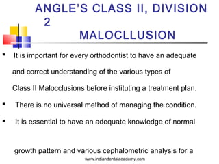
- 1. ANGLE’S CLASS II, DIVISION 2 MALOCLLUSION It is important for every orthodontist to have an adequate and correct understanding of the various types of Class II Malocclusions before instituting a treatment plan. There is no universal method of managing the condition. It is essential to have an adequate knowledge of normal growth pattern and various cephalometric analysis for a www.indiandentalacademy.com
- 2. INDIAN DENTAL ACADEMY Leader in continuing dental education www.indiandentalacademy.com www.indiandentalacademy.com
- 3. Angle’s Class II div 2 malocclusion is characterized by Class II Molar relation. The classic feature of this malocclusion is the presence of lingually inclined upper central incisors and labially inclined upper lateral incisors overlapping the central incisors. Incidence : 5 – 10 % Variations in this form exist. www.indiandentalacademy.com
- 4. INCISAL INTER ARCH RELATIONSHIP Anteroposterior incisor relatioships The British Standard Classification of incisor relationships has been widely adopted. • CLASS I INCISOR RELATIONSHIP The lower incisal edges occlude with or lie immediately below the cingulum plateaux (middle part of the palatal surfaces) of the upper central incisors. www.indiandentalacademy.com
- 5. CLASS II INCISOR RELATIONSHIP The lower incisal edges lie posterior to the cingulum plateaux of the upper incisors. DIVISION 1 – The upper central incisors are proclined or of average inclination and there is an increase in overjet. DIVISION 2 – The upper central incisors are retroclined ; the overjet is minimal but may be increased. www.indiandentalacademy.com
- 6. • CLASS III INCISOR RELATIONSHIP The lower incisal edges lie anterior to the cingulum plateaux of the upper incisors ; the overjet is reduced or reversed. www.indiandentalacademy.com
- 7. Van der Linden classification of Class II Div 2 depending on the spatial conditions in the maxillary dental arch. Type A- The upper central and lateral incisors are retroclined. It is of less severe in nature. www.indiandentalacademy.com
- 8. Type B- The central incisors are retroclined and overlapped by the lateral incisors. Type C- The central and lateral incisors are retroclined and overlapped by the canines. www.indiandentalacademy.com
- 9. FEATURES OF CLASS II DIV 2 • Molars in disto-occlusion. • The classic feature of the upper incisors. • Deep overbite. • Pleasing straight profile. • Broad square face. • Backward path of closure. • Deep mento-labial sulcus. • Absence of abnormal muscle activity. www.indiandentalacademy.com
- 10. CLINICAL FEATURES OF CLASS II DIV 2 EXTRAORAL • Squarish face (Brachycephalic). • Upper lip is invariably short and positioned high with respect to the upper anteriors. • Lower lip is thick flabby covering the upper incisors and exhibiting www.indiandentalacademy.com
- 11. • Usually straight to mildly convex profile because of less skeletal discrepancy and the retroclined incisors. • Usually straight face. • Deep mentolabial sulcus. www.indiandentalacademy.com
- 12. INTRAORAL CHARACTERISTICS • Class II molar relation indicating distal relation of mandible to the maxilla. • Decreased overjet, an increased overbite. • Deep bite usually traumatic. www.indiandentalacademy.com
- 13. • The upper arch is usually broad, ‘U’ shaped. • The palatal vault is usually deep. • An exaggerated curve of spee. www.indiandentalacademy.com
- 14. Aetiological considerations for class II div 2 Class II molar relationship 1. Dentoalveolar factors – Loss or absence of mesial teeth in maxilla. 2. Skeletal factors – Class II skeletal pattern. www.indiandentalacademy.com
- 15. Class II div 2 incisor realtionship 1. Dentoalveolar factors – Underdeveloped incisal cingulae. Lid Effect. 2. Skeletal factors – Class II skeletal pattern (often mild) ; Decreased lower facial height. 3. Neuromuscular factors – High lower lip line ; Over closure and under eruption of posterior teeth related to lack of inter incisal contact. www.indiandentalacademy.com
- 16. MANAGEMENT OF CLASS II DIV 2 Three important factors to consider in the management are : 1. The AGE at which the patient is seen. 2. The NATURE AND SEVEARITY of the problem. 3. The UNDERLYING ETIOLOGIC FACTORS as seen from the diagnostic aids and clinical and functional examination. www.indiandentalacademy.com
- 17. Mandible is usually guided posteriorly due to premature contact from the retroclined incisors and thereby restricting its growth. The treatment sequence remains the same except that for any form of treatment modality to be instituted the retroclined teeth have to be aligned in a proper labiolingual direction. www.indiandentalacademy.com
- 18. 1. Mixed dentition phase – Use of functional appliances after proclining the maxillary anteriors. Results are good even after the eruption of permanent teeth. The maxillary first premolars are extracted generally to create space for aligning crowded maxillary anterior segment. www.indiandentalacademy.com
- 19. 2. After the cessation of growth – The need for orthognathic surgery increases with the increase in the severity of the problem. The surgical procedures are also the same but the use of presurgical orthodontics becomes imperative to achieve stable results. Overall the treatment results are better after the resolution of class II div 2 malocclusion as compared to class II div 1 www.indiandentalacademy.com malocclusion.
- 22. DEEP BITE DEFINITION – GRABER has defined deep bite as a condition of excessive overbite, where the vertical measurement between the maxillary and mandibular incisal margins is excessive when the mandible is brought into habitual or centric occlusion. www.indiandentalacademy.com
- 23. Deep over bite can be of two types : Incomplete over bite – Its an incisor relationship in which the lower incisors fail to occlude with either the upper incisors or the mucosa of the palate when the teeth are occluded. Complete over bite – Its an incisor relationship where the lower incisors contact the palatal surface of the upper incisors or the palatal tissue when the teeth are in centric occlusion. This kind of deep bite often results in truama. www.indiandentalacademy.com
- 24. Classification – Deep bite can be broadly classified into two types . 1. Skeletal deep bite 2. Dental deep bite www.indiandentalacademy.com
- 25. Skeletal deep bite - Usually of Genetic origin. - Caused by upward and forward rotation of mandible. - Worsened in cases of forward inclination of maxilla. - Characterized by the presence of following features : • Horizontal growth pattern. • Reduced anterior facial height. • Reduced inter-occlusal clearance. • Mandibular plane, FH plane, SN plane etc. parallel to each other. www.indiandentalacademy.com
- 26. Dental deep bite Dental deep bite occur due to over – eruption of anteriors or infra – occlusion of molars. Deep bite due to over - eruption of anteriors : • Due to over-eruption of lower incisors usually seen class II malocclusions. • Patient exhibits an excessive curve of spee. • Inter- occlusal clearance is usually normal as the molars are fully erupted. www.indiandentalacademy.com
- 27. Deep bite due to infra-occlusion of molars : • Presence of lateral tongue posture or lateral tongue thrust may prevent the molars from erupting to their normal occlusal level. • Characterized by the presence of partially erupted molars (i,e reduced crown height ) and large inter- occlusal clearance. www.indiandentalacademy.com
- 28. Diagnosis Routine Diagnostic aids , 1. Clinical Examination 2. Study Models 3. Lateral Cephalogram – Helps to differentiate skeletal deep bite from dental deep bite. The patients with skeletal deep bite show a reduced mandibular plane angle as well as www.indiandentalacademy.com reduced anterior facial height.
- 29. Factors to consider in treatment of deep bite Deep bites are usually corrected by intrusion of anterior teeth or by extrusion of posterior teeth. There are certain factors that help an orthodontist to decide which of the two modalities is indicated for a given patient. • Lip relationship. • Consideration of vertical facial relationship. • Consideration of inter-occlusal space. www.indiandentalacademy.com
- 30. Treatment Deep bites can be treated by using Removable, Fixed or myofunctional appliances. Romovable appliances • Anterior bite plane Myofunctional Fixed appliance therapy appliances • Activator • Use of anchorage bends • Bionator • Use of archwires with reverse curve of spee • Use of utility arches www.indiandentalacademy.com
- 31. OPEN BITE Malocclusions can occur in three planes i,e. sagittal, transverse and in the vertical plane. Open bite is a malocclusion in the vertical plane, characterized by lack of vertical overlap between the maxillary and mandibular dentition. It may be an anterior or a posterior open bite. www.indiandentalacademy.com
- 33. Anterior open bite – Is a condition where there is no vertical overlap between the upper and lower incisors. Posterior open bite – Is a condition characterized by lack of contact Between the posteriors when the teeth are in centric occlusion. www.indiandentalacademy.com
- 34. Etiology The etiology is multifactorial. No single factor can account for most open bites. Can occur due to a variety of hereditary and non-hereditary factors. www.indiandentalacademy.com
- 35. Etiologic considerations of open bite Some of the etiologic factors responsible for anterior open bite : 1. Prolonged Thumb-sucking. 2. Tongue thrusting. 3. Nasopharyngeal airway obstruction and associated mouth breathing. 4. Inherited factors such as increased tongue size, and abnormal skeletal growth pattern of the maxilla and mandible. www.indiandentalacademy.com
- 36. Posterior open bites are very rare. The etiologic factors responsible for posterior open bite : 1. Mechanical interferences with the tooth eruption, either before or after the tooth emerges from the alveolar bone. 2. Failure of the eruptive mechanism of the tooth so that the expected amount of eruption does not occur. www.indiandentalacademy.com
- 37. Features of skeletal anterior open bite : • Increased lower anterior facial height. • Decreased upper anterior facial height. • Increased anterior and decreased posterior facial height. • A steep mandibular plane angle. • Small mandibular body and ramus. www.indiandentalacademy.com
- 38. • The patient may have a short upper lip with excessive maxillary incisor exposure. • The patient often has a long and narrow face. • Divergent cephalometric planes. • Steep anterior cranial base. • Downward and forward rotation of mandible. www.indiandentalacademy.com
- 39. Features of dental anterior open bite : • Proclined upper anterior teeth. • Upper and lower anteriors fail to fail to overlap resulting in a space. • Patient may have a narrow maxillary arch due to lowered tongue posture due to a habit. www.indiandentalacademy.com
- 40. Treatment Anterior open bite Posterior open bite • Removal of cause • Removal of cause Removable or fixed type Lateral tongue spikes for habit breaking appliance. lateral tongue thrust. • Myofunctional appliances Skeletal anterior open bite – F.R.IV or a modified activator • Fixed Orthodontic therapy • Surgical correction • If due to infra occlusion of ankylosed teeth, it is best treated by crowns. www.indiandentalacademy.com
- 41. www.indiandentalacademy.com Leader in continuing dental education www.indiandentalacademy.com