![Rigaku Journal, 30(1), 2014 27
Sample preparation for X-ray fluorescence analysis I
is important to determine the extent of the grinding
to balance the required analysis precision against the
preparation time needed to obtain that level of precision.
Examples of ground sample surface are shown in Fig. 2.
2.2. Powder
2.2.1. Powder method
Powder samples need to be pulverized when their
particle sizes are coarse. The samples are pressed after
pulverization to make pellets (Fig. 3). Analysis errors
in the measurement of powder samples come mostly
from heterogeneity effects. The heterogeneity effects can
be classified into mineralogical effect, segregation and
grain size effect.
[Mineralogical effect]
This is a phenomenon showing an analysis error due
to the variation in the mineral components of a sample,
when an element in the mineral sample containing a
number of different types of minerals is analyzed. The
explanation of this effect is described using the case of a
sample with aluminum oxide (Al2O3) and silicon dioxide
(SiO2) as an example.
Simplified two models are shown in Fig. 4. The
particle sizes of both models are the same and the
concentration of each component is 50mass%. Fig.
4(a) shows the model of mixture of particles of Al2O3
and SiO2 (Mixture), and Fig. 4(b) shows the model
of compound particles consisting of Al2O3 and SiO2
(Compound). Intensities of Si-Kα X-ray emitted from
the sample are shown by the width of the arrow.
The result is that the Fig. 4(a) mixture and the Fig.
4(b) compound differ in the effects of absorption of
fluorescence X-rays by co-existing elements, and X-ray
intensities are not the same even if their concentrations
are the same. This is called the mineralogical effect.
[Segregation]
There are two cases of segregation, namely, non-
uniformity of mixture of particles in sample and
non-uniformity of concentration in particles. A coated
particle is given as an example of the case that
concentration distribution exists in the particle. If such
a sample is analyzed using semi-quantitative analysis
by the fundamental parameter method (FP method),
after making pressed pellet without any preparation,
the analysis error of this sample due to the segregation
is large. In the case of the coated particle, pulverization
of the sample powder can reduce the segregation degree
reducing the analysis error as the coating layer also
becomes micro-particles.
[Grain-size effect]
This is a phenomenon showing the change of X-ray
intensity of the analyzed element due to the size of
particles. The effect is pronounced for X-rays having
low energies originating from light elements. Their
analysis is easily affected by the particle size as the
analysis depth is shallow and the analysis area is only
on or in the proximity of the sample surface. On the
other hand, as the X-rays of heavy elements have higher
energies and the analysis depth is deeper, the effect of
particle size on the X-rays is relatively small compared
with the effect on light elements. Figure 5(a) shows the
difference between the irradiated area of primary X-rays
and the detected area of fluorescence X-rays, and there
is a portion of area not irradiated by the primary X-rays
when the sample is viewed from the detecting angle of
fluorescence X-rays. On the other hand, Fig. 5(b) shows
the entire area is irradiated by the primary X-rays and
there is no part in the shadow, the fluorescence X-rays
are generated uniformly.
Heterogeneity effects can be reduced by
pulverization, but it cannot be eliminated entirely. It
is important for the powder method to minimize the
heterogeneity effect and to make standard samples and
unknown samples under the same condition (having the
same heterogeneity effect).
2.2.2. Fusion bead method
When materials like rocks, mineral ores, etc. are
analyzed with the pressed powder pellet method,
analysis errors due to the heterogeneity effect
mentioned above are generally present. To eliminate
the heterogeneity effect, there is a sample preparation
method called the fusion bead method. This is a method
Lathe Belt sander Grinder
Fig. 2. Examples of ground sample surface.
Fig. 3. Example of pressed pellet sample.](data:image/gif;base64,R0lGODlhAQABAIAAAAAAAP///yH5BAEAAAAALAAAAAABAAEAAAIBRAA7)
Recommended
Recommended
More Related Content
What's hot
What's hot (20)
Similar to Rigaku journal xrf
Similar to Rigaku journal xrf (20)
Recently uploaded
Recently uploaded (20)
Rigaku journal xrf
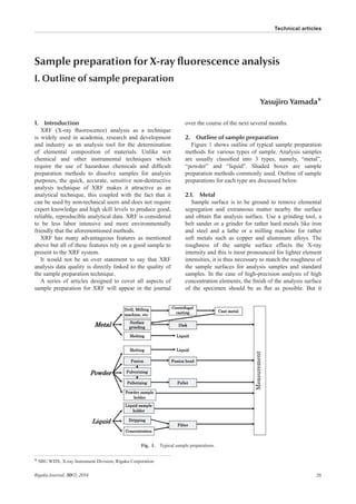
- 1. Rigaku Journal, 30(1), 2014 26 Sample preparation for X-ray fluorescence analysis I. Outline of sample preparation Yasujiro Yamada* 1. Introduction XRF (X-ray fluorescence) analysis as a technique is widely used in academia, research and development and industry as an analysis tool for the determination of elemental composition of materials. Unlike wet chemical and other instrumental techniques which require the use of hazardous chemicals and difficult preparation methods to dissolve samples for analysis purposes, the quick, accurate, sensitive non-destructive analysis technique of XRF makes it attractive as an analytical technique, this coupled with the fact that it can be used by non-technical users and does not require expert knowledge and high skill levels to produce good, reliable, reproducible analytical data. XRF is considered to be less labor intensive and more environmentally friendly that the aforementioned methods. XRF has many advantageous features as mentioned above but all of these features rely on a good sample to present to the XRF system. It would not be an over statement to say that XRF analysis data quality is directly linked to the quality of the sample preparation technique. A series of articles designed to cover all aspects of sample preparation for XRF will appear in the journal over the course of the next several months. 2. Outline of sample preparation Figure 1 shows outline of typical sample preparation methods for various types of sample. Analysis samples are usually classified into 3 types, namely, “metal”, “powder” and “liquid”. Shaded boxes are sample preparation methods commonly used. Outline of sample preparations for each type are discussed below. 2.1. Metal Sample surface is to be ground to remove elemental segregation and extraneous matter nearby the surface and obtain flat analysis surface. Use a grinding tool, a belt sander or a grinder for rather hard metals like iron and steel and a lathe or a milling machine for rather soft metals such as copper and aluminum alloys. The roughness of the sample surface effects the X-ray intensity and this is most pronounced for lighter element intensities, it is thus necessary to match the roughness of the sample surfaces for analysis samples and standard samples. In the case of high-precision analysis of high concentration elements, the finish of the analysis surface of the specimen should be as flat as possible. But it * SBU WDX, X-ray Instrument Division, Rigaku Corporation. Fig. 1. Typical sample preparations. Technical articles
- 2. Rigaku Journal, 30(1), 2014 27 Sample preparation for X-ray fluorescence analysis I is important to determine the extent of the grinding to balance the required analysis precision against the preparation time needed to obtain that level of precision. Examples of ground sample surface are shown in Fig. 2. 2.2. Powder 2.2.1. Powder method Powder samples need to be pulverized when their particle sizes are coarse. The samples are pressed after pulverization to make pellets (Fig. 3). Analysis errors in the measurement of powder samples come mostly from heterogeneity effects. The heterogeneity effects can be classified into mineralogical effect, segregation and grain size effect. [Mineralogical effect] This is a phenomenon showing an analysis error due to the variation in the mineral components of a sample, when an element in the mineral sample containing a number of different types of minerals is analyzed. The explanation of this effect is described using the case of a sample with aluminum oxide (Al2O3) and silicon dioxide (SiO2) as an example. Simplified two models are shown in Fig. 4. The particle sizes of both models are the same and the concentration of each component is 50mass%. Fig. 4(a) shows the model of mixture of particles of Al2O3 and SiO2 (Mixture), and Fig. 4(b) shows the model of compound particles consisting of Al2O3 and SiO2 (Compound). Intensities of Si-Kα X-ray emitted from the sample are shown by the width of the arrow. The result is that the Fig. 4(a) mixture and the Fig. 4(b) compound differ in the effects of absorption of fluorescence X-rays by co-existing elements, and X-ray intensities are not the same even if their concentrations are the same. This is called the mineralogical effect. [Segregation] There are two cases of segregation, namely, non- uniformity of mixture of particles in sample and non-uniformity of concentration in particles. A coated particle is given as an example of the case that concentration distribution exists in the particle. If such a sample is analyzed using semi-quantitative analysis by the fundamental parameter method (FP method), after making pressed pellet without any preparation, the analysis error of this sample due to the segregation is large. In the case of the coated particle, pulverization of the sample powder can reduce the segregation degree reducing the analysis error as the coating layer also becomes micro-particles. [Grain-size effect] This is a phenomenon showing the change of X-ray intensity of the analyzed element due to the size of particles. The effect is pronounced for X-rays having low energies originating from light elements. Their analysis is easily affected by the particle size as the analysis depth is shallow and the analysis area is only on or in the proximity of the sample surface. On the other hand, as the X-rays of heavy elements have higher energies and the analysis depth is deeper, the effect of particle size on the X-rays is relatively small compared with the effect on light elements. Figure 5(a) shows the difference between the irradiated area of primary X-rays and the detected area of fluorescence X-rays, and there is a portion of area not irradiated by the primary X-rays when the sample is viewed from the detecting angle of fluorescence X-rays. On the other hand, Fig. 5(b) shows the entire area is irradiated by the primary X-rays and there is no part in the shadow, the fluorescence X-rays are generated uniformly. Heterogeneity effects can be reduced by pulverization, but it cannot be eliminated entirely. It is important for the powder method to minimize the heterogeneity effect and to make standard samples and unknown samples under the same condition (having the same heterogeneity effect). 2.2.2. Fusion bead method When materials like rocks, mineral ores, etc. are analyzed with the pressed powder pellet method, analysis errors due to the heterogeneity effect mentioned above are generally present. To eliminate the heterogeneity effect, there is a sample preparation method called the fusion bead method. This is a method Lathe Belt sander Grinder Fig. 2. Examples of ground sample surface. Fig. 3. Example of pressed pellet sample.
- 3. Rigaku Journal, 30(1), 2014 28 Sample preparation for X-ray fluorescence analysis I to make a glass disk which is a uniform sample by fusing powder sample with a flux such like lithium tetra-borate at 1000–1200°C. In general, the mixing ratio of sample and flux in weight is sample 1: flux 10. It is important for the fusion bead method to pay attention to trace component analysis due to the sensitivity drop of the dilution effect. Another advantage of the fusion bead method is that standard samples for calibration can be made with pure chemical reagents. A procedure for making a fusion bead sample is shown in Fig. 6. 2.3. Liquid There are two ways to analyze liquid samples, namely, to pour the liquid into the sample cell having a sample film which constitutes the liquid sample holder (Liquid method) or to measure the filter paper on which liquid is deposited then dried (Micro-droplet method). For the analysis of trace metal components, there is a method to concentrate metallic components using chemical reagents (Concentration method). 2.3.1. Liquid method The liquid sample is poured directly into the sample cell that has a sample film fixed to it and elements heavier than Na are analyzed directly in the liquid. Polypropylene-type film is used for acid and alkaline solutions, and polyester-type film is used for lubricant oil, fuel oil etc. It is important to check the impurities in the film used for the analysis beforehand as the polyester-type film can contain impurities that may be elements of interest in the analysis. The preparation of the liquid sample is easier for a tube below type instruments where the analysis sample surface faces downward, than for a tube above type instrument where Model (a): Mixture Model (b): Compound Fig. 4. Mineralogical effect. Model (a) Model (b) Fig. 5. Grain size effect. Fig. 6. Preparation procedure of fusion bead sample.
- 4. Rigaku Journal, 30(1), 2014 29 Sample preparation for X-ray fluorescence analysis I the analysis sample surface faces upward. The typical sample cells for the tube below type and the tube above type are shown in Fig. 7. 2.3.2. Micro-droplet method This is the method to measure a filter paper or the like on which liquid has been pipetted and dried. It is possible to measure these samples under vacuum atmosphere without sample film, thus light elements such as Mg, Na, F, etc. in liquids can be analyzed with higher sensitivity. The filter paper for exclusive use of the deposition method (Micro-carry) which has slits to control the expansion of liquid samples is shown in Fig. 8, and it makes quantitative analysis possible by pipetting a fixed amount of liquid on the filter. For greater sensitivity, special films (Ultra-carry, Ultra-carry light) have been developed to hold a larger volume of liquid for higher sensitivity measurements of elements in aqueous solution type sample (Fig. 9). 2.3.3. Concentration method This is a method to measure the collected precipitate produced by the reaction between the metal ion in the liquid and a chelate reagent. The precipitate is collected using a filtration technique and the resultant filter is dried and the precipitate collected on the filter is measured by XRF. The technique is useful for the analysis of trace metal elements in aqueous solutions. The outline of the typical sample preparation methods is discussed in this article. In-depth sample preparation methods for powder samples will be discussed in the next issue. Liquid sample cell for tube below system (Analysis surface down) Liquid sample cell for tube above system (Analysis surface up) Fig. 7. Liquid sample cell. Fig. 8. Filter-paper (MicroCarry). Fig. 9. High sensitivity. Sample carrier film (UltraCarry Light).