
Recommended
More Related Content
What's hot
What's hot (20)
Similar to Examination of bone in general
Similar to Examination of bone in general (20)
Recently uploaded
Recently uploaded (20)
Examination of bone in general
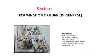
- 1. Semina r PRESENTED BY- ASHEESH BABU PATEL (PG RESIDENT- 1st YEAR) DATE- 02/11/2020 DEPT. OF FORENSIC MEDICINE AND TOXICOLOGY, LATE. B.R.K.M.G.M.C. JAGDALPUR, C.G. 1 EXAMINATION OF BONE (IN GENERAL) 1
- 2. CONTENTS • Introduction • Source of bone for medicolegal examination • Anatomy of bone • Information that can be sought from examination of skeletal remains • Aids and Tools for examination of bone • Medicolegal importance 2
- 3. INTRODUCTION Forensic osteology is a sub-speciality of forensic medicine and deals with examination and assessment of human skeleton. The assessment includes both- the identification of victims characteristics and cause and manner of death from skeleton.1 Sometimes bones are found disposed off in jungle, in the open, in the ditches or rubbish dumps, etc., or may be found while digging foundation for buildings or skeleton may be exhumed. In case of mass disaster, where many persons die in the same area at the same time from fire, air crashes, etc., the help of the anthropologist is sought in identification, if the remains are skeletonised, badly burnt or mostly destroyed.2 3
- 4. SOURCE OF BONES FOR MEDICOLEGAL EXAMINATION • Police/magistrate requisition for medicolegal examination, panchnama, • Scene/photograph from where bone is recovered, map. • source of bone - • In the open, in jungle • In rubbish dumps • Exhumated • Charred bone etc. 4
- 6. Given article is bone or not ? • The bone can be differentiated from other object that look like bone or dummy bone by- • external appearance- muscular marking, presence of growth plate, growth plate fusion marking etc. • By X-ray examination – showing trabeculation, bone marrow, dense bone which are feature of original bone. • On cut section original bone show haversian system in diaphysis and cancellus bone in epiphysis or cancellous bone between two layers of compact bone in case of flat bones. • Precipitation test can differentiate between human bone and other bone like objects. 6
- 7. INFORMATION THAT CAN BE SOUGHT FROM EXAMINATION OF SKELETAL REMAINS 2. Belong to human being ? • By anatomy(gross and microscopic features- larger haversian system and canal) • Serological examination - Precipitin test with specific antisera of human 1.Bone or not ? by- 1. gross examination 2. microscopic examination - Haversian system and presence of osteons 7
- 8. a b 8
- 9. 4. Race 1. cephalic index – skull bone 2. from teeth features 3. features and indices of long bones 3. One individual or more ? 1. if belong to same individual - race, age, sex, stature, time since death and fit snugly and nicely at their corresponding joints. 2. mixed agglutination test 9
- 10. 5. sex • All bone- 100% • Pelvis + skull- 98% • Pelvis- 95% • Skull- 90 % • Long bones- 80% Can be determined by - Morphological examination Morphometry or osteometry Multivariate discrimination function analysis Demonstration of Y- chromosome DNA profiling 10
- 11. 6. AGE Age at the time of death from bone/skeleton is determined by noting the- • Ossification data- appearance and fusion of ossification centers • Age related changes in individual bones • Radiographic method • Histological method – size and number of osteon increases with age 11
- 12. 7. STATURE • For estimation of stature long bone are more reliable than flat and irregular bones.1 • When stature is estimated from a bone , an allowance of 2.5- 4 cm is added to the calculated stature in order to compensate the loss of soft tissues.1 • Different formulae are used to estimate stature-1 1. Karl pearson 2. Trotter and glesser 3. Dupertuis haden 4. Pan 5. Nat 6. Shah and siddiqui For fragmented bone-1 1. Muller 2. Steele 3. Steele and McKern 12
- 13. 8. ADDITIONAL IDENTIFICATION FEATURES2 • Special feature in teeth- torsion, angulation, staining, cracks, caries, sealings etc. • Features in bone- bony deformities, healed fracture, malunion etc. • In some cases – DNA testing may be helpful. 13
- 14. 9. CAUSE OF DEATH • In most cases not possible,3 • Ante-mortem injury to bones covering the vital organs • Ante-mortem fracture of bones of non-vital parts suggest serious assault • Some poisons like As can be detected In some cases cause of death can be ascertained-3 14
- 15. 10. TYPE OF WEAPON • Hard blunt weapon • Light heavy sharp cutting • Pointed or firearm injury • tuberculosis • Sarcoma • Thalassemia, pagets disease of bone, congenital disease and deformities, metabolic bone disease etc. • helps in identification and may indicate COD By examination of injury over skeleton type of weapon used can be known- 11. Disease or pathology 15
- 16. 12. TIME SINCE DEATH • Time of death can only be roughly ascertained3 Condition of bone Time since death Bone + soft tissue(fascia, ligaments) 2 weeks – 2 months2 Bone ( wet, no soft tissue attached) 1 to 3 months2 Bone (dry with putrid smell) Within 3 months2 Bone ( dry, no putrid smell, retained normal colour) 3 months – 1 year2 • After some years unpreserved bone get destroyed and gradually reduce to dust, but exact ageing of skeletonization is not possible. 2 16
- 17. the Exclusion of a Forensically Relevant Lay Time 17
- 18. 18
- 19. 13. By site and type of fracture or detection of any poison or pathology , nature of death may be said - ex. Deposition of heavy metal Pb in the growing part of bone at metaphysis. 14. Mode and place of disposal - • Deep grave – skeletonize slowly • Open air – dries up early • In forest/water – bone may be partly eaten by animal • Stains on bone – gives idea about place of disposal 19
- 20. 15. MANNER OF SEPARATION • Natural separation • Unnatural separation- assault, accident, by animal etc. 20
- 21. Aids/Tools for examination of bone • Osteometric board • Weighing scale for bone • Bone chart • Microscopy – haversian system • UV fluorescence • Specific gravity and density test- decreases with bone become older • Benzidine test and Kastel-Mayer test – detects presence of blood and may be positive upto 100 years. • Precipitation test, gel diffusion or Coomb’s test may be positive for 5-10 years • DNA analysis • Nitrogen content and amino acid content- decreases with time • X-ray ,CT-scan (radiographic techniques) • Carbon-14 dating • Strontium and plutonium dating etc. 21
- 23. Bone chart 23
- 24. a b 24
- 25. MEDICOLEGAL IMPORTANCE • Identification of victim • Identification of missing person • Antemortem vs postmortem trauma • Cause of death, manner of death can be known • Time since death. 25
- 26. bibliography 1. Bardale R., Forensic Osteology, Principles of forensic medicine and toxicology, 1st edition 2011. 2. Reddy K.S.N. ,Murthy O.P., Medicolegal Autopsy, The essential of forensic medicine and toxicology, 24th edition 2017. 3. Nandy A. ,Death and Post-Mortem changes, Principles of Forensic Medicine including Toxicology, 3rd edition 2010. 4. Black S.M., Bone Pathology and Antemortem Trauma, Encyclopedia of Forensic and Legal Medicine, 2005 Elsevier Ltd. P. 105-112 5. Verhoff M.A. and Kreutz K., Macroscopical Findings on Soil-Embedded Keletal Remains Allowing the Exclusion of a Forensically Relevant Lay Time, Forensic Pathology Reviews, vol-3, Edited by: M Tsokos, 2005, p244-247. 26
- 27. THANK YOU 27