Rheumatic fever in pediatrics
•Download as DOCX, PDF•
35 likes•18,187 views

Tikrit university collage of medicine pediatric lecture dr.Emad Alsadoon

Recommended
More Related Content
What's hot
What's hot (20)
Viewers also liked
Similar to Rheumatic fever in pediatrics
Similar to Rheumatic fever in pediatrics (20)
Recently uploaded
Recently uploaded (20)
Rheumatic fever in pediatrics
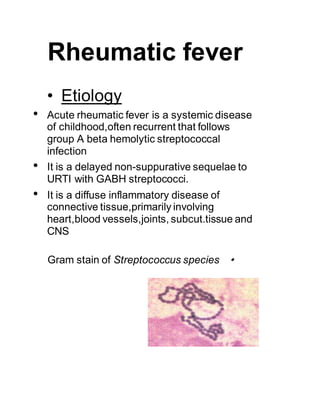
- 1. Rheumatic fever • Etiology • Acute rheumatic fever is a systemic disease of childhood,often recurrent that follows group A beta hemolytic streptococcal infection • It is a delayed non-suppurative sequelae to URTI with GABH streptococci. • It is a diffuse inflammatory disease of connective tissue,primarily involving heart,blood vessels,joints, subcut.tissue and CNS •Gram stain of Streptococcus species
- 2. • Epidemiology • Ages 5-15 yrs are most susceptible • Rare <3 yrs • Girls>boys • Common in 3rd world countries • Environmental factors-- over crowding, poor sanitation, poverty, • Incidence more during fall ,winter & early spring • Pathogenesis • Delayed immune response to infection with group.A beta hemolytic streptococci. • After a latent period of 1-3 weeks, antibody induced immunological damage occur to heart valves,joints, subcutaneous tissue & basal ganglia of brain • evidence for Strep pyogenes as a cause of ARF: – close temporal relationship between epidemics of streptococcal pharyngitis / scarlet fever & epidemics of ARF
- 3. – most patients with ARF give a history of pharyngitis, & if absent serology supports this – continuous antibiotic prophylaxis that prevents Strep pyogenes pharyngitis also prevents ARF – M protein share epitiopes which ARF patients mount IgG response – serotypes vary in their ability cause ARF, certain M types are strongly associated with ARF while others are not • Exact mechanism that lead to the development of ARF not completely understood • Requirements – Strep pharyngitis – Host susceptibility – NO evidence that streptococcal toxins are important
- 4. • Group A Beta Hemolytic Streptococcus Strains that produces rheumatic fever - M types l, 3, 5, 6,18 & 24 • Pharyngitis- produced by GABHS can lead to- acute rheumatic fever , rheumatic heart disease & post strept. Glomerulonepritis • Skin infection- produced by GABHS leads to post streptococcal glomerulo nephritis only. It will not result in Rh.Fever or carditis as skin lipid cholesterol inhibit antigenicity
- 5. • M proteins • consist of two chains with a-helical structure, very rare in bacteria, but a common in mammalian proteins • > 80 distinct M proteins • Antigenic variation is due primarily to single amino acid substitutions • the M protein types of GAS associated with impetigo and other skin infections are different from those associated with clinical pharyngitis • Rheumatic fever always follows pharyngitis and is more commonly associated with certain M types (1,3,5,16,18) • Glomerulonephritis can follow pharyngitis (commonly M- types 1,4,12,15) or impetigo (M-types 49,52,55,59-61) and appears to be restricted to a smaller list of "nephritogenic" strains • Pathologic Lesions • Fibrinoid degeneration of connective tissue,inflammatory edema, inflammatory cell infiltration & proliferation of specific cells resulting in formation of Ashcoff nodules, resulting in- -Pancarditis in the heart -Arthritis in the joints -Ashcoff nodules in the subcutaneous tissue -Basal gangliar lesions resulting in chorea
- 6. • Rheumatic Carditis Histology (40X) • Clinical Features 1.Arthritis • Flitting & fleeting migratory polyarthritis, involving major joints • Commonly involved joints-knee,ankle,elbow & wrist • Occur in 80%,involved joints are exquisitely tender • In children below 5 yrs arthritis usually mild but carditis more prominent • Arthritis do not progress to chronic disease
- 7. • 2.Carditis Manifest as pancarditis(endocarditis, myocarditis and pericarditis),occur in 40-50% of cases • Carditis is the only manifestation of rheumatic fever that leaves a sequelae & permanent damage to the organ • Valvulitis occur in acute phase • Chronic phase- fibrosis,calcification & stenosis of heart valves(fishmouthvalves) • Rheumatic heart disease. Abnormal mitral valve. Thick, fused chordate • Another view of thick and fused mitral valves in Rheumatic heart disease
- 8. • 3.Sydenham Chorea • Occur in 5-10% of cases • Mainly in girls of 1-15 yrs age • May appear even 6/12 after the attack of rheumatic fever • Clinically manifest as-clumsiness, deterioration of handwriting,emotional lability or grimacing of face • Clinical signs- pronator sign, jack in the box sign , milking sign of hands • 4.Erythema Marginatum • Occur in <5%. • Unique,transient,serpiginous-looking lesions of 1-2 inches in size • Pale center with red irregular margin • More on trunks & limbs & non-itchy • Worsens with application of heat • Often associated with chronic carditis
- 9. 5.Subcutaneous nodules • Occur in 10% • Painless,pea-sized,palpable nodules • Mainly over extensor surfaces of joints,spine,scapulae & scalp • Associated with strong seropositivity • Always associatedwith severe carditis Other features (Minor features) • Fever-(upto 101 degree F) • Arthralgia • Pallor • Anorexia • Loss of weight
- 10. • Laboratory Findings • High ESR • Anemia, leucocytosis • Elevated C-reactive protien • ASO titre >200 Todd units. (Peak value attained at 3 weeks,then comes down to normal by 6 weeks) • Anti-DNAse B test • Throat culture-GABHstreptococci • ECG- prolonged PR interval, 2nd or 3rd degree blocks,ST depression,T inversion • 2D Echo cardiography- valve edema,mitral regurgitation, LA & LV dilatation,pericardial effusion,decreasedcontractility • Diagnosis • Rheumatic fever is mainly a clinical diagnosis • No single diagnostic sign or specific laboratory test available for diagnosis • Diagnosis based on MODIFIED JONES CRITERIA
- 11. •Jones Criteria (Revised) for Guidance in the Diagnosis of Rheumatic Fever* •*The presence of two major criteria, or of one major and two minor criteria, indicates a high probabilityof acute rheumatic fever, if supported by evidence of Group A streptococcal nfection. • Exceptions to Jones Criteria Chorea alone, if other causes have been excluded Insidious or late-onset carditis with no other explanation Patients with documented RHD or prior rheumatic fever,one major criterion,or of fever,arthralgia or high CRP suggests recurrence Major Manifestation Minor Manifestations Supporting Evidence of StreptococalInfection Carditis Polyarthritis Chorea Erythema Marginatum Subcutaneous Nodules Clinical Laboratory Increased Titer of Anti- Streptococcal Antibodies ASO (anti-streptolysin O), others Positive Throat Culture for Group A Streptococcus Recent Scarlet Fever Previous rheumatic fever or rheumatic heart disease Arthralgia Fever Acute phase reactants: Erythrocyte sedimentation rate, C-reactive protein, leukocytosis Prolonged P- R interval
- 12. • Differential Diagnosis • Juvenile rheumatiod arthritis • Septic arthritis • Sickle-cell arthropathy • Kawasaki disease • Myocarditis • Scarlet fever • Leukemia • Treatment • Step I - primary prevention (eradication of streptococci) • Step II - anti inflammatory treatment (aspirin,steroids) • Step III- supportive management & management of complications • Step IV- secondary prevention (prevention of recurrent attacks)
- 13. • STEP I: Primary Prevention of Rheumatic Fever (Treatmentof Streptococcal Tonsillopharyngitis) • Agent Dose Mode Duration • Benzathine penicillin G 600 000 U for patients Intramuscular Once 27 kg (60 lb) 1 200 000 U for patients >27 kg or • Penicillin V Children: 250 mg 2-3 times daily Oral 10 d (phenoxymethyl penicillin) Adolescents and adults: 500 mg 2-3 times daily • For individuals allergic to penicillin • Erythromycin: 20-40 mg/kg/d 2-4 times daily Oral 10 d Estolate (maximum 1 g/d) • or • Ethylsuccinate 40 mg/kg/d 2-4 times daily Oral 10 d (maximum 1 g/d)
- 14. • Step II: Anti inflammatory treatment Clinical condition Drugs • 3.Step III: Supportive management & management of complications • Bed rest • Treatment of congestive cardiac failure: - digitalis,diuretics • Treatment of chorea: -diazepam or haloperidol • Rest to joints & supportive splinting Arthritis only Aspirin 75-100 mg/kg/day,give as 4 divided doses for 6 weeks (Attain a blood level 20- 30 mg/dl) Carditis Prednisolone 2-2.5 mg/kg/day, give as two divided doses for 2 weeks Taper over 2 weeks & while tapering add Aspirin 75 mg/kg/day for 2 weeks. Continue aspirin alone 100 mg/kg/day for another 4 weeks
- 15. • STEP IV : Secondary Prevention of Rheumatic Fever (Prevention of Recurrent Attacks) • Agent Dose Mode • Benzathine penicillin G 1 200 000 U every 4 weeks* Intramuscular or • Penicillin V 250 mg twice daily Oral or • Sulfadiazine 0.5 g once daily for patients 27 kg (60 lb Oral 1.0 g once daily for patients >27 kg (60 lb) • For individuals allergic to penicillin and sulfadiazine • Erythromycin 250 mg twice daily Oral • *In high-risk situations, administration every 3 weeks is justified and recommended
- 16. • Duration of Secondary Rheumatic Fever Prophylaxis • Categor Duration • Rheumatic fever with carditis and residual heart disease until (persistent valvar disease*) lifelong At least 10 y since last episode and at least age 40 y, sometimes prophylaxis Rheumatic fever with carditis, but no residual heart disease (no valvar disease*) 10 y or into adulthood well whichever is longer • Rheumatic fever without carditis 5 y or until age 21 y, whichever is longer • *Clinical or echocardiographic evidence. • Prognosis • Rheumatic fever can recur whenever the individual experience new GABH streptococcal infection,if not on prophylactic medicines • Good prognosis for older age group & if no carditis during the initial attack • Bad prognosis for younger children & those with carditis with valvar lesions