1. ASIA-PACIFIC JOURNAL OF ONCOLOGY & HEMATOLOGY ORIGINAL RESEARCH
Management of Ocular Posterior Segment Hemorrhages
in Patients With Hemophilia
Teng-Yi Wang1, Chi-Ting Horng2, Yeu-Chin Chen3, Yung-Hsiang Chang1, Ke-Hung Chien1, Da-Wen Lu1, Jiann-Torng Chen1 and
Ming-Ling Tsai1,4
Affiliations: : 1Department of Ophthalmology, Tri-Service General Hospital, Taipei, Taiwan; 2Department of Ophthalmology,
Kaohsiung Armed Force General Hospital, Kaohsiung, Taiwan; 3Division of Hematology-Oncology, Department of Internal Medicine,
Tri-Service General Hospital, Taipei, Taiwan; 4Department of Ophthalmology, Veterans General Hospital, Taichung, Taiwan
Submission date: 10th May 2009, Revision date: 15th June 2009, Acceptance date: 1st July 2009
A B S T R A C T
Hemophilia is a bleeding disorder characterized by a deficiency of clotting factors. This disease can have a profound
and debilitating effect on the gastrointestinal, neurologic, and musculoskeletal systems. Ophthalmologists may encounter
patients with hemophilia and ocular hemorrhages ranging from subconjunctival hemorrhages to ocular posterior segment
hemorrhages that may lead to significant visual impairment. The role of the ophthalmologist in the care of a hemophilia
patient usually consists of controlling the bleeding source and establishing a clear optic medium to restore visual acuity.
Factor replacement therapy is essential for gaining rapid control of bleeding diatheses in these patients. Furthermore,
surgical intervention may be necessary to remove residual hemorrhages and recover vision. In this study, we analyzed the
available reports on ocular posterior segment hemorrhages developing in patients with hemophilia, focusing on patient
characteristics, mechanisms, and treatments.
Keywords: hemophilia, retinal hemorrhage, disc hemorrhage, ocular surgery
Correspondence: Ming-Ling Tsai, M.D., Ph.D., Department of Ophthalmology, Tri-Service General Hospital, No.
325, Sec. 2, Chenggong Rd, Taipei 114, Taiwan, Republic of China. Tel: +886-9-68392245; fax: +886-2-28388737; e-mail:
doc30845@yahoo.com.tw
INTRODUCTION therapy rapidly led to medical and social improvements,
Hemophilia is an unusual bleeding disorder caused by with a decrease in the frequency of hemorrhages and
an acquired or hereditary deficiency of clotting factors considerably improved life expectancy of patients with
in the blood. Acquired hemophilia, much rarer than the hemophilia. However, there are still case reports show-
hereditary type, is a rare autoimmune bleeding disorder, ing that ocular hemorrhages such as subconjunctival,
resulting from the presence of autoantibodies directed macular, and optic disc hemorrhages occur in hemo-
against coagulation factors. The etiology of the disor- philiacs. Of these conditions, ocular posterior segment
der remains obscure, although approximately half of all hemorrhages (retina, choroids, optic nerve, etc) always
cases are associated with underlying conditions, such as draw the attention of ophthalmologists and hematolo-
lupus and rheumatoid arthritis. Hereditary hemophilia gists because they can lead to significant visual impair-
is an X-linked genetic bleeding disease caused by a defi- ment. Therefore, it is necessary to improve our under-
ciency of coagulation factor VIII (hemophilia A) and fac- standing of the diagnosis and management of patients
tor IX (hemophilia B), or an autosomal recessively inher- with ocular posterior segment hemorrhages associated
ited factor XI deficiency (hemophilia C). Because of the with hemophilia. Moreover, ocular surgical intervention
hereditary pattern of hemophilia, patients are almost may be necessary when surgical conditions developed
invariably male, while women can be carriers of the dis- secondary to posterior segment hemorrhages such as
ease. Before the introduction of clotting factor replace- glaucoma, cataract, and vitreous hemorrhages.
ment therapy, the mean life expectancy of patients with In this investigation, we analyzed the previous reports
hemophilia was commonly less than 30 years, and pa- describing the characteristics of ocular posterior seg-
tients mostly died of intracranial or other hemorrhages. ment hemorrhages developing in patients with hemo-
Since the 1960s, a series of coagulation factor prepa- philia. In addition, we also reviewed the characteristics
rations including cryoprecipitates, plasma-derived fac- of hemophilia patients who underwent ocular surgery,
tors, and recombinant factors have been available for because ocular posterior segment hemorrhages may
the treatment of hemophilia. This factor replacement lead to many conditions that require ocular surgery.
APJOH 2009; 1: (3). September 2009 1 www.slm-oncology.com
2. Asia-Pacific Journal of Oncology & Hematology
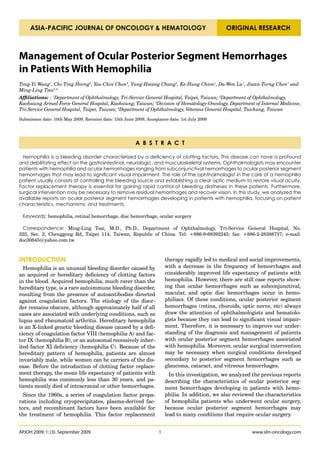
Table 1. Demographics of Patients With Ocular Hemorrhages Associated With Hemophilia in the Literature
Case no.
1 2 3
Authors Hon et al Maguluri et al Wang et al
Age (years) 80 52 13
Ocular findings
Visual acuity 6/60 20/Hand motion 6/6
Posterior
Macular hemorrhage Hyphema
segment
Vitreous hemorrhage Disc hemorrhages
Laboratory findings
PT (s) and normal range 12.2 (11.2–13.2) 11.3 (10–13) n/a
PTT (s) and normal range 86.2 (27.6–37.6) 45.1 (24–35) n/a
Factor status VIII inhibitor VIII 11% VIII 1.9%
Inhibitor status Present, 57 BU None None
Diagnosis Acquired hemophilia A Mild hemophilia A Moderate hemophilia A
Macular hemorrhage,
Choroidal hemorrhage,
Hemorrhagic complications choroidal hemorrhage with Disc hemorrhages
retinal detachment
acute angle closure glaucoma
Mechanisms Spontaneous After surgery Spontaneous
Treatments
Aspiration of hyphema,
Ocular management None vitrectomy, lensectomy, None
enucleation
For intraocular pressure
reduction: acetazolamide,
mannitol, pilocarpine
For hemorrhages: Perioperative rFVIII Infusion of factor VIII
Medical therapy
prednisolone 40 mg daily, infusion for enucleation concentrates: 21000 U in total
factor VIII concentrate
For laser peripheral iridec-
tomy: rFVIIa
Loss of vision, enucleation
Died of intracranial
Prognosis without bleeding Visual acuity: 6/6
hemorrhage
complications
BU, Bethesda units; rFVIIa, activated recombinant factor VII; rFVIII, Recombinant factor VIII.
METHODS RESULTS
We searched PubMed and Medline databases using We identified 45 references through electronic and
keywords including hemorrhage, hemophilia, vitreous, manual searches. After reading the titles and abstracts,
retina, choroids, optic disc, ocular surgery, ocular poste- we excluded 23 irrelevant references and selected 22 for
rior segment, and related terms. We excluded patients further assessment. Only 21 references met the crite-
with ocular posterior segment hemorrhages resulting ria. Because some previous treatments such as cryopre-
cipitate would not be used nowadays by a hematologist,
from other systemic diseases such as diabetes mellitus,
only cases that had been documented for more than 10
hypertension, and vessel embolization. Patients with
years were listed in the references [4-7, 9]. In this in-
prior ocular diseases such as optic disc edema, papillitis, vestigation, all of the relevant data are shown in Ta-
ischemic optic neuropathy, drusen, and juxtapapillary ble 1, which documents the features of ocular posterior
subretinal neovascular membranes were also excluded. segment hemorrhage in patients with hemophilia, and
Medical and ocular history, systemic condition, ocular Table 2, which summarizes the characteristics of hemo-
examinations, and ancillary testing were reviewed ret- philia patients who underwent ocular surgery. Moreo-
rospectively for each patient. ver, these selected cases are described below.
APJOH 2009; 1: (3). September 2009 2 www.slm-oncology.com
3. Management of Ocular Posterior Segment Hemorrhages in Patients With Hemophilia
Table 2. Demographics of Hemophilia Patients Scheduled for Ophthalmic Surgery in the Literature
Case no.
1 2 3 4 5 6 7
Authors Jijina et al Jijina et al Jijina et al Jijina et al Jijina et al Biron-An- Pekçelen
dréani et al et al
Age (years) 60 8 9 75 56 40 36
Ocular Cataract Trabeculec- Hyphema Cataract Cataract Cataract Cataract
surgery extraction tomy aspiration, extraction extraction extraction extraction
vitrectomy
Laboratory
findings
Factor status VIII 9.5% IX <1% VIII 32% IX 26% VIII 12% VIII 1% VIII <1%
Inhibitor None None None None None Present: Present:
status OD 3.5 BU >600 BU
OS 1.8 BU
Diagnosis Mild hemo- Severe he- Mild hemo- Hemophilia Mild hemo- Acquired Acquired
philia A mophilia B philia A B philia A hemophilia hemophilia
A A
Preventive Preop: 2000 Preop: 1800 Preop: Preop: 3000 Preop: OD: 150 IU/ Preop: 90
treatment U factor U factor IX DDAVP U factor IX DDAVP and kg of Auto- μg/kg (7.2
VIII concen- concentrate and factor Postop: ep- 3000 U of plex (total mg) of
trate raised Postop: VIII 1800 silon amino factor VIII 12000 IU) rFVIIa and
factor level 4800 U in U raised caproic acid raised factor OS: 90 μg/ tranexamic
to 80% total factor level 1 g q8 h for level to 62% kg (8.1 mg) acid 20 mg/
Postop: to 15% for 10 days Postop: of rFVIIa kg q8 h
1000 U q8 aspiration factor VIII preopera- Postop: 90
h for 48 h of hyphema; 3000 U, tively; total μg/kg of
(total 7000 factor VIII DDAVP 0.3 48.6 mg rFVIIa q12
U) 1500 U for μg/kg qod h twice
vitrectomy for 4 times,
Postop: and epsilon
DDAVP 0.3 amino
μg/kg qd for caproic acid
3 days for 1 g q8 h for
aspiration 10 days
of hyphema;
750 U factor
VIII qd for
5 days for
vitrectomy
Total 6750
U of factor
VIII
BU, Bethesda units; DDAVP, desmopressin; OD, right eye; OS, left eye; rFVIIa, activated recombinant factor VII.
Patients With Hemophilia relatively less common in these cases. One of the three
and Intraocular Hemorrhage cases completely lost vision, whereas one recovered to
The demographic characteristics of the three patients normal vision. One patient died of intracranial hemor-
with hemophilia complicated with intraocular hemor- rhage. In the early years, cryoprecipitates and epsilon
rhage are shown in Table 1. The age range was wide, amino caproic acids were used for hemostasis before and
from 13 to 80 years of age. Ocular posterior segment during operations. With the advent of new coagulation
hemorrhages may develop in both hereditary and ac- products, factor concentrates and recombinant factors
quired hemophilia. All levels of severity of hemophilia have remained among the mainstream treatments. In
were associated with posterior segment hemorrhages. cases where factor inhibitors were present, steroids
One of the three patients had hemorrhages after sur- were also added to the treatment formula to optimize
gery and trauma, while in two of the other patients, it the treatment outcome.
had occurred spontaneously. Two of the three patients Case 1. Hon et al [1] reported a rare case of nontrau-
had choroidal detachment with hemorrhage and poor matic macular hemorrhage. The patient initially pre-
visual outcome. Disc and macular hemorrhages were sented with monocular blindness due to gross macular
www.slm-oncology.com 3 APJOH 2009; 1: (3). September 2009
4. Asia-Pacific Journal of Oncology & Hematology
hemorrhage, and returned because of painful loss of vi- Jijina et al [8] carried out ophthalmic surgery in five
sion. A choroidal hemorrhagic detachment detected by patients (cases 1 to 5) during the period between 1994
ultrasound B scan resulted in secondary glaucoma that and 2000. The surgeries performed included cataract
did not respond to medical therapy. The laboratory ex- extraction with intraocular lens implantation, trab-
aminations later revealed the diagnosis of acquired fac- eculectomy, vitrectomy, and hyphema aspiration. These
tor VIII inhibitor. Despite treatment with prednisolone, patients received factor VIII or IX before and after the
factor VIII concentrates and laser iridotomy, the patient operations with or without addition of desmopressin
died of a sudden cerebral hemorrhage. (DDAVP).
Case 2. Maguluri et al [2] described an undiagnosed Case 1. The preoperative factor VIII level was increased
hemophilia patient with delayed suprachoroidal hemor- from 9.5% to 80% with the use of 2000 units of factor
rhage after head injury. The patient had hyphema and VIII concentrate. A total of 7000 units of factor VIII was
vitreous hemorrhage and received an anterior cham- used for the cataract extraction procedure, and no post-
ber washout, but the hyphema recurred together with operative complications were noted.
choroidal detachment and suprachoroidal hemorrhage.
Case 2. The preoperative level of factor IX was <1%.
Accordingly, he received pars plana lensectomy and vit-
Preoperative use of 1800 units of factor IX resulted in
rectomy, but the bleeding persisted, resulting in a dis-
100% correction. The trabeculectomy was successfully
organized retinal anatomy with poor visual outcome.
performed, and a total of 4800 units of factor IX was
The hematology service was then consulted because of
used.
the persistent bleeding despite surgical intervention,
and laboratory evaluation revealed a factor VIII level of Case 3. The patient underwent aspiration of hyphema
11%. With perioperative recombinant factor VIII infu- owing to trauma. The preoperative level of factor VIII
sion, enucleation for pain relief was performed without was 32%. He received DDAVP and 1800 units of factor
,
complications. VIII.
Case 3. Wang et al [3] reported a 13-year-old boy with Case 4. The preoperative level of factor IX was 26%.
known hemophilia A who suffered a black shadow in The patient underwent trabeculectomy and bilateral
front of his left eye. Vision was 6/6 in both eyes. Fun- cataract extraction with intraocular lens implantation
dal examination revealed disc hemorrhages of the left followed by a single postoperative injection of 3000 units
eye. Laboratory examination showed a factor VIII ac- of factor IX concentrate together with oral epsilon ami-
tivity of 1.9%. The hematologists treated him with re- no caproic acid.
combinant factor VIII concentrate for 8 weeks, approxi- Case 5. Cataract surgery was performed under the cov-
mately 21000 units in total. His vision remained 6/6 at 3 er of a preoperative infusion of DDAVP and factor VIII
month follow-up, and the hemorrhage was significantly concentrate. A total of 6000 international units (IU) of
absorbed. factor VIII was used.
Ocular Surgery in Hemophilia Patients In general, major operations such as vitrectomy and
trabeculectomy require a longer duration of postopera-
In Table 2, we summarize the patient characteristics,
tive infusion of factors. Preoperative factors infused
elective surgeries performed, and preventive treatments
range from 1500 units to 3000 units, and postoperative
for 7 hemophilia patients that are reported in the litera-
factors from 3000 to 7000 units. The basal levels of fac-
ture.
tor VIII ranged from 9.5% to 32%, and of factor IX from
Again, the age range was wide (8 years to 75 years of less than 1% to 26%.
age). All levels of severity of hemophilia were encoun-
tered in ophthalmic surgery. Early cases received whole Use of recombinant FVIIa in acquired hemo-
blood, cryoprecipitates, porcine antihemophilic globulin, philia (cases 6 and 7)
and epsilon amino caproic acid for preventive treatment
Recombinant FVIIa (rFVIIa) has been implicated in
before surgery with acceptable results [4–7]. Most of
bleeding prevention in ophthalmic surgery, especially
the surgeries carried out in hemophilia patients were
in cases where factor inhibitors are present. In cases of
cataract extractions (five cases), which are classified as
acquired hemophilia with the presence of inhibitors, pr-
anterior segment surgeries. Only one posterior segment
eoperative preventive measures other than factor con-
surgery (vitrectomy) was known to have been performed
in a hemophilia patient. In hereditary hemophilia cases centrates have been used [9–11].
(cases 1 to 5), the calculated preoperative factor levels Case 6. Biron-Andréani et al [10] describe a 40-year-old
achieved by factor treatment were between 80% and patient with factor VIII inhibitor. The preoperative level
150%, whereas acquired hemophilia patients (cases 6 of factor VIII was below 1 IU/dL with an inhibitor titer
and 7) requiring ophthalmic surgeries were given Auto- of 1.8 Bethesda units. rFVIIa was given immediately
plex (prothrombin complex concentrate) or activated re- before the surgery to control bleeding by binding to ac-
combinant factor VII (rFVIIa) to provide adequate coag- tivated platelets, thereby increasing thrombin genera-
ulation. Only well described cases are presented below. tion. The cataract surgery was done under peribulbar
APJOH 2009; 1: (3). September 2009 4 www.slm-oncology.com
5. Management of Ocular Posterior Segment Hemorrhages in Patients With Hemophilia
Figure 1. (A) Normal appearance of optic disc, vessels, and macular area on fundus. (B) Examination using optical coherence tomog-
raphy can produce cross-sectional images in vivo and provide the thickness of the macula
anesthesia without bleeding or postoperative complica- translates an optical image into neural impulses, and
tions. sends these to the brain via the optic nerve. The macula
Case 7. Pekçelen et al [11] performed a cataract extrac- is located roughly in the center of the retina, temporal
tion under topical anesthesia in a 36-year-old patient to the optic nerve (Figure 1A). It is a highly sensitive
with hemophilia A and inhibitor. They used less rFVIIa part of the retina, responsible for detailed central vision.
than did Biron-Andréani et al because of the different In this investigation, two of three patients developed
type of anesthesia. hemorrhages located at the choroid that required enu-
cleation. The suprachoroidal space is normally a virtual
DISCUSSION space situated between the choroid and the sclera. The
choroid is firmly attached to the sclera at the ampullae
As shown in Table 1, we found that only three cases of of the vortex veins, and the posterior ciliary arteries lie
hemophilia developed ocular posterior segment hemor- within this space and are encased by collagenous tissue.
rhages. All had a deficiency of coagulation factor VIII, Previous studies reported that trauma-induced hypot-
which resulted from either hereditary hemophilia (fac- ony appears to be the major precipitating factor, result-
tor levels ranging from 1.9% to 11%) or acquired factor
ing in stretching of the long or a short posterior ciliary
VIII inhibitor. We also observed that the acquired type
artery. When a deficiency of coagulation occurs, the re-
commonly occurred in old age, whereas the hereditary
sult is an extravasation of blood from the stretched ves-
type occurred at a young age. These hemorrhages may
sels, and suprachoroidal hemorrhage develops.
be caused by trauma, or can occur spontaneously. The
cases with poor visual acuity mentioned above remind Controversy still exists regarding the best course of
us of the importance of laboratory examinations for treatment for these patients. Although the introduction
evaluation of coagulation status in patients with hem- of perfluorocarbon liquids as a surgical adjunct during
orrhagic events. The success of treatment for patients vitrectomy may assist in the prevention of suprachoroi-
with hemophilia relies on accurate diagnosis, coopera- dal hemorrhage, the visual outcomes remain disap-
tion with the hematologists, and appropriate coagula- pointing. The macula is also a site at risk of bleeding in
tion products. Delay in diagnosis and treatment of ocu- patients with hemophilia. It may develop vascular ab-
lar posterior segment hemorrhages often results in loss normalities which could result from ocular or systemic
of vision, and sometimes even death. conditions including age-related macular degeneration,
high myopia, diabetic retinopathy, and uncontrolled hy-
In our investigation, we observed that ocular posterior
pertension. Moreover, vitreous may gradually shrink
segment hemorrhages in these patients commonly de-
and separate from the macula with age. The vascular
veloped at the macula, disc, and suprachoroidal space.
The ocular posterior segment consists of the vitreous abnormalities and vascular stretching effect may lead to
body, retina, choroid, posterior sclera and optic nerve. macular hemorrhage, especially in patients with blood
The retina is a thin disc-shaped layer of light-sensitive coagulation diseases [12].
tissue on the back wall of the eye. The outer retina is Kokame et al [13] suggested that the optic disc is an-
supplied by the choroid, which is supplied by the various other potential bleeding site in the ocular posterior seg-
ciliary arteries. The inner retina is supplied by the cen- ment because the vitreoretinal dragging of retinal and
tral retinal artery and related branch vessels. The retina choroidal tissues over and around the elevated edge may
www.slm-oncology.com 5 APJOH 2009; 1: (3). September 2009
6. Asia-Pacific Journal of Oncology & Hematology
Figure 2. Absorption of optic disc hemorrhage after treatment with factor VIII concentrates in a boy with hemophilia A
put the capillaries at risk for bleeding. Sibony et al [14] A, the PTT can be normal under conditions of stress,
proposed that discs with a compressed or tilted shape which can result in failure to diagnose the condition.
have a high risk of developing peripapillary hemorrhag- Thus, if there is a family history of hemophilia A, it may
es. Compressed optic discs may compromise vessel circu- be prudent to directly measure the factor VIII level in a
lation. Tilted optic discs may change the optic vessel di- patient with ocular hemorrhages, as a factor VIII defi-
rection into the retina along an acutely angled pathway. ciency alters the course of management.
When vitreous traction occurs, vessels running out of The diagnosis of hemophilia with inhibitors and ac-
these discs may be stretched or compressed, predispos- quired hemophilia is suggested when a patient with he-
ing to hemorrhages. Previous studies also report that mophilia begins to lose his or her hemostatic response to
myopic eyes have thinner and more distensible sclera, factor supplementation, and when a bleeding tendency
and that the discs may also suffer a stretching effect. develops in a previously healthy person repetitively. The
These observations may partly explain why, in our pre- possibility of this diagnosis is further increased when a
vious case of a 13-year-old boy with moderate hemophil- patient has a prolonged PTT and measurably decreased
ia A, spontaneous peripapillary hemorrhage occurred factor VIII levels.
when the coagulation factor level was low (1.9%) [3]. As shown in Table 2, we analyzed ophthalmic surgery
The patient had a spontaneous optic disc hemorrhage in seven previously reported cases with known hemo-
with peripapillary retinal and subretinal hemorrhages philia. Most of the surgeries were cataract extraction
in his left eye (Figure 2). The hemorrhages resolved af- through avascular cornea. About 40 years ago, Ruben-
ter the administration of factor VIII concentrate. stein et al [15] reported four hemophilia patients with
The diagnosis of ocular posterior segment hemorrhage postoperative ocular hemorrhages with poor visual out-
is made by an ophthalmologist after dilated fundoscopic come. These authors suggested that surgery should be
examination. Sonography is applied to detect posterior avoided whenever possible in people with hemophilia.
segment hemorrhages when opacities occur in the ante- However, there have been successful cataract extrac-
rior segment, such as corneal lesions and cataract. Opti- tions performed with adequate preoperative prepara-
cal coherence tomography (Figure 1B) is also used to tions including whole blood transfusion, use of steroids,
evaluate retinal thickness, and to identify microscopic antihemophilic globulin, and epsilon amino caproic acid
retinal hemorrhages. A differential diagnosis of ocular [4, 5]. Nowadays, ophthalmic surgery on a known he-
posterior segment hemorrhage depends on a detailed mophiliac with preventive antihemophilic treatment
systemic survey of the underlying medical and ocular (factor concentrates and rFVIIa) and improved surgical
conditions such as diabetes mellitus, hypertension, and techniques often results in a good surgical outcome.
glaucoma. In cases with hemophilia and prior systemic The management principles of ocular posterior seg-
diseases, the absence of pathological changes of the fun- ment hemorrhages in patients with congenital hemo-
dus in previous assessments and in the other eye should philia include stabilization of the bleeding tendency
prompt suspicion of a bleeding diathesis. Most of the pa- and management of hemorrhages. Stabilization of the
tients with ocular trauma were not diagnosed as hemo- bleeding tendency has improved tremendously over the
philiacs until uncontrolled postoperative hemorrhages years because of the ready availability of high purity
or delayed spontaneous hemorrhages led to laboratory plasma-derived recombinant factor concentrates and
tests of prothrombin time (PT), partial thromboplastin other medications. Previous studies suggest keeping the
time (PTT), and factor evaluation that resulted in the factor VIII activity level above 30% and the factor IX
diagnosis of hemophilia. However, in mild hemophilia
APJOH 2009; 1: (3). September 2009 6 www.slm-oncology.com
7. Management of Ocular Posterior Segment Hemorrhages in Patients With Hemophilia
activity level above 20% for mild hemorrhages (such as tality from hemorrhages. Treatment of acquired hemo-
early hemarthrosis, epistaxis and gingival bleeding); the philia involves three aspects to achieve proper coagu-
factor VIII activity level above 50% and the factor IX lation status, including elevation of circulating FVIII
activity level above 40% for major hemorrhages; and the level, supplementing of bypassing agents, and eradica-
factor VIII activity level above 80% to 90% and the fac- tion of inhibitor. However, human FVIII concentrate is
tor IX activity level above 60% to 80% for life-threaten- usually an inadequate hemostatic therapy unless the
ing bleeding episodes (such as major trauma or surgery inhibitor titer is low, less than 5 BU/mL. Thus, the use
and intracranial hemorrhages) [16]. However, there are of bypassing agents and strategies to raise the level of
no definitive guidelines with regard to handle intraocu- circulating FVII is required. Bypassing agents are cur-
lar hemorrhages in patients with congenital hemophilia. rently the most used approach and both recombinant
It is reasonable to keep hemostatic levels of the deficient factor VIIa (rFVIIa) and factor eight inhibitor bypassing
factor above 60% until the process potentially triggering activity (FEIBA) have been demonstrated to be effective
bleeding is well under control. If bleeding does not re- in the treatment of acquired hemophilia. The eradica-
solve despite adequate treatment, clotting factor levels tion of inhibitor may also be achieved with immuno-
should be monitored and inhibitors should be checked to
suppressive agents including corticosteroids, cytotoxic
determine whether the level is unexpectedly low.
drugs, and anti-CD20 monoclonal antibody. Although
Variations in responses related to patient or product about 25% of patients will achieve remission spontane-
parameters make determinations of factor levels impor- ously without immunosuppressive treatment, patients
tant. These determinations should be performed imme- still remain at risk of severe or life-threatening bleeding
diately after infusions and continued thereafter to en- until the inhibitor is eradicated. Therefore, the choice of
sure an adequate response and maintenance levels. For the most appropriate therapeutic strategy will depend
dosage calculations, the general guidelines suggest that on patient characteristics and the site and severity of
for factor VIII, 1 unit/kg increases factor VIII plasma the bleed [18].
levels by 2%. The reaction half-life is 8 to 12 h. In addi-
tion, administration of DDAVP can raise the factor VIII The protocol for prevention of surgical bleeding in pa-
level sufficiently high in patients with mild hemophilia tients with acquired hemophilia has also been hampered
A. Antifibrinolytic drugs (e.g. tranexamic acid, epsilon by the lack of prospective studies due to the rare na-
amino caproic acid) are effective as adjunct treatments ture of this disorder. Lak et al reported that hemostasis
to decrease the use of coagulation products. could be controlled in 5 out of 7 operation procedures by
administering immunosuppressor and coagulation by-
Some hemorrhages absorb spontaneously when the
passing agents (rFVIIa, NovoSeven, and aPCC; FEIBA).
bleeding tendency is stabilized. Surgical intervention
Moreover, the protocol for administration of rFVIIa
may be necessary if hemorrhages persist or related com-
plications such as glaucoma, cataract, or vitreous hem- was suggested at a dose of 90 μg/kg intravenously (IV),
orrhage develop. Ophthalmic intervention in congeni- 2 hourly for 12 doses (24 h) and 4 hourly for 8 doses
tal hemophiliacs has been reported to have reasonable (32 h). It was followed by 90 μg/kg 4 to 6 hourly until
outcomes under adequate preventative cover. However, the bleeding resolved and the wound healed (range 8 h
there is no consensus of opinion regarding the minimum to 12 days treatment). They observed that there were
safe factor levels during surgery and in the postoperative no intraoperative bleeds in the 7 procedures and only
period. According to guidelines provided by the World 2 patients required blood transfusions. The efficacy of
Federation of Hemophilia, the dosage and duration of rFVIIa therapy in that study was also revealed by dem-
clotting factor concentrate coverage vary. For hemo- onstrating a shortening of the PT and by the increased
philia A, the desired preoperative level is 80% to 100%, level of FVII activity [19].
while for hemophilia B a 60% to 80% level is desirable. Dealing with ocular hemorrhages in patients with he-
The desirable postoperative levels for hemophilia A are mophilia remains a challenge for both hematologists
60% to 80% for 1 to 3 days, 40% to 60% for 4 to 6 days, and ophthalmologists. Accurate diagnosis and prompt
and 30% to 50% for 7 to 14 days. The desirable postop- treatment are necessary. Furthermore, further investi-
erative levels for hemophilia B are 40% to 60% for 1 to gation is essential to identify the appropriate protocol to
3 days, 30% to 50% for 4 to 6 days, and 20% to 40% for 7 handle ocular posterior ocular segment hemorrhages in
to 14 days [17]. Previous studies reported that for gen-
patients with hemophilia.
eral surgery, preoperative factor levels should be raised
to 80% to 100% of normal, and levels of >50% main- Disclosure: The authors have no financial interests to dis-
tained for 7 to 10 days [8]. Furthermore, the eyeball is close related to the contents of this article.
a delicate structure and as transparency of the media is
ACKNOWLEDGMENTS: This study was supported by Na-
an absolute requirement for good visual recovery, every
tional Science Council grant NSC-91-2314-B-016-080; the
attempt should be made to ensure adequate hemostasis.
Research & Development Fund of the Tri-Service General
Bleeding in acquired hemophilia needs aggressive Hospital (TSGH-C97-69; TSGH-C98-69), and the Chen-Han
treatment, since there is significant morbidity and mor- Foundation.
www.slm-oncology.com 7 APJOH 2009; 1: (3). September 2009
8. Asia-Pacific Journal of Oncology & Hematology
REFERENCES
1. Hon C, Liu H, Chan J. Acquired factor VIII inhibitor presenting
as macular haemorrhage. Haemophilia. 2005;11:164–166.
2. Maguluri S, Bueno CL, Fuller IB, Eagle RC, Spell DW. Delayed
suprachoroidal hemorrhage and factor VIII deficiency. Am J
Ophthalmol. 2005;139:195–197.
3. Wang TY, Horng CT, Cheng SN, Li CH, Chen JT, Tsai ML. Optic
disc hemorrhages in a patient with hemophilia A. Int J Hematol.
2008;87:550–552.
4. Degrosz I, Borbely L, Szabados D, Rado JP Successful extraction
.
of complicated cataract in a patient suffering from haemophilia
B. Acta Ophthalmol. 1965;43:574–578.
5. Osterlind G, Nilsson IM. Extraction of cataract in a patient with
severe haemophilia A. Acta Ophthalmol. 1968;46:176–181.
6. Strauss L, Ramsell TG. Successful cataract extraction in a severe
haemophiliac. Br J Ophthalmol. 1968;52:242–244.
7. Rothkoff L, Biedner B, Shoham K. Bilateral cataract: extraction
in classic haemophilia with retrobulbar anaesthesia and periph-
eral iridectomy. Br J Ophthalmol. 1977;61:765–766.
8. Jijina F, Ghosh K, Madkaikar M, Mohanty D. Ophthalmic surgery
in haemophilia. Haemophilia. 2001;7:464–467.
9. Tourbaf KD, Dunlap BE, Ambrus JL, Rodman DJ, Atwal AJ.
The use of FEIBA (factor eight inhibitor bypassing activity) in
cataract extraction in hemophilia A patient with inhibitor. J Med.
1982;13:399–410.
10. Biron-Andréani C, Dupeyron G, Mainemer M, Schved JF. Suc-
cessful use of recombinant factor VIIa in a haemophiliac with
inhibitor undergoing cataract surgery. Blood Coagul Fibrinolysis.
2001;4:215–216.
11. Pekçelen Y, Yavuz AS, Namli S, Gücükoglu A. Short-course use of
recombinant factor VIIa in a haemophilia patient with inhibi-
tor undergoing cataract surgeries. Blood Coagul Fibrinolysis.
2005;16:445–446.
12. Miller NR. Walsh and Hoyt’s Clinical Neuroophthalmology. Vol.
4. 4th ed. Baltimore: Williams & Wilkins; 1982.
13. Kokame GT, Yamamoto I, Kishi S, Tamura A, Drouilhet JH.
Intrapapillary hemorrhage with adjacent peripapillary subretinal
hemorrhage. Ophthalmology. 2004;111:926–930.
14. Sibony P Fourman S, Honkanen R, El Baba F. Asymptomatic
,
peripapillary subretinal hemorrhage: a study of 10 cases. J Neu-
roophthalmol. 2008;28:114–119.
15. Rubenstein RA, Albert DM, Scheie HG. Ocular complications of
hemophilia. Arch Ophthalmol. 1966;76:231–232.
16. Manco-Johnson MJ. Update on treatment regimens: Prophylaxis
versus on-demand therapy. Semin Hematol. 2003;40:3–9.
17. World Federation of Hemophilia. Guidelines for the Management
of Hemophilia. Canada: World Federation of Hemophilia; 2005.
18. Grünewald M, Beneke H, Güthner C, Germowitz A, Brommer
A, Griesshammer M. Acquired haemophilia: experiences with a
standardized approach. Haemophilia. 2001;7:164-169.
19. Lak M, Sharifian RA, Karimi K, Mansouritorghabeh H. Acquired
hemophilia A: Clinical features, surgery and treatment of 34
cases, and experience of using recombinant factor VIIa. Clin Appl
Thromb Hemost. 2009 Feb 11. [Epub ahead of print]
APJOH 2009; 1: (3). September 2009 8 www.slm-oncology.com
![Asia-Pacific Journal of Oncology & Hematology
Table 1. Demographics of Patients With Ocular Hemorrhages Associated With Hemophilia in the Literature
Case no.
1 2 3
Authors Hon et al Maguluri et al Wang et al
Age (years) 80 52 13
Ocular findings
Visual acuity 6/60 20/Hand motion 6/6
Posterior
Macular hemorrhage Hyphema
segment
Vitreous hemorrhage Disc hemorrhages
Laboratory findings
PT (s) and normal range 12.2 (11.2–13.2) 11.3 (10–13) n/a
PTT (s) and normal range 86.2 (27.6–37.6) 45.1 (24–35) n/a
Factor status VIII inhibitor VIII 11% VIII 1.9%
Inhibitor status Present, 57 BU None None
Diagnosis Acquired hemophilia A Mild hemophilia A Moderate hemophilia A
Macular hemorrhage,
Choroidal hemorrhage,
Hemorrhagic complications choroidal hemorrhage with Disc hemorrhages
retinal detachment
acute angle closure glaucoma
Mechanisms Spontaneous After surgery Spontaneous
Treatments
Aspiration of hyphema,
Ocular management None vitrectomy, lensectomy, None
enucleation
For intraocular pressure
reduction: acetazolamide,
mannitol, pilocarpine
For hemorrhages: Perioperative rFVIII Infusion of factor VIII
Medical therapy
prednisolone 40 mg daily, infusion for enucleation concentrates: 21000 U in total
factor VIII concentrate
For laser peripheral iridec-
tomy: rFVIIa
Loss of vision, enucleation
Died of intracranial
Prognosis without bleeding Visual acuity: 6/6
hemorrhage
complications
BU, Bethesda units; rFVIIa, activated recombinant factor VII; rFVIII, Recombinant factor VIII.
METHODS RESULTS
We searched PubMed and Medline databases using We identified 45 references through electronic and
keywords including hemorrhage, hemophilia, vitreous, manual searches. After reading the titles and abstracts,
retina, choroids, optic disc, ocular surgery, ocular poste- we excluded 23 irrelevant references and selected 22 for
rior segment, and related terms. We excluded patients further assessment. Only 21 references met the crite-
with ocular posterior segment hemorrhages resulting ria. Because some previous treatments such as cryopre-
cipitate would not be used nowadays by a hematologist,
from other systemic diseases such as diabetes mellitus,
only cases that had been documented for more than 10
hypertension, and vessel embolization. Patients with
years were listed in the references [4-7, 9]. In this in-
prior ocular diseases such as optic disc edema, papillitis, vestigation, all of the relevant data are shown in Ta-
ischemic optic neuropathy, drusen, and juxtapapillary ble 1, which documents the features of ocular posterior
subretinal neovascular membranes were also excluded. segment hemorrhage in patients with hemophilia, and
Medical and ocular history, systemic condition, ocular Table 2, which summarizes the characteristics of hemo-
examinations, and ancillary testing were reviewed ret- philia patients who underwent ocular surgery. Moreo-
rospectively for each patient. ver, these selected cases are described below.
APJOH 2009; 1: (3). September 2009 2 www.slm-oncology.com](data:image/gif;base64,R0lGODlhAQABAIAAAAAAAP///yH5BAEAAAAALAAAAAABAAEAAAIBRAA7)