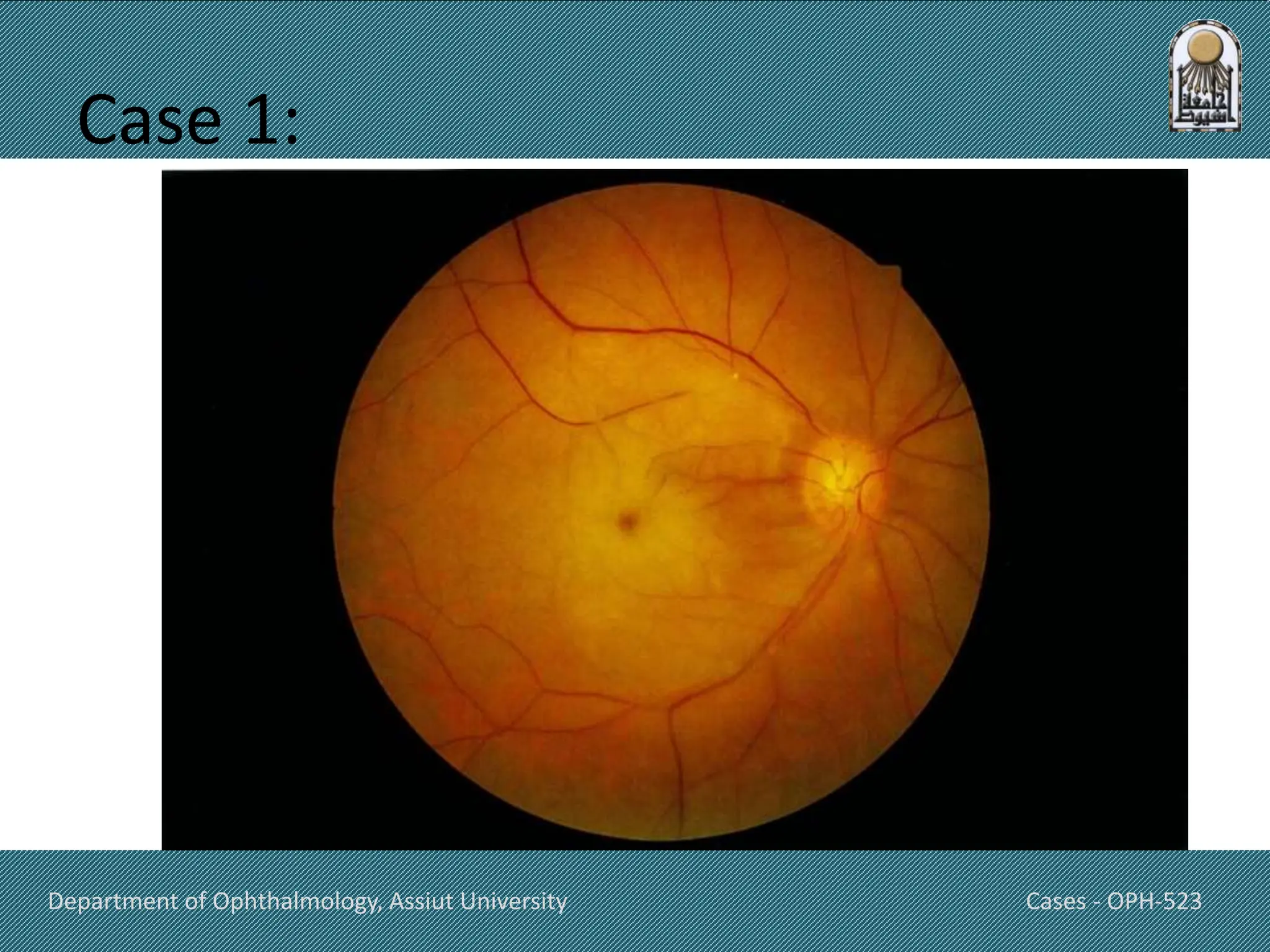
Retina
OPH-523
Cases Department of Ophthalmology
Assiut University
Pic
Week 10
cases
Department of Ophthalmology, Assiut University Cases - OPH-523
Case 1:
• A 68-year-old man is brought to the emergency department
by his wife because of sudden loss of vision in his right eye
that occurred 1 hour ago. He does not have eye pain. He had
several similar episodes of loss of vision; as these episodes all
resolved after some minutes, he did not follow up on them.
He has a history of hypertension, coronary artery disease,
and type 2 diabetes mellitus. His father died from myocardial
infarction at the age of 70 years. He has smoked up to one
pack of cigarettes daily for 43 years and drinks one alcoholic
beverage 3–5 days per week.
Department of Ophthalmology, Assiut University Cases - OPH-523
• Current medications include aspirin,
nitroglycerin, lisinopril, metoprolol,
atorvastatin, and metformin. His pulse is
78/min, and blood pressure is 148/84 mm Hg.
• Ophthalmologic examination shows no
perception of light in the right eye and 20/40
vision in the left eye. Corneal reflexes are
brisk. The swinging flashlight test
shows dilation of both pupils when the right
eye is illuminated.
Department of Ophthalmology, Assiut University Cases - OPH-523
• Intraocular pressure is 17 mm Hg in the right
eye and 16 mm Hg in the left eye.
• . On auscultation, there is a bruit over the
right carotid artery. Fundoscopy of the right
eye shows the findings in the photograph.
Department of Ophthalmology, Assiut University Cases - OPH-523
Case 1:
Department of Ophthalmology, Assiut University Cases - OPH-523
• Which of the following is the most likely
diagnosis?
A.Optic neuritis
B.Central retinal vein occlusion
C.Central retinal artery occlusion
D. Acute angle-closure glaucoma
E. Anterior ischemic optic neuropathy
F. Vitreous hemorrhage
G. Retinal detachment
H. Age-related macular degeneration
Department of Ophthalmology, Assiut University Cases - OPH-523
The answer is C
• Central retinal artery occlusion is
characterized by sudden, painless loss
of vision and a relative afferent pupillary
defect, which manifests with no perception of
light in the affected eye and an
abnormal swinging flashlight test. A past
history of amaurosis fugax is also common.
This patient has multiple risk factors for
retinal artery disease, including diabetes
mellitus, hypertension, and atherosclerosis.
Department of Ophthalmology, Assiut University Cases - OPH-523
• Along with the multiple cardiovascular risk
factors, a carotid
bruit suggests carotid artery atherosclerosis,
which can lead to the release of
a thrombus and subsequent occlusion of
the central retinal artery; this is the most
common cause of central retinal artery
occlusion.
Department of Ophthalmology, Assiut University Cases - OPH-523
• Retinal artery occlusion is an ophthalmologic emergency.
Therapeutic measures include eyeball massage, inhalation of
a mixture of 95% oxygen and 5% carbon dioxide, and
arterial vasodilators (e.g., sublingual nitroglycerine). A delay in
the initiation of therapy can result in permanent loss of vision.
Department of Ophthalmology, Assiut University Cases - OPH-523
Case 2:
• A 60-year-old woman is brought to the
emergency department because of sudden,
painless loss of vision in her right eye that
occurred 30 minutes ago while watching TV. She
has coronary artery disease, hypertension,
and type 2 diabetes mellitus, and has had trouble
adhering to her medication regimen. Her blood
pressure is 160/85 mm Hg. Examination
shows 20/50 vision in the left eye and no
perception of light in the right eye. Direct
pupillary reflex is present in the left eye but
absent in the right eye.
Department of Ophthalmology, Assiut University Cases - OPH-523
• Intraocular pressure is 16 mm Hg in the left
eye and 18 mm Hg in the right eye.
Fundoscopic examination of the right eye
shows a pale, white retina with a bright red
area within the macula. The optic disc appears
normal. Which of the following is the most
likely diagnosis?
Department of Ophthalmology, Assiut University Cases - OPH-523
A. Central retinal vein occlusion
B. Retinal detachment
C. Central retinal artery occlusion
D. Acute angle-closure glaucoma
E. Anterior ischemic optic neuropathy
Department of Ophthalmology, Assiut University Cases - OPH-523
The answer is C
• The patient has multiple risk factors for retinal
artery disease, including diabetes, hypertension,
and atherosclerosis. Central retinal artery
occlusion (CRAO) is characterized by sudden,
painless loss of vision and an afferent pupillary
defect. Ophthalmoscopy findings of CRAO include
retinal pallor and a cherry-red spot in the foveal
region. Narrow retinal vessels with a cattle-truck
appearance are another classic finding of retinal
artery occlusion.
Department of Ophthalmology, Assiut University Cases - OPH-523
Case 3:
• A 62-year-old woman comes to the physician
because of increasing blurring of vision in
both eyes. The patient says that the blurring
has made it difficult to read, although she has
noticed that she can read a little better when
holding the book below or above eye level.
She also requires a bright light to see objects
more clearly. She reports that her symptoms
began 8 years ago and have gradually gotten
worse over time. She has hypertension and
type 2 diabetes mellitus.
Department of Ophthalmology, Assiut University Cases - OPH-523
• Current medications include glyburide and
lisinopril. When looking at an Amsler grid, she
says that the lines in the center appear wavy
and bent. The results of a funduscopic
examination are shown. Which of the
following is the most likely diagnosis?
Department of Ophthalmology, Assiut University OPH-523
Department of Ophthalmology, Assiut University Cases - OPH-523
A. Central serous retinopathy
B. Hypertensive retinopathy
C. Diabetic retinopathy
D. Cystoid macular edema
E. Senile cataract
F. Presbyopia
G. Honeycomb macular dystrophy
H. Age-related macular degeneration
Department of Ophthalmology, Assiut University Cases - OPH-523
The answer is H:
• The presence of soft drusen near the macula is
consistent with age-related macular
degeneration (AMD). AMD is the most
common cause of impaired vision in the
elderly in developed countries. Cardiovascular
diseases such as hypertension increase the risk
of AMD. Nonexudative AMD (dry AMD) is the
most common type of AMD and leads to a
slowly progressive central or pericentral loss of
vision.
Department of Ophthalmology, Assiut University Cases - OPH-523
• Exudative AMD (wet AMD) makes up 10% of
cases and is associated with retinal
neovascularization, retinal hemorrhage,
and/or loss of vision that progresses more
rapidly.
Department of Ophthalmology, Assiut University Cases - OPH-523
Case 4:
• A 72-year-old woman comes to the emergency
department because of a 5 days history of
reduced vision in her eye. She says that she can
see black moving spots and flashes of light in her
right eye. The symptoms started when she was
jumping on a trampoline with her grandchild. She
does not report pain. Her medical history is
significant for type 2 diabetes mellitus and atrial
fibrillation. Since childhood, she has worn glasses
for progressive myopia
Department of Ophthalmology, Assiut University Cases - OPH-523
• Her medications are metformin, metoprolol,
and apixaban.
• Examination shows a visual acuity in the left
eye of 15/20 and counting fingers at 2 meters
in the right eye. A photograph of the
fundoscopic examination of the right eye is
shown. Fundoscopic examination of the left
eye shows no abnormalities.
Department of Ophthalmology, Assiut University OPH-523
Department of Ophthalmology, Assiut University Cases - OPH-523
Which of the following is the most likely cause
of the patient's vision loss?
A. Occlusion of central retinal artery
B. Choroidal neovascularization with
intraretinal bleeding
C. Retinal tear with detachment of the retina.
D. Impaired drainage of aqueous humor from
the eye
E. Detachment of the posterior vitreous.
Department of Ophthalmology, Assiut University Cases - OPH-523
The answer is C
• A retinal tear with detachment of the
neurosensory retina from the retinal pigment
epithelium is the underlying mechanism of
retinal detachment (RD), which most
commonly manifests with sudden, painless
vision loss and symptoms such as spots and
flashes of light in the visual field of the
affected eye. The most frequent causes of RD
are tears or holes in the retina
(rhegmatogenous RD);
Department of Ophthalmology, Assiut University Cases - OPH-523
• less commonly ;RD can occur without a retinal
tear (nonrhegmatogenous RD). Risk factors
include high myopia, trauma (in this case, likely
caused by jumping on a trampoline), previous
intraocular surgery, and posterior vitreous
detachment.
• The prognosis in RD is good if surgery is
performed at an early stage and if there is no
macular involvement. Without treatment,
progressive RD causes blindness, especially if the
macula is involved.
Department of Ophthalmology, Assiut University Cases - OPH-523
Thank You

Common retinal diseases, case discussion.pptx

  • 1.
    Retina OPH-523 Cases Department ofOphthalmology Assiut University Pic Week 10 cases
  • 2.
    Department of Ophthalmology,Assiut University Cases - OPH-523 Case 1: • A 68-year-old man is brought to the emergency department by his wife because of sudden loss of vision in his right eye that occurred 1 hour ago. He does not have eye pain. He had several similar episodes of loss of vision; as these episodes all resolved after some minutes, he did not follow up on them. He has a history of hypertension, coronary artery disease, and type 2 diabetes mellitus. His father died from myocardial infarction at the age of 70 years. He has smoked up to one pack of cigarettes daily for 43 years and drinks one alcoholic beverage 3–5 days per week.
  • 3.
    Department of Ophthalmology,Assiut University Cases - OPH-523 • Current medications include aspirin, nitroglycerin, lisinopril, metoprolol, atorvastatin, and metformin. His pulse is 78/min, and blood pressure is 148/84 mm Hg. • Ophthalmologic examination shows no perception of light in the right eye and 20/40 vision in the left eye. Corneal reflexes are brisk. The swinging flashlight test shows dilation of both pupils when the right eye is illuminated.
  • 4.
    Department of Ophthalmology,Assiut University Cases - OPH-523 • Intraocular pressure is 17 mm Hg in the right eye and 16 mm Hg in the left eye. • . On auscultation, there is a bruit over the right carotid artery. Fundoscopy of the right eye shows the findings in the photograph.
  • 5.
    Department of Ophthalmology,Assiut University Cases - OPH-523 Case 1:
  • 6.
    Department of Ophthalmology,Assiut University Cases - OPH-523 • Which of the following is the most likely diagnosis? A.Optic neuritis B.Central retinal vein occlusion C.Central retinal artery occlusion D. Acute angle-closure glaucoma E. Anterior ischemic optic neuropathy F. Vitreous hemorrhage G. Retinal detachment H. Age-related macular degeneration
  • 7.
    Department of Ophthalmology,Assiut University Cases - OPH-523 The answer is C • Central retinal artery occlusion is characterized by sudden, painless loss of vision and a relative afferent pupillary defect, which manifests with no perception of light in the affected eye and an abnormal swinging flashlight test. A past history of amaurosis fugax is also common. This patient has multiple risk factors for retinal artery disease, including diabetes mellitus, hypertension, and atherosclerosis.
  • 8.
    Department of Ophthalmology,Assiut University Cases - OPH-523 • Along with the multiple cardiovascular risk factors, a carotid bruit suggests carotid artery atherosclerosis, which can lead to the release of a thrombus and subsequent occlusion of the central retinal artery; this is the most common cause of central retinal artery occlusion.
  • 9.
    Department of Ophthalmology,Assiut University Cases - OPH-523 • Retinal artery occlusion is an ophthalmologic emergency. Therapeutic measures include eyeball massage, inhalation of a mixture of 95% oxygen and 5% carbon dioxide, and arterial vasodilators (e.g., sublingual nitroglycerine). A delay in the initiation of therapy can result in permanent loss of vision.
  • 10.
    Department of Ophthalmology,Assiut University Cases - OPH-523 Case 2: • A 60-year-old woman is brought to the emergency department because of sudden, painless loss of vision in her right eye that occurred 30 minutes ago while watching TV. She has coronary artery disease, hypertension, and type 2 diabetes mellitus, and has had trouble adhering to her medication regimen. Her blood pressure is 160/85 mm Hg. Examination shows 20/50 vision in the left eye and no perception of light in the right eye. Direct pupillary reflex is present in the left eye but absent in the right eye.
  • 11.
    Department of Ophthalmology,Assiut University Cases - OPH-523 • Intraocular pressure is 16 mm Hg in the left eye and 18 mm Hg in the right eye. Fundoscopic examination of the right eye shows a pale, white retina with a bright red area within the macula. The optic disc appears normal. Which of the following is the most likely diagnosis?
  • 12.
    Department of Ophthalmology,Assiut University Cases - OPH-523 A. Central retinal vein occlusion B. Retinal detachment C. Central retinal artery occlusion D. Acute angle-closure glaucoma E. Anterior ischemic optic neuropathy
  • 13.
    Department of Ophthalmology,Assiut University Cases - OPH-523 The answer is C • The patient has multiple risk factors for retinal artery disease, including diabetes, hypertension, and atherosclerosis. Central retinal artery occlusion (CRAO) is characterized by sudden, painless loss of vision and an afferent pupillary defect. Ophthalmoscopy findings of CRAO include retinal pallor and a cherry-red spot in the foveal region. Narrow retinal vessels with a cattle-truck appearance are another classic finding of retinal artery occlusion.
  • 14.
    Department of Ophthalmology,Assiut University Cases - OPH-523 Case 3: • A 62-year-old woman comes to the physician because of increasing blurring of vision in both eyes. The patient says that the blurring has made it difficult to read, although she has noticed that she can read a little better when holding the book below or above eye level. She also requires a bright light to see objects more clearly. She reports that her symptoms began 8 years ago and have gradually gotten worse over time. She has hypertension and type 2 diabetes mellitus.
  • 15.
    Department of Ophthalmology,Assiut University Cases - OPH-523 • Current medications include glyburide and lisinopril. When looking at an Amsler grid, she says that the lines in the center appear wavy and bent. The results of a funduscopic examination are shown. Which of the following is the most likely diagnosis?
  • 16.
    Department of Ophthalmology,Assiut University OPH-523
  • 17.
    Department of Ophthalmology,Assiut University Cases - OPH-523 A. Central serous retinopathy B. Hypertensive retinopathy C. Diabetic retinopathy D. Cystoid macular edema E. Senile cataract F. Presbyopia G. Honeycomb macular dystrophy H. Age-related macular degeneration
  • 18.
    Department of Ophthalmology,Assiut University Cases - OPH-523 The answer is H: • The presence of soft drusen near the macula is consistent with age-related macular degeneration (AMD). AMD is the most common cause of impaired vision in the elderly in developed countries. Cardiovascular diseases such as hypertension increase the risk of AMD. Nonexudative AMD (dry AMD) is the most common type of AMD and leads to a slowly progressive central or pericentral loss of vision.
  • 19.
    Department of Ophthalmology,Assiut University Cases - OPH-523 • Exudative AMD (wet AMD) makes up 10% of cases and is associated with retinal neovascularization, retinal hemorrhage, and/or loss of vision that progresses more rapidly.
  • 20.
    Department of Ophthalmology,Assiut University Cases - OPH-523 Case 4: • A 72-year-old woman comes to the emergency department because of a 5 days history of reduced vision in her eye. She says that she can see black moving spots and flashes of light in her right eye. The symptoms started when she was jumping on a trampoline with her grandchild. She does not report pain. Her medical history is significant for type 2 diabetes mellitus and atrial fibrillation. Since childhood, she has worn glasses for progressive myopia
  • 21.
    Department of Ophthalmology,Assiut University Cases - OPH-523 • Her medications are metformin, metoprolol, and apixaban. • Examination shows a visual acuity in the left eye of 15/20 and counting fingers at 2 meters in the right eye. A photograph of the fundoscopic examination of the right eye is shown. Fundoscopic examination of the left eye shows no abnormalities.
  • 22.
    Department of Ophthalmology,Assiut University OPH-523
  • 23.
    Department of Ophthalmology,Assiut University Cases - OPH-523 Which of the following is the most likely cause of the patient's vision loss? A. Occlusion of central retinal artery B. Choroidal neovascularization with intraretinal bleeding C. Retinal tear with detachment of the retina. D. Impaired drainage of aqueous humor from the eye E. Detachment of the posterior vitreous.
  • 24.
    Department of Ophthalmology,Assiut University Cases - OPH-523 The answer is C • A retinal tear with detachment of the neurosensory retina from the retinal pigment epithelium is the underlying mechanism of retinal detachment (RD), which most commonly manifests with sudden, painless vision loss and symptoms such as spots and flashes of light in the visual field of the affected eye. The most frequent causes of RD are tears or holes in the retina (rhegmatogenous RD);
  • 25.
    Department of Ophthalmology,Assiut University Cases - OPH-523 • less commonly ;RD can occur without a retinal tear (nonrhegmatogenous RD). Risk factors include high myopia, trauma (in this case, likely caused by jumping on a trampoline), previous intraocular surgery, and posterior vitreous detachment. • The prognosis in RD is good if surgery is performed at an early stage and if there is no macular involvement. Without treatment, progressive RD causes blindness, especially if the macula is involved.
  • 26.
    Department of Ophthalmology,Assiut University Cases - OPH-523 Thank You