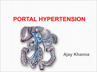
Portal Hypertension: Causes, Symptoms, and Treatment
•Download as PPT, PDF•
1 like•357 views

This document provides information on portal hypertension, including its definition, anatomy, pathophysiology, etiology, clinical features, investigations, treatment of variceal bleeding, ascites, encephalopathy, Budd-Chiari syndrome, and various surgical procedures. Portal hypertension is defined as a portal venous pressure greater than 12 mmHg and is characterized by the development of portosystemic collaterals. Common causes include liver cirrhosis, portal vein thrombosis, and Budd-Chiari syndrome. Treatment involves reducing portal pressure, treating complications, and addressing the underlying liver disease.

Recommended
More Related Content
What's hot
What's hot (20)
Similar to Portal Hypertension: Causes, Symptoms, and Treatment
Similar to Portal Hypertension: Causes, Symptoms, and Treatment (20)
Recently uploaded
Recently uploaded (20)
Portal Hypertension: Causes, Symptoms, and Treatment
- 2. DEFINITION Normal Portal Pressure : 5-10 mm Pressure of > 12 mm : Portal Hypertension Portal Hypertension is characterized by gradient of >5 mm Hg between portal venous and central venous pressure. At gradient of 8-10 mm Hg – Varices develops If gradient >12 mm Hg – Bleeding from varices
- 3. ANATOMY • Hepatic blood flow • 2/3rd – Portal vein • 1/3rd – Hepatic artery • Portal vein length – 6-8 Cm • Diameter - 13-16 mm 1/3 2/3
- 4. Portosystemic Collateral Formation Lower end of esophagus Azygos vein Lt gastric V Umblicus Ant abd. veins Paraumblical Lower end Rectum Middle and Inf Superior Haem. Haemmorrhoidal V Retroperitoneum Bare area of liver
- 5. Porta Systemic Collaterals esophageal and gastric veins inferior rectal-anal veins anterior abdominal wall veins retroperitoneal venous plexus
- 6. PATHOPHYSIOLOGY Portal pressure = resistant X flow Resistance to portal flow Rise in portal pressure Development of portasystem collaterals Hyperdynamic circulation
- 7. ETIOLOGY Extrahepatic (Prehepatic/ EHPVO) Portal cavernoma Portal vein thrombosis (most common cause) Splenic vein thrombosis Compression from outside Intrahepatic Presinusoidal Schistosomiasis (most common cause) Primary biliary cirrhosis Sinusoidal Alcoholic cirrhosis (most common cause) Postsinusoidal Alcoholic cirrhosis (most common cause) Veno-occlusive disease Non Cirrhotic Portal Fibrosis
- 8. ETIOLOGY (Contd) Posthepatic Budd-Chiari syndrome (most common cause) Right sided heart failure Constrictive pericarditis High-flow portal hypertension Hepatic artery-portal vein fistula Splenic arteriovenous fistula Massive splenomegaly Left Sided Portal Hypertension (Sinistral)
- 9. Clinical features Bleeding (Upper GI) Splenomegaly Jaundice Encephalopathy Ascites Growth Retardation Coagulopathy Recurrent Infection
- 10. Examination Jaundice Hepatomegaly Splenomegaly Ascites Veins on abdominal wall Oedema Encepahlopathy Hernia
- 11. INVESTIGATIONS • Hematological: CBC, Platelet, • Biochemical: LFT, PT • Radiological: Triple Phase Portography, MRI, CT • Ultrasonography/Doppler • Endoscopy • Pressure studies • Liver Biopsy
- 12. Barium swallow multiple irregular filling defects as “string of beads” or “earthworm”
- 13. ESOPHAGOGASTRO DUODENOSCOPY Grading as per Endoscopy Paqet Grading Grade I: Small varices without luminal prolapse Grade II: Mod. Size varices showing luminal prolapse along with minimal obscuring of gastro esophageal junction. Grade III: Large varices showing luminal prolapse substantially obscuring the gastro esophageal junction. Grade IV: Very large varices completely obscuring gastroesophageal junction.
- 14. USG/Doppler USG: Portal vein diameter, thrombus, collaterals , splenomegaly & ascites. Portal flow direction and velocity – Hepatopetal/Fugal Portosytemic surgical shunt follow up To evaluate patency of shunt To evaluate direction of flow
- 15. Portography: Splenoportovenography Transhepatic/transjugular Umblical portography Triple Phase Portography (CT) MRI Portograophy
- 16. Portal Pressure Measurement Wedged hepatic venous pressure Occluded Pressure (SOPP/HOPP) Splenic pulp pressure Catheterization of umbilical vein Liver Biospy Laparoscopy
- 17. Child’s grade (Pugh Modification) Point 1 Point 2 Point 3 Bilirubin mgm/dl <2 2-3 >3 Albumin (gm/dl) >3.5 3-3.5 <3 Ascites None Controlled Uncontrolled Encephalopathy None Mild Mod-severe Prothrombin Time* <3 Sec prolonged 3-6 Sec prolonged >6 Sec prolonged Total : 5-15 Child A < 6 Child B 7-9 Child C 10-15 * In Original Child score it was nutritional status
- 18. TREATMENT Portal hypertension treatment involves three level of management Reduction of portal pressure Treatment of complications arising from portal hypertension Variceal bleeding Hypersplenism Treatment of underlying liver disease and its complications
- 19. TREATMENT OF VARICEAL BLEEDING Primary Prophylaxis : No prior H/O bleed Propranolol EST Secondary Prophylaxis Propranolol EST Surgery
- 20. Emergency Treatment of Bleed Resuscitation Pharmacological therapy Endoscopic therapy Balloon Temponade Transjugular intrahepatic portosystemic shunt (TIPS) Surgical treatment
- 21. PHARMACOLOGICAL THERAPY Beta blockers Vasopressin with or without nitroglycerine Glypressin Somatostatin / octreotide Metoclopramide Long acting nitrates
- 22. Beta blockers bleeding by cardiac output. Dose 20-60 mg bid 25% in HR. Reduces 40% of bleeding episodes
- 23. Vasopressin vasoconstriction of the splanchnic circulation. Dose 0.2 unit/kg wt, dissolved in 200 ml of 5% dextrose, over 20 minutes. Disadvantages colicky abdominal pain, & diarrhoea . Anginal pains, so it is contraindicated in the elderly. Produce temporary control of bleeding in about 80% of cases. To prolong its action it is combined with glycine (Glypressin).
- 24. ENDOSCOPIC THERAPY Injection sclerotherapy Variceal band ligation Gastric variceal injection Thrombin injections
- 25. EST Techniques: Intravariceal injection Paravariceal injection Combination of both intravariceal + paravariceal injection
- 26. Sclerosants : 5% Ethanolamine oleate 0.5% sodium polidocanol 5% sodium morrhuate 1% to 3% sodium tetradecyl sulphate High concentration of alcohol 3% phenol
- 27. ENDOSCOPIC VARICEAL LIGATION (EVL) Occludes venous channels Sessions < sclerotherapy Same results as sclerotherapy complications vs sclerotherapy Endoscopic treatment of choice
- 28. BALLOON TEMPONADE Temporary measure Hazardous in infants and small children Success rate is 80-90% Complications : Aspiration pneumonia, pressure necrosis , obstruction of pharynx, ulceration of esophagus, rupture of balloon, re-bleeding after deflation (60%).
- 29. TYPES OF BALLOON TEMPONADE TUBE: Sengstaken Black’s more tube Linton-Nicholos tube for gastric variceal haemorrhage Minnesota tube – 4 lumen tube
- 30. TIPS(Transjugular Intrahepatic Porta systemic shunt) • Shunt between portal vein and hepatic vein • Can be used as short term bridge to liver transplantation • Goal – To reduce portosystemic gradient < 12 mm Hg
- 32. SURGICAL TREATMENT SHUNT SURGERY Prophylactic Emergency shunt surgery Reduced with advent of other non surgical procedure End to side porto-caval shunt preferred Elective shunt surgery
- 33. Types of Shunt Surgery Non Selective Portacaval (end to side/ side to side) Mesocaval (With or without graft) Proximal Leino Renal Shunt Spleen preserving Side to side Splenorenal shunt (Mitra) Selective Shunt Distal Leino Renal Shunt (Warren) Coronary Caval (Inokuchi) Porta Caval (SARFEH SHUNT) 8 mm graft
- 35. Warren Distal Leino Renal Shunt
- 36. Types of Operation Non Shunt Surgery: Stapler transection, (Johnston) Boerema, Milnes Walker Suigura and Futagawa, Tanner, Hassab, Liver Transplantation
- 37. Non Selective shunt operations 1- Porta-caval operation End to side Side to side
- 38. Porta-caval operation very efficient in lowering the portal pressure no bleeding occurs from the varices. disadvantages: deprives the liver of portal blood flow accelerates the onset of liver failure. Recurrent hepatic encephalopathy in 30-50% of patients.
- 39. Proximal spleno-renal shunt indicated if the portal vein is thrombosed or if splenectomy is indicated due to hypersplenism . The incidence of encephalopathy is less than after porta caval shunt. it is less effective In preventing further bleeding. If the splenic vein is less than 1 cm the anastomosis is liable to thrombosis.
- 40. Mesocaval (Drapanas) shunt insertion of a a synthetic graft as dacron, or autogenic vein between the superior mesenteric vein and inferior vena cava. The incidence of thrombosis is high
- 41. Selective shunt (Warren shunt) The Rt and Lt gastric vessels are ligated. The proximal end of splenic vein is ligated while the distal end is anastomosed to the left renal vein. The short gastric veins are preserved and will selectively decompress the lower end of the oesophagus. The incidence of encephalopathy is low, and the liver functions remain normal.
- 43. Non Shunt Surgery Stapler Transection (Johnston) Boerema Crile Milnes Walker Suigura Futagawa Tanner Hassab
- 44. Sugiura and Futagawa procedure (1973) Transthoracic paraesophageal devascularization and esophageal transaction combined with an abdominal component consisting of splenectomy, devascularization of upper stomach, vagotomy and pyeloroplasty Modified Sugiura procedure (Ginsberg 1982) – Abdominal approach. Splenectomy, paraesophageal and gastric devascularization, vagotomy, stapler esophageal transaction via gastrostomy and pyeloroplasty.
- 45. HYPERSPLENISM SELECTIVE SPLENIC EMBOLIZATION : Splenic immune function is conserved, size of spleen is reduced and portal flow is maintained. Aim of procedure is to infarct 70-80% of splenic tissue. Pneumococcal vaccination is given with this therapy and sometimes long term antibiotics prophylaxis is needed. SPLENECTOMY: Usually not advocated Indicated in isolated portal vein thrombosis with recurrent or life threatening haemorrhage and a massive spleen. Shunt Procedures
- 46. Ascites Disordered albumin synthesis and decreased plasma colloid osmotic pressure caused by hepatocellular function damage Increased capillary filter pressure due to increased portal hypertension Lymph liquid leakage into abdominal cavity from surface of the liver because of lymph back-flow obstruction Salt and water retention by aldosterone and antidiuretic hormones deactivation disturbance
- 47. Complications of ascites Respiratory embarrasement Spontaneous bacterial peritonitis Hernia Investgations: SAAG > 1.1 (Serum Ascitic Albumin Gradient) Treatment : Salt Restriction Diuretics Paracentesis Shunt - Le Veen Shunt, Denver Shunt TIPS Liver Transplant
- 48. Encephalopathy Lactulose Stop bleeding Neomycin Enemas
- 49. Budd Chiari syndrome Syndrome due to obstruction of hepatic veins Causes : Spontaneous thrombosis Myeloproliferative disorders Web Neoplasms Polycyathemia Contraceptive pills
- 50. Presentation of Budd Chiari Syndrome Acute : Nausea, vomiting, severe pain in Rt hypochondrium, rapid enlargement of liver, hypotension, death Sudden onset Rapid development of Ascites, hepatic insufficiency, oedema, coma Chronic Cirrhosis, Ascites, Varices, infection, hepatic failure Classic Triad Abdominal Pain Ascites Hepatomegaly
- 51. Treatment of Budd Chiari Syndrome Anticoagulation Membrane IVC : Tranatrial Meatotomy Meso atrial Shunt : Shunt between Rt. Atrium and Sup Mesenteric vein