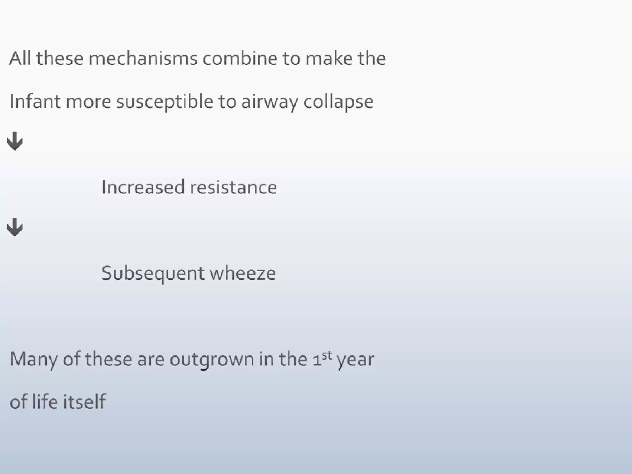
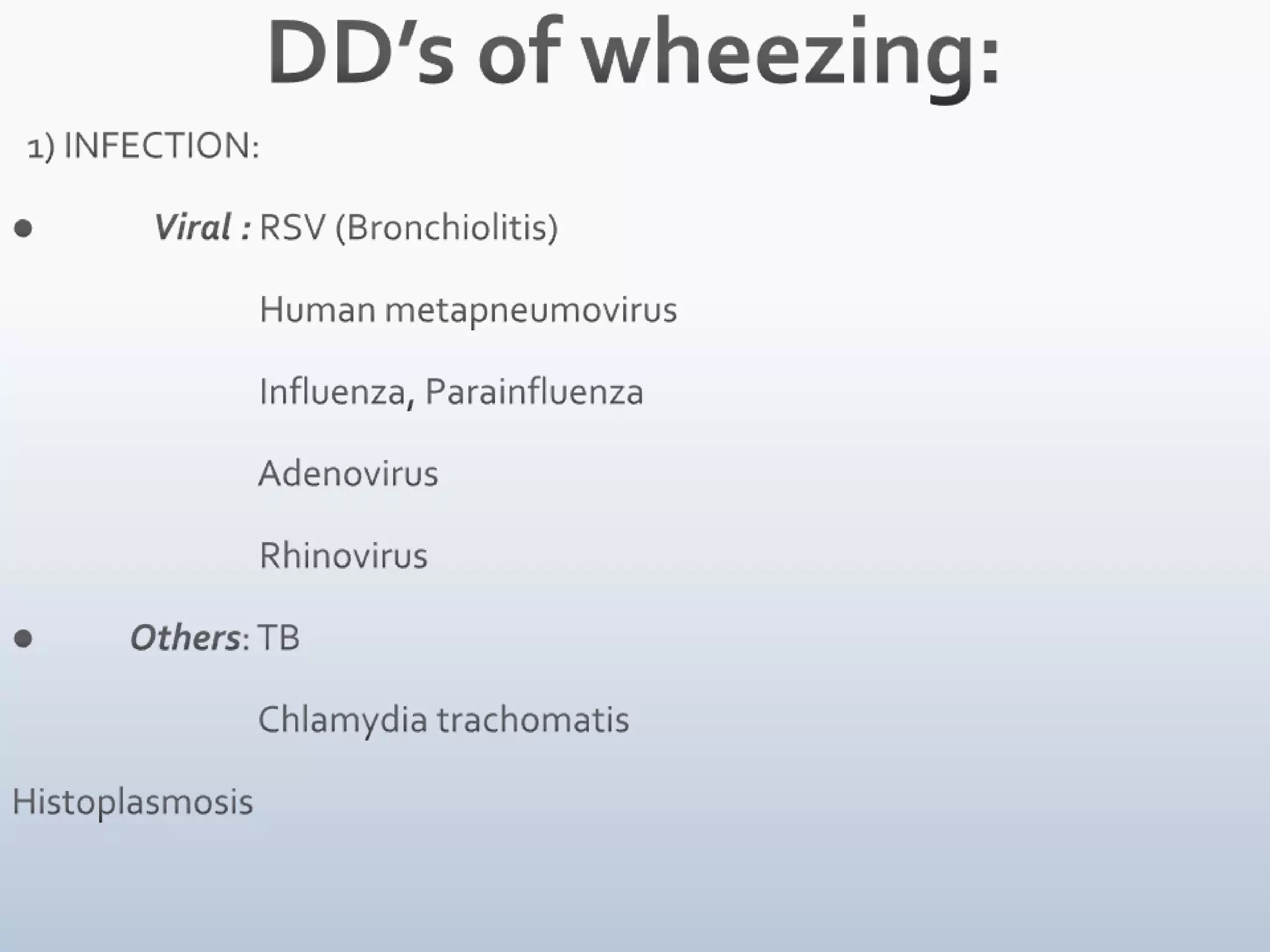
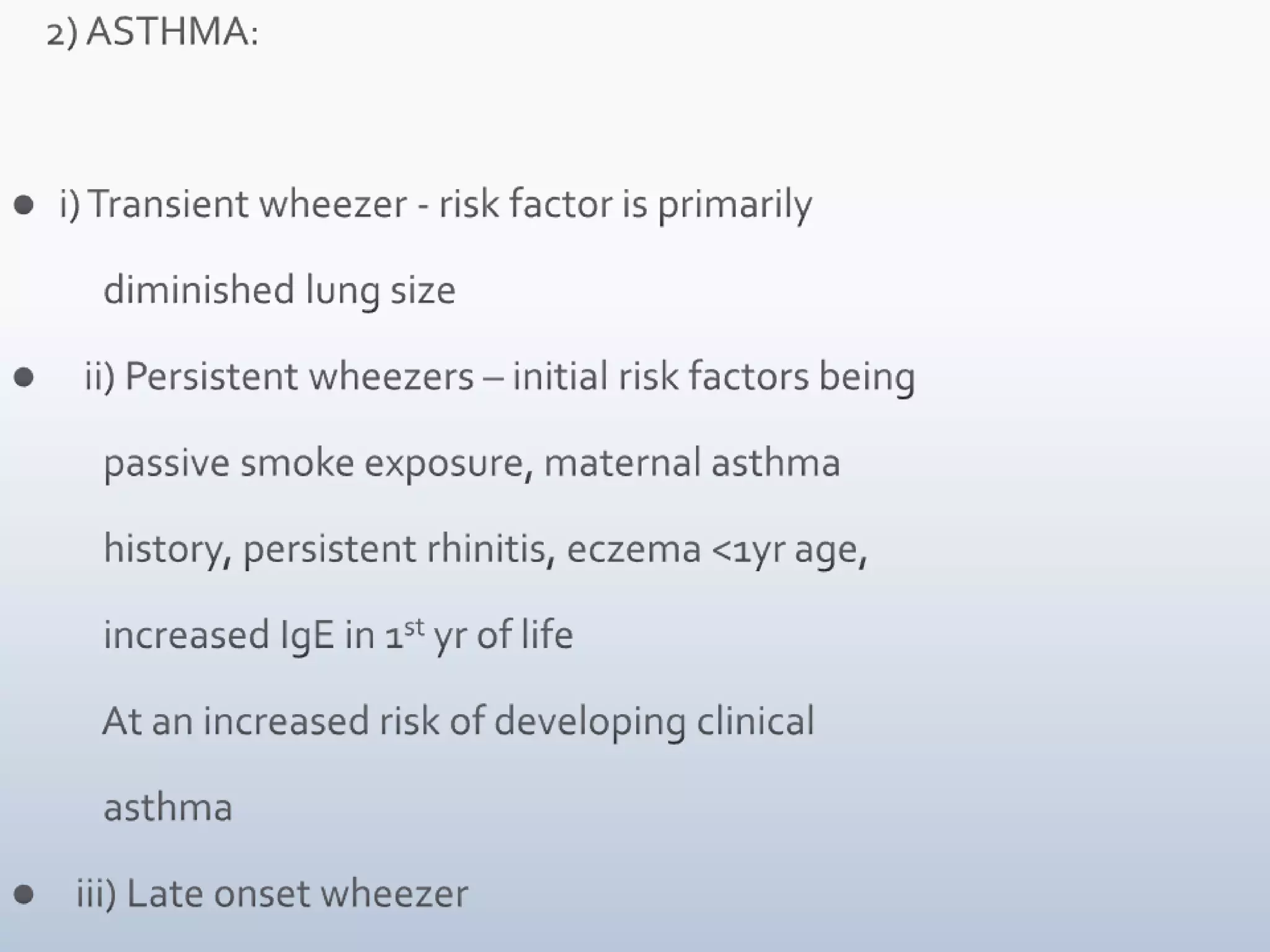
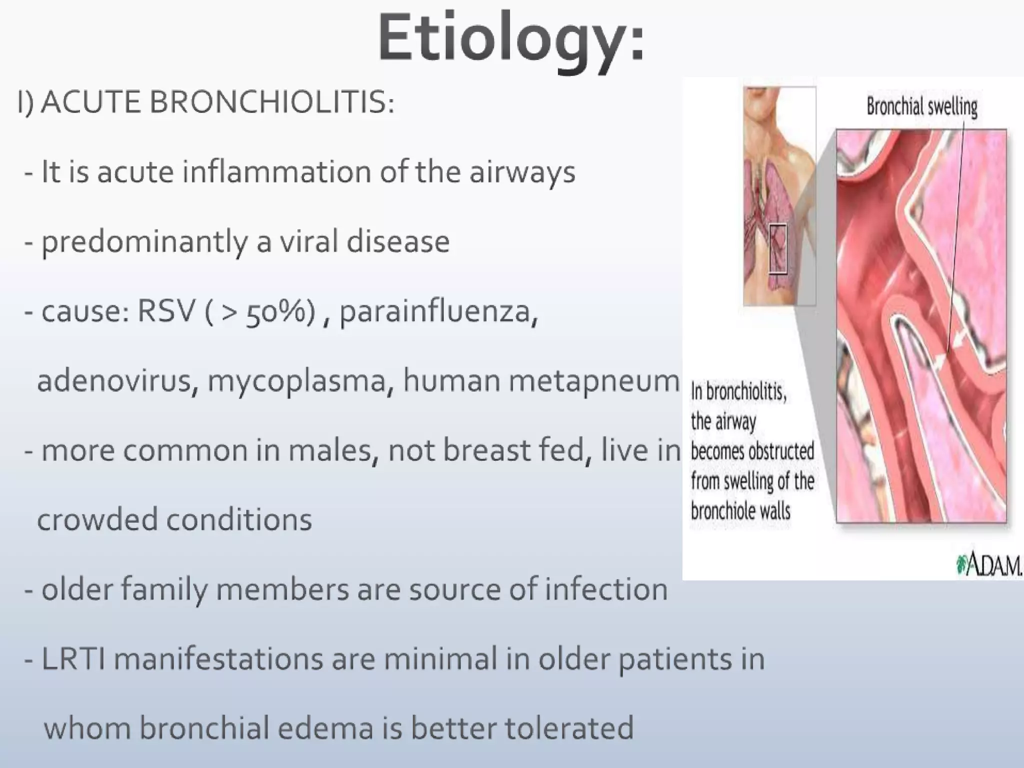
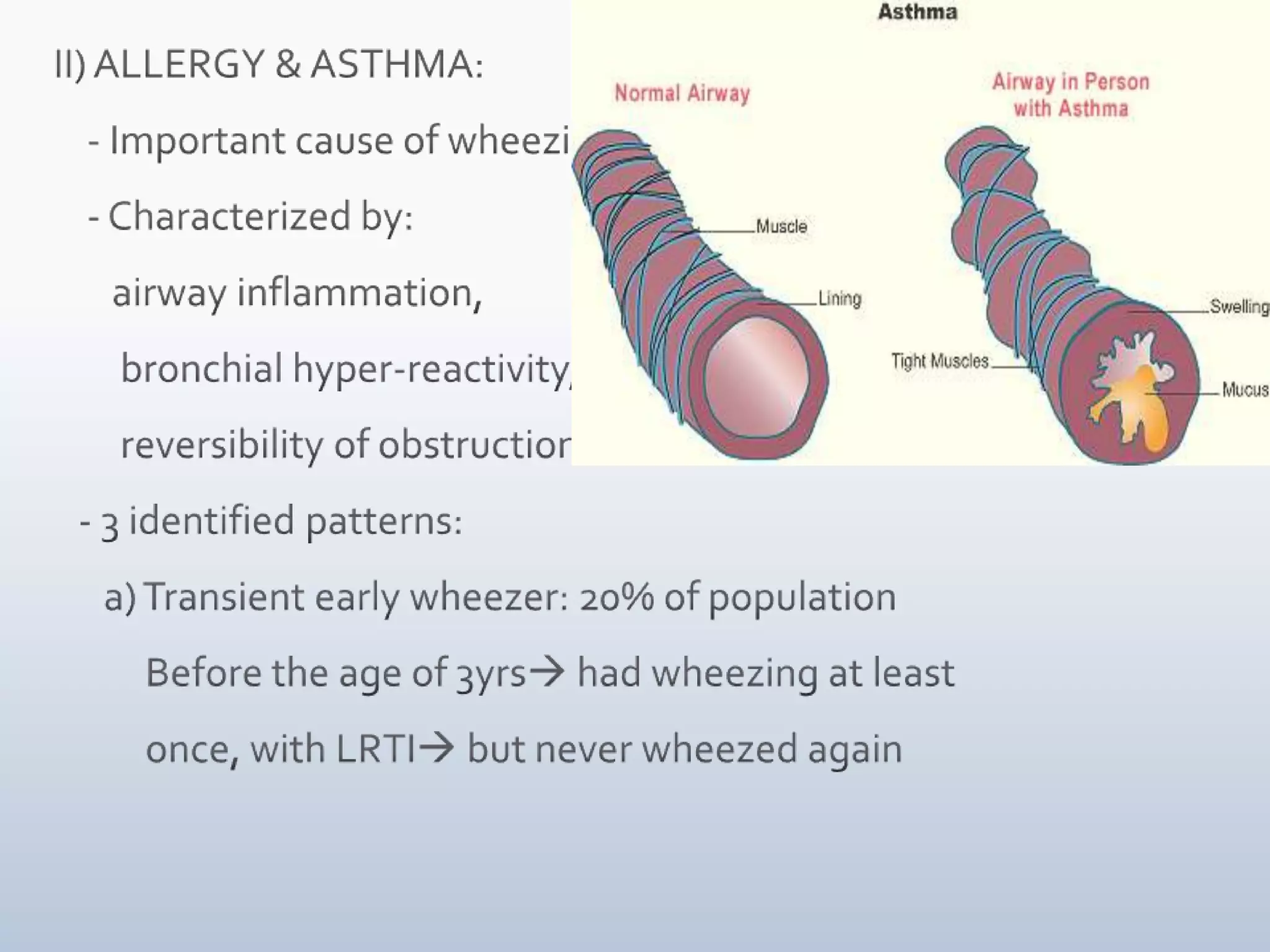
Wheezing in childrenDr Divya NairDepartment of Pediatrics,Mahavir Hospital & Research Centre,Hyderabad.
IntroductionWheeze is a continuous & musical sound that      originates from oscillations in narrowed     airwaysMostly heard in expiration due to critical     airway obstructionSign of lower (intra-thoracic) airway     obstruction
If there is widespread narrowing of airways      leading to various levels of obstruction to     airflow (eg. asthma), polyphonic wheeze is     heard i.e. sounds of various pitchesMonophonic wheeze (single pitch) is produced     in larger airways during expiration eg. distal tracheomalacia, bronchomalacia
Infants & children are prone to wheeze due to different set of lung mechanics ( as compared to  older children & adults)Obstruction to airflow  airway caliber  compliance of lung     1) Resistance  =  1/ ( radius of tube)4         In children < 5 years, small caliber peripheral             airways can contribute upto 50% of airway           resistanceMarginal additional narrowing can cause further           flow limitation & subsequent wheeze
2) Compliant chest walls, especially in newborns,      leads to intra-thoracic airway collapse due to      inward pressure produced in expiration3) Differences in tracheal cartilage composition &       airway muscle tone causes further increase in      airway compliance
 All these mechanisms combine to make the  Infant more susceptible to airway collapse                       Increased resistance                       Subsequent wheeze        Many of these are outgrown in the 1st year  of life itself
4) Immunologic & molecular influences:Infants have increased levels of lymphocytes & neutrophils in BAL fluidVariety of inflammatory mediators have been     implicated eg. Histamine, leukotrienes,     interleukins, etcFetal & early post-natal “programming” affects the      structure & function of fetal lung by factors      including fetal nutrition, fetal & neonatal exposure     to maternal smoking
DD’s of wheezing:  1) INFECTION:            Viral : RSV (Bronchiolitis)                                Human metapneumovirus                                Influenza, Parainfluenza                                Adenovirus                                Rhinovirus         Others: TB                                Chlamydia trachomatisHistoplasmosis
    2) ASTHMA:i) Transient wheezer - risk factor is primarily            diminished lung size   ii) Persistent wheezers – initial risk factors being             passive smoke exposure, maternal asthma            history, persistent rhinitis, eczema <1yr age,            increased IgE in 1st yr of life           At an increased risk of developing clinical            asthma  iii) Late onset wheezer
  3) Anatomic abnormalities:            a) Central airway abnormalities:           - Malacia of larynx, trachea, bronchi           - Tracheoesophageal fistula ( H type)           - Laryngeal cleft (leading to aspiration)b) Extrinsic airway anomalies (leading to com- pression):           - Vascular ring/ sling           - MediastinalLN’pathy (infection/ tumor)           - Esophageal foreign body
c) Intrinsic airway anomalies:        - Airway hemangioma       - Cystic adenomatoid malformation       - Bronchial/ lung cyst       - Congenital lobar emphysema       - Aberrant tracheal bronchus       - Sequestration       - CHD with L R shunt ( pulmonary edema)       - Foreign body
 4) Immunodeficiency states:     - IgA deficiency     - B cell defiency     - AIDS     - Bronchiectasis 5) Mucociliary clearance disorders:     - Cystic fibrosis     - Primary ciliarydyskinesias     - Bronchiectasis
 6) BronchopulmonaryDysplasia  7) Aspiration Syndromes      - GERD      - Pharyngeal/ swallow dysfunction   8) Interstitial lung disease  9) Heart Failure 10) Anaphylaxis 11) Inhalation Injury – Burns12) WALRTI, Wheeze a/w URTI13) Drugs: Ibuprofen, Aspirin, Rifampicin, Erythromycin
Etiology: I) ACUTE BRONCHIOLITIS:  - It is acute inflammation of the airways   - predominantly a viral disease  - cause: RSV ( > 50%) , parainfluenza,     adenovirus, mycoplasma, human metapneumovirus  - more common in males, not breast fed, live in     crowded conditions  - older family members are source of infection  - LRTI manifestations are minimal in older patients in       whom bronchial edema is better tolerated
  - RSV infection leads to a complex immune responsei) Eosinophilsdegranulate release Eosinophilic        Cationic Protein cytotoxic to airway epithelium    ii) IgE antibody release may be related to wheezing   iii) Other mediators: Chemokines (IL-8, Macrophage          Inflammatory Protein); Leukotrienes , IF gamma  - Characterized by bronchiolar obstruction with      edema, mucus & cellular debris
 - Resistance in small airway is increased more in exhalation than      inspiration  respiratory obstruction  early air trapping & overinflation complete obstruction atelectasis - Hypoxemia occurs early in the course due to     VQ mismatch - Hypercapnia develops due to severe    obstructive disease and respiratory fatigue
II) ALLERGY & ASTHMA:    - Important cause of wheezing    - Characterized by:        airway inflammation,        bronchial hyper-reactivity,        reversibility of obstruction   - 3 identified patterns:       a) Transient early wheezer: 20% of population           Before the age of 3yrs had wheezing at least            once, with LRTI but never wheezed again
b) Persistent wheezer: 14% of population       Before 3yrs age had wheezing episodes       still wheezing at 6yrs agec) Late onset wheezer: 15% of population      No wheezing by 3yrs but wheezing by       6yrs - The remaining 50% of children had never     wheezed by 6yrs of age
 III) CHRONIC INFECTIONS:      - Should be considered in infants who seem         to fall out of range of a normal clinical         course      - Cystic fibrosis is a common cause      - Persistent respiratory symptoms, digital         clubbing, malabsorption, FTT, electrolyte         abnormalities, resistance to bronchodilator            therapy
 IV) CONGENITAL MALFORMATIONS:       - Causes wheezing in early infancy       - Findings can be diffuse or focal       - Can be from an ext compression/ intrinsic abn       - External vascular compression: vascular ring/           sling compressing trachea/ esophagus       -  CVS causes: massive cardiomegaly, LA            enlargement, dilated pulmonary arteries       - Pulmonary edema d/t CHF  lymphatic &           bronchial vessel engorgement obstruction &           edema of bronchioles Wheeze
V) FOREIGN BODY ASPIRATION:     - Can cause acute/ chronic wheezing     - Common between 2mths- 4 yrs of age     - Infants may present with atypical histories or        misleading radiological/ clinical findings     - D/d: asthma, other obstructive disorders     - Esophageal FB can transmit pressure to        membranous trachea  compromises the          airway lumen
VI) GASTROESOPHAGEAL REFLUX:       - can cause direct aspiration into tracheo-         bronchial tree       - may trigger a vagal/ neural reflex                   increased airway resistance                          airway reactivity
VII) TRAUMA & TUMORS:        - rare causes of wheezing in children        - trauma of any type to TB tree (aspiration/ burns/ scalds)  inflammation of the airways  subsequent wheeze        - SOL (lung/ extrinsic)  compression           obstruction to airway
Clinical ManifestationsHISTORY & PHYSICAL EXAMINATION   - ODP & associated factors of wheezing:   - Birth history: weeks of gestation, NICU admission, h/o intubation/ O2 requirement, maternal         complications eg. Infn- HSV, HIV; prenatal smoke       exposure    - Past medical history: co-morbid conditions eg.        syndromes or association
   - Social history:        Environmental history of smokers at home,        number of siblings, occupation of inhabitants at        home, pets, TB exposure   - Family history:       of CF, immuno-deficeincy, asthma in 1st degree       relatives OR       any other recurrent respiratory conditions should       be obtained
RISKS OF FAMILY HISTORY OF ATOPYNo family history	:16%
Single parent atopy	:	22%Maternal Atopy		:	32 %
Both parents atopic	:	50%(Aberdeen Study 1994)
Pertinent medical history in wheezing infant:Did the onset of symptoms begin at birth or thereafter?Is the infant a noisy breather & when is it most prominent?Is there a history of cough apart from wheezing?Was there an earlier LRTI?Have there been any emergency department visits, hospitalizations, or ICU admission for RD?Is there a history of eczema?
How is the infant growing & developing?Is there associated failure to thrive?Is there failure to thrive without feeding difficulties?Are there s/o intestinal malabsorption including frequent , greasy, or oily stools?Is there a maternal history of genital HSV infection?What was the gestational age at delivery?Was the patient intubated as neonate?Does the infant bottle feed in the bed or crib, especially in propped position?
Are there any feeding difficulties including choking, gagging, arching, or vomiting with feeds?Any new food exposure?Is there a toddler in the home or lapse in supervision in which foreign body aspiration could have happened?Change in caregivers or chance or non accidental trauma?
Physical examination:    - Vitals especially RR, SPO2    - Growth charts for s/o FTT   - Upper airway s/oatopy: boggy turbinates ,       posterior oropharynxcobblestoning   - Evaluate skin for eczema, hemangioma   - Midline lesions may be associated with intrathoracic lesions   - Clubbing
   - S/O RD- Tachypnea, nasal flaring, tracheal      tugging, SCR/ICR, excessive use of accessory      muscles   - Prolonged expiratory time, expiratory whistling        sounds   - Auscultation: aeration to be noted, expiratory        wheeze, lack of audible wheeze due to complete       airway obstruction   - Trial of bronchodilators to evaluate change of        wheezing    - Stridor +/-.
Air TrappingHyperinflated chest Barrel shaped Loss of cardiac dullness Liver pushed down Hoover sign           Normal diagphragm movement           Hyperinflation = diaphragm flattened           Diaphragm contraction = paradoxical inward            movement of lower interrcostal area during            inspiration
In Acute Bronchiolitis:     - h/o exposure to older contact with URTI       infant 1st develops mild URTI with sneezing & rhinorrhea may be associated with       decreased appetite & fever  gradually RD       ensues with paroxysmal wheezy cough, dyspnea, irritability
  - Apnea may be more prominent than      wheezing early in the course of disease esp    with very young infants ( < 2 mths ) or former     premature infants - Degree of tachypnea doesn’t always correlate     with degree of hypoxemia/ hypercarbia - Fine crackles/ overt wheezes present - Hyperinflation of lungs palpation of liver &     spleen
Diagnostic evaluationInitial evaluation depends on likely etiology    1. Chest Xray: hyperinflation, SOL, s/o chronic diseases like bronchiectasis, focal infiltrates    2. Trial of bronchodilators-        diagnostic & therapeutic in bronchiolitis & asthma, won’t effect fixed obstruction        May worsen wheezing in tracheal/ bronchomalacia    3. Baseline immunity in complicated cases
Exclude other conditions 4) Structural problems: bronchoscopy 5) URTD : Polysomnography 6) Esophageal disease: Barium swallow, pH probes, Upper GI scopy 7) Primary ciliarydyskinesia: nasal ciliary motility, Exhaled NO,      Electron Microscopy, saccharine test  8) TB: mantoux, induced sputum/ gastric lavage/ BAL = Culture,       microscopy & PCR  9) Bronchiectasis: HRCT scan, BAL
  10) CF: sweat test, nasal potentials, genotypes  11) Systemic immune deficiency: Ig subtypes,          lymphocytes & neutrophil function, HIV  12) Cardiovascular disease: echo, angiography  13) Viral testing (PCR, viral culture) is helpful if diagnosis          is uncertain.
Treatment1) Comfort the child –      Try to keep your baby calm. Having a cough and a        noisy wheeze frightens children and breathing is      more difficult when they are upset. 2) Offer frequent liquids –     Drinking less but more often may be easier
3) Bronchodilators:     - administer inhaled short acting beta-2 agonist         (egsalbutamol) & observe the response     - Children < 3 yrs: inhaled medications by MDI         with mask & spacer, if therapeutic benefit         demonstrated    -  response is unpredictable     - Therapy to be continued in all asthma          patients with exacerbations with viral          illness
 4) Ipratropium bromide:     - can be used as adjunct therapy     - also useful in patients with significant tracheal or  bronchomalacia     - Anticholinergic agent5) Oral/ IV steroids:      - used for atopic wheezing infants thought to have         asthma i.e. refractory to other medications
6) Inhaled steroids:      - appropriate for maintenance therapy in known reactive          airways but not useful in acute illness      - to be used if significant h/oatopy ( food allergy, eczema)         present      - maintenance treatment with inhaled steroids is          recommended for multiple-trigger wheeze.
 7) In acute bronchiolitis:     - hospitalze     - mainstay of treatment is supportive     - hypoxemic child: cool humidified oxygen     - avoid sedatives     - keep head & chest elevated at 30 degree angle     - NG tube feeds to avoid aspiration     - respdecompensation tracheal intubation     - Bronchodilators show modest short term          improvement in clinical features     - Nebulized epinephrine more effective
  8) Montelukast is recommended for the treatment of       episodic (viral) wheeze,to be started when symptoms of a       viral cold develop 9) Ribavarine: antiviral administered by aerosol     - Used for children with CHD/ CLD10) No role of antibiotics unless secondary bacterial         infection
Prevention 1) Reduction in severity & incidence of ac. bronchiolitis due to       RSV is possible through administration of pooled Hyperimmune      RSV Intravenous Immunoglobulin (RSV IVIg, Respigam) and 2) Palivizumab, a monoclonal antibody to the RSV F protein, before &       during RSV season      It is recommended for children < 2yrs age with chronic lung disease      (BPD) or prematurity3)  Inhaled corticosteroids and montelukast may be      considered in preschool child with recurrent wheeze.
4) Avoid smoking –    Smoking in the home or car increases the risk of respiratory     problems in children5) Educating parents regarding causative factors and      treatment is useful. 6) Allergen avoidance may be considered when sensitisation     has been established7) Meticulous handwashing is the best measure     to prevent nosocomial infection

Wheeze in Children

  • 1.
    Wheezing in childrenDrDivya NairDepartment of Pediatrics,Mahavir Hospital & Research Centre,Hyderabad.
  • 2.
    IntroductionWheeze is acontinuous & musical sound that originates from oscillations in narrowed airwaysMostly heard in expiration due to critical airway obstructionSign of lower (intra-thoracic) airway obstruction
  • 3.
    If there iswidespread narrowing of airways leading to various levels of obstruction to airflow (eg. asthma), polyphonic wheeze is heard i.e. sounds of various pitchesMonophonic wheeze (single pitch) is produced in larger airways during expiration eg. distal tracheomalacia, bronchomalacia
  • 4.
    Infants & childrenare prone to wheeze due to different set of lung mechanics ( as compared to older children & adults)Obstruction to airflow  airway caliber compliance of lung 1) Resistance = 1/ ( radius of tube)4 In children < 5 years, small caliber peripheral airways can contribute upto 50% of airway resistanceMarginal additional narrowing can cause further flow limitation & subsequent wheeze
  • 5.
    2) Compliant chestwalls, especially in newborns, leads to intra-thoracic airway collapse due to inward pressure produced in expiration3) Differences in tracheal cartilage composition & airway muscle tone causes further increase in airway compliance
  • 6.
    All thesemechanisms combine to make the Infant more susceptible to airway collapse Increased resistance Subsequent wheeze Many of these are outgrown in the 1st year of life itself
  • 7.
    4) Immunologic &molecular influences:Infants have increased levels of lymphocytes & neutrophils in BAL fluidVariety of inflammatory mediators have been implicated eg. Histamine, leukotrienes, interleukins, etcFetal & early post-natal “programming” affects the structure & function of fetal lung by factors including fetal nutrition, fetal & neonatal exposure to maternal smoking
  • 8.
    DD’s of wheezing: 1) INFECTION: Viral : RSV (Bronchiolitis) Human metapneumovirus Influenza, Parainfluenza Adenovirus Rhinovirus Others: TB Chlamydia trachomatisHistoplasmosis
  • 9.
    2) ASTHMA:i) Transient wheezer - risk factor is primarily diminished lung size ii) Persistent wheezers – initial risk factors being passive smoke exposure, maternal asthma history, persistent rhinitis, eczema <1yr age, increased IgE in 1st yr of life At an increased risk of developing clinical asthma iii) Late onset wheezer
  • 10.
    3)Anatomic abnormalities: a) Central airway abnormalities: - Malacia of larynx, trachea, bronchi - Tracheoesophageal fistula ( H type) - Laryngeal cleft (leading to aspiration)b) Extrinsic airway anomalies (leading to com- pression): - Vascular ring/ sling - MediastinalLN’pathy (infection/ tumor) - Esophageal foreign body
  • 11.
    c) Intrinsic airwayanomalies: - Airway hemangioma - Cystic adenomatoid malformation - Bronchial/ lung cyst - Congenital lobar emphysema - Aberrant tracheal bronchus - Sequestration - CHD with L R shunt ( pulmonary edema) - Foreign body
  • 12.
    4) Immunodeficiencystates: - IgA deficiency - B cell defiency - AIDS - Bronchiectasis 5) Mucociliary clearance disorders: - Cystic fibrosis - Primary ciliarydyskinesias - Bronchiectasis
  • 13.
    6) BronchopulmonaryDysplasia 7) Aspiration Syndromes - GERD - Pharyngeal/ swallow dysfunction 8) Interstitial lung disease 9) Heart Failure 10) Anaphylaxis 11) Inhalation Injury – Burns12) WALRTI, Wheeze a/w URTI13) Drugs: Ibuprofen, Aspirin, Rifampicin, Erythromycin
  • 14.
    Etiology: I) ACUTEBRONCHIOLITIS: - It is acute inflammation of the airways - predominantly a viral disease - cause: RSV ( > 50%) , parainfluenza, adenovirus, mycoplasma, human metapneumovirus - more common in males, not breast fed, live in crowded conditions - older family members are source of infection - LRTI manifestations are minimal in older patients in whom bronchial edema is better tolerated
  • 15.
    -RSV infection leads to a complex immune responsei) Eosinophilsdegranulate release Eosinophilic Cationic Protein cytotoxic to airway epithelium ii) IgE antibody release may be related to wheezing iii) Other mediators: Chemokines (IL-8, Macrophage Inflammatory Protein); Leukotrienes , IF gamma - Characterized by bronchiolar obstruction with edema, mucus & cellular debris
  • 16.
    - Resistancein small airway is increased more in exhalation than inspiration  respiratory obstruction  early air trapping & overinflation complete obstruction atelectasis - Hypoxemia occurs early in the course due to VQ mismatch - Hypercapnia develops due to severe obstructive disease and respiratory fatigue
  • 17.
    II) ALLERGY &ASTHMA: - Important cause of wheezing - Characterized by: airway inflammation, bronchial hyper-reactivity, reversibility of obstruction - 3 identified patterns: a) Transient early wheezer: 20% of population Before the age of 3yrs had wheezing at least once, with LRTI but never wheezed again
  • 18.
    b) Persistent wheezer:14% of population Before 3yrs age had wheezing episodes still wheezing at 6yrs agec) Late onset wheezer: 15% of population No wheezing by 3yrs but wheezing by 6yrs - The remaining 50% of children had never wheezed by 6yrs of age
  • 19.
    III) CHRONICINFECTIONS: - Should be considered in infants who seem to fall out of range of a normal clinical course - Cystic fibrosis is a common cause - Persistent respiratory symptoms, digital clubbing, malabsorption, FTT, electrolyte abnormalities, resistance to bronchodilator therapy
  • 20.
    IV) CONGENITALMALFORMATIONS: - Causes wheezing in early infancy - Findings can be diffuse or focal - Can be from an ext compression/ intrinsic abn - External vascular compression: vascular ring/ sling compressing trachea/ esophagus - CVS causes: massive cardiomegaly, LA enlargement, dilated pulmonary arteries - Pulmonary edema d/t CHF  lymphatic & bronchial vessel engorgement obstruction & edema of bronchioles Wheeze
  • 21.
    V) FOREIGN BODYASPIRATION: - Can cause acute/ chronic wheezing - Common between 2mths- 4 yrs of age - Infants may present with atypical histories or misleading radiological/ clinical findings - D/d: asthma, other obstructive disorders - Esophageal FB can transmit pressure to membranous trachea  compromises the airway lumen
  • 22.
    VI) GASTROESOPHAGEAL REFLUX: - can cause direct aspiration into tracheo- bronchial tree - may trigger a vagal/ neural reflex  increased airway resistance  airway reactivity
  • 23.
    VII) TRAUMA &TUMORS: - rare causes of wheezing in children - trauma of any type to TB tree (aspiration/ burns/ scalds)  inflammation of the airways  subsequent wheeze - SOL (lung/ extrinsic)  compression  obstruction to airway
  • 24.
    Clinical ManifestationsHISTORY &PHYSICAL EXAMINATION - ODP & associated factors of wheezing: - Birth history: weeks of gestation, NICU admission, h/o intubation/ O2 requirement, maternal complications eg. Infn- HSV, HIV; prenatal smoke exposure - Past medical history: co-morbid conditions eg. syndromes or association
  • 25.
    - Social history: Environmental history of smokers at home, number of siblings, occupation of inhabitants at home, pets, TB exposure - Family history: of CF, immuno-deficeincy, asthma in 1st degree relatives OR any other recurrent respiratory conditions should be obtained
  • 26.
    RISKS OF FAMILYHISTORY OF ATOPYNo family history :16%
  • 27.
  • 28.
  • 29.
    Pertinent medical historyin wheezing infant:Did the onset of symptoms begin at birth or thereafter?Is the infant a noisy breather & when is it most prominent?Is there a history of cough apart from wheezing?Was there an earlier LRTI?Have there been any emergency department visits, hospitalizations, or ICU admission for RD?Is there a history of eczema?
  • 30.
    How is theinfant growing & developing?Is there associated failure to thrive?Is there failure to thrive without feeding difficulties?Are there s/o intestinal malabsorption including frequent , greasy, or oily stools?Is there a maternal history of genital HSV infection?What was the gestational age at delivery?Was the patient intubated as neonate?Does the infant bottle feed in the bed or crib, especially in propped position?
  • 31.
    Are there anyfeeding difficulties including choking, gagging, arching, or vomiting with feeds?Any new food exposure?Is there a toddler in the home or lapse in supervision in which foreign body aspiration could have happened?Change in caregivers or chance or non accidental trauma?
  • 32.
    Physical examination: - Vitals especially RR, SPO2 - Growth charts for s/o FTT - Upper airway s/oatopy: boggy turbinates , posterior oropharynxcobblestoning - Evaluate skin for eczema, hemangioma - Midline lesions may be associated with intrathoracic lesions - Clubbing
  • 33.
    - S/O RD- Tachypnea, nasal flaring, tracheal tugging, SCR/ICR, excessive use of accessory muscles - Prolonged expiratory time, expiratory whistling sounds - Auscultation: aeration to be noted, expiratory wheeze, lack of audible wheeze due to complete airway obstruction - Trial of bronchodilators to evaluate change of wheezing - Stridor +/-.
  • 34.
    Air TrappingHyperinflated chestBarrel shaped Loss of cardiac dullness Liver pushed down Hoover sign Normal diagphragm movement Hyperinflation = diaphragm flattened Diaphragm contraction = paradoxical inward movement of lower interrcostal area during inspiration
  • 35.
    In Acute Bronchiolitis: - h/o exposure to older contact with URTI  infant 1st develops mild URTI with sneezing & rhinorrhea may be associated with decreased appetite & fever  gradually RD ensues with paroxysmal wheezy cough, dyspnea, irritability
  • 36.
    -Apnea may be more prominent than wheezing early in the course of disease esp with very young infants ( < 2 mths ) or former premature infants - Degree of tachypnea doesn’t always correlate with degree of hypoxemia/ hypercarbia - Fine crackles/ overt wheezes present - Hyperinflation of lungs palpation of liver & spleen
  • 37.
    Diagnostic evaluationInitial evaluationdepends on likely etiology 1. Chest Xray: hyperinflation, SOL, s/o chronic diseases like bronchiectasis, focal infiltrates 2. Trial of bronchodilators- diagnostic & therapeutic in bronchiolitis & asthma, won’t effect fixed obstruction May worsen wheezing in tracheal/ bronchomalacia 3. Baseline immunity in complicated cases
  • 38.
    Exclude other conditions4) Structural problems: bronchoscopy 5) URTD : Polysomnography 6) Esophageal disease: Barium swallow, pH probes, Upper GI scopy 7) Primary ciliarydyskinesia: nasal ciliary motility, Exhaled NO, Electron Microscopy, saccharine test 8) TB: mantoux, induced sputum/ gastric lavage/ BAL = Culture, microscopy & PCR 9) Bronchiectasis: HRCT scan, BAL
  • 39.
    10)CF: sweat test, nasal potentials, genotypes 11) Systemic immune deficiency: Ig subtypes, lymphocytes & neutrophil function, HIV 12) Cardiovascular disease: echo, angiography 13) Viral testing (PCR, viral culture) is helpful if diagnosis is uncertain.
  • 40.
    Treatment1) Comfort thechild – Try to keep your baby calm. Having a cough and a noisy wheeze frightens children and breathing is more difficult when they are upset. 2) Offer frequent liquids – Drinking less but more often may be easier
  • 41.
    3) Bronchodilators: - administer inhaled short acting beta-2 agonist (egsalbutamol) & observe the response - Children < 3 yrs: inhaled medications by MDI with mask & spacer, if therapeutic benefit demonstrated - response is unpredictable - Therapy to be continued in all asthma patients with exacerbations with viral illness
  • 42.
    4) Ipratropiumbromide: - can be used as adjunct therapy - also useful in patients with significant tracheal or bronchomalacia - Anticholinergic agent5) Oral/ IV steroids: - used for atopic wheezing infants thought to have asthma i.e. refractory to other medications
  • 43.
    6) Inhaled steroids: - appropriate for maintenance therapy in known reactive airways but not useful in acute illness - to be used if significant h/oatopy ( food allergy, eczema) present - maintenance treatment with inhaled steroids is recommended for multiple-trigger wheeze.
  • 44.
    7) Inacute bronchiolitis: - hospitalze - mainstay of treatment is supportive - hypoxemic child: cool humidified oxygen - avoid sedatives - keep head & chest elevated at 30 degree angle - NG tube feeds to avoid aspiration - respdecompensation tracheal intubation - Bronchodilators show modest short term improvement in clinical features - Nebulized epinephrine more effective
  • 45.
    8)Montelukast is recommended for the treatment of episodic (viral) wheeze,to be started when symptoms of a viral cold develop 9) Ribavarine: antiviral administered by aerosol - Used for children with CHD/ CLD10) No role of antibiotics unless secondary bacterial infection
  • 46.
    Prevention 1) Reductionin severity & incidence of ac. bronchiolitis due to RSV is possible through administration of pooled Hyperimmune RSV Intravenous Immunoglobulin (RSV IVIg, Respigam) and 2) Palivizumab, a monoclonal antibody to the RSV F protein, before & during RSV season It is recommended for children < 2yrs age with chronic lung disease (BPD) or prematurity3) Inhaled corticosteroids and montelukast may be considered in preschool child with recurrent wheeze.
  • 47.
    4) Avoid smoking– Smoking in the home or car increases the risk of respiratory problems in children5) Educating parents regarding causative factors and treatment is useful. 6) Allergen avoidance may be considered when sensitisation has been established7) Meticulous handwashing is the best measure to prevent nosocomial infection
  • 48.
    PrognosisApprox 60% ofinfants who wheeze, will stop wheezingAc. Bronchiolitis: highest risk in 1st 2-3 days ; Case Fatality Rate < 1% Death  apnea, severe dehydration, uncompensated resp acidosis Mean duration of symp - 12 days High incidence of wheezing & asthma in children with h/obronchiolitisCHD/ BPD/ Immuno-deficiency: More severe disease, higher morbidity & mortality
  • 49.
    THANK YOU