
Recommended
More Related Content
What's hot
What's hot (20)
Similar to galctomannan detection in aspergillus by deepankar nov. 2018
Similar to galctomannan detection in aspergillus by deepankar nov. 2018 (20)
Recently uploaded
Recently uploaded (20)
galctomannan detection in aspergillus by deepankar nov. 2018
- 1. GALACTOMANNAN DETECTION IN ASPERGILLUS SUBMITTED BY: DEEPANKAR MSC(H)-| SUBMITTED TO: DR. VIJAY PRABHA
- 2. ASPERGILLOSIS • Aspergillosis is an infection caused by a type of mold. The illnesses resulting from aspergillosis usually affect the respiratory system, but their signs and severity vary greatly. • The mold that triggers the illnesses, aspergillus, is everywhere — indoors and outdoors. Most strains of this mold are harmless, but a few can cause serious illnesses when people with weakened immune systems, underlying lung disease or asthma inhale their spores.
- 3. • In some people, the spores trigger an allergic reaction. Other people develop mild to serious lung infections. The most serious form of aspergillosis — invasive aspergillosis — occurs when the infection spreads to blood vessels and beyond. • Invasive aspergillosis • This is the most severe form of aspergillosis. It occurs when the infection spreads rapidly from the lungs to the brain, heart, kidneys or skin. Invasive pulmonary aspergillosis occurs only in people whose immune systems are weakened as a result of cancer chemotherapy, bone marrow transplantation or a disease of the immune system. Untreated, this form of aspergillosis may be fatal.
- 4. INTRODUCTION • The diagnosis of invasive aspergillosis (IA) has posed major challenges to the medical community. • The overall problem has stemmed from poor standardization and correlation between the microbiological, radiological and clinical findings of invasive aspergillosis.
- 5. • The primary purpose of this review is to qualify the detection of galactomannan (GM), in serum, by Platelia Aspergillus EIA as a biomarker for diagnosis of invasive aspergillosis (IA)
- 6. Galactomannan and Biology of Aspergillosis • Galactomannan is a hetero-polysaccharide composed of a mannan core and lactofuransyl side-chain (Figure 1) and found in the cell wall primarily of mold-like fungi especially in Aspergillus spp. and Penicillium spp. but is also found in other species of fungi.
- 7. • The genus Aspergillus comprises more than 250 species. The species most commonly implicated in aspergillosis are A. fumigatus(a), A. flavus(b), A. niger(c), and A. terreus(d) of which A. fumigatus is the most common accounting for over 50% of infections by this genus. • Colonies of four species of Aspergillus are shown in figure. (a) (b) (c) (d)
- 8. DIAGNOSIS • Early clinical manifestations include signs and symptoms of pneumonia, such as cough, sputum production, hemoptysis, pleuritic pain, or pleural friction rub, or signs and symptoms of sinusitis, such as nasal discharge, nasal bleeding, pain, or orbital swelling. The clinical symptoms of invasive Aspergillus infection (IA) can mimic tuberculosis and other infections.
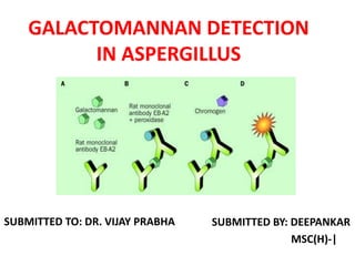
- 9. • Although, more definitive indicators in the diagnosis of IA can possibly be had from histological and bronchiolar lavage (BAL) samples for culture. GALACTOMANAN EIA: • Each GM molecule has as many as ten epitopes. Both capture and detector antibodies specific for the epitopes can be attached to the molecule. The Platelia Aspergillus EIA comprises of a rat monoclonal antibody (MAb) EB-A2 that reacts with the specific epitope of GM.
- 10. • It is an IgM antibody and binds to an epitope located on the β (1→5) galactofuranose- containing side chain of the GM molecule. A similar epitope seems to be present in other fungi. The epitope recognized by the EB-A2 MAb, is a common oligosaccharide moiety of a wide range of intracellular and extracellular glycoproteins of Aspergillus species and therefore, detection of GM can possibly be used as a biomarker for the diagnosis of IA.
- 11. GRAPHIC PRESENTATION OF THE PRINCIPLE OF THE GM EIA TEST:
- 12. BioRad Platelia Aspergillus EIA : • The principle of the PlateliaTM Aspergillus EIA, currently in use, is the same as the original assay. EB-A2 monoclonal antibody directed against Aspergillus GM is absorbed on the inner surface of a 96 well micro-titer plate. • There are two major phases to the assay (a) the extraction phase (b) the test phases
- 13. (A)Extraction phase: With the use of separate pipette tips 300 µL of each control or test serum are pipetted into separate polypropylene tubes, then 100 µL of ethylenediamine-tetraacetic acid (EDTA) solution, the serum treatment solution, are added and the two solutions are mixed vigorously then heated at 120^C for six minutes. The heated mixture is then centrifuged at 10,000 x g for 10 minutes. The supernatant of the mixture is used for GM testing. It can be used immediately or stored at 2-8^C for up to 48 hours.
- 14. (B)Testing phase of EIA: With all reagents at room temperature and mixed appropriately 50 µL of conjugate (antibody label) are added to each test /control designated well. This is followed by the addition of 50 µL of treated sample. The plate is covered and incubated at 37^C for 90 (±5) minutes. After incubation, the contents of each well are aspirated separately and the antigen-antibody complexes at the bottom of each well are washed five times with a washing solution containing tris NaCl buffer, tween 20 and thimerosal. The wells are drained and then 200 µL of Substrate-Chromogen reagent reaction solution are added to each well and the mixture is incubated at room temperature in the dark for 30 minutes. The reaction is stopped by the addition of a stop solution. The contents of the wells are mixed and the optical density of each well is read at 450 nm.
- 15. • The classification of a sample as to the presence or absence of GM is determined by its index which is calculated by dividing the OD of the sample by the mean OD of the cut-off control. The test brochure suggests cut-off index for serum GM as ≥0.5. • Samples with an index ≥ 0.5, i.e., positive for GM. • Sera with an index < 0.5 are considered negative for GM. https://www.youtube.com/watch?v=IxnvYQuxF6s
- 16. THANK YOU!