2. DIPHTHERIA
• Is an acute infectious disease
caused by toxigenic strains of
Cornybacterium diphtheriae.
3. • The bacilli multiply locally, usually in the throat, and
elaborate a powerful exotoxin which is responsible for
the following pathology.
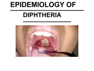
• The formation of grayish or yellowish membrane (false
membrane) commonly over the tonsils, pharynx, with
well defined edges and the membrane cannot be wiped
away.
• Marked congestion, edema or local tissue
destruction.
• Enlargement of the regional lymph nodes.
• Signs and symptoms of toxemia.
6. • The causative agent, C diphtheriae is
a gram-positive, non motile organism.
• It has no invasive power, but
produces a powerful exotoxin which
can affect the heart leading to
myocarditis.
7. • Four types of Diphtheria
bacilli are differentiated.
• gravis, mitis, belfanti and
intermedius, all
pathogenic to man.
9. CASE
• Cases range from subclinical infection to
clinical cases.
• Mild or silent infections may exhibit a mere
running nose or sore throat.
• These cases pose more threat in the spread
than the active cases.
10. CARRIER
• Carriers are common sources of
infection.
• Carriers may be temporary or
chronic nasal or throat carriers.
12. INFECTIVITY
• The period of infectivity may vary from
14 to 28 days from the onset of the
disease.
• But carriers may remain infective for
much longer periods.
13. • A case or a carrier may be considered non
communicable, when at least 2 cultures
properly obtained from nose and throat, 24
hours apart are negative for diphtheria bacilli.
14. HOST FACTORS
• AGE : Diphtheria particularly affects children
(1-5 yrs).
• GENDER : Both genders are affected.
15. IMMUNITY
• Infants born of immune mothers are
relatively immune during the first few
months of life.
• Children in developing countries seem to
acquire active immunity through active
infection.
17. MODE OF TRANSMISSION
• Droplet infection.
• Transmitted directly to susceptible persons
from infected cutaneous lesions.
• Transmission by objects (cups,
thermometers, toys, pencils) contaminated by
the nasopharyngeal secretions of the
patients is possible, but only for short
periods.
18. PORTAL OF ENTRY
• RESPIRATORY ROUTE : Commonly the portal of
entry is the respiratory tract.
• NON RESPIRATORY ROUTE : The portal of entry
sometimes may be the skin where cuts, wounds
and ulcers not properly attended to, may get
infected with diphtheria bacilli.(umbilical cord)
INCUBATION PERIOD
• 2 To 6 days.
20. Pharyngo-tonsillar diphtheria
• Sore throat
• Difficulty in swallowing
• Low grade fever at presentation
• Presence of pseudo membrane
over tonsils
• Oedema in sub mandibular
region
• Bull necked appearance
21. • Examination of the throat may show only mild
erythema, localized exudate or a pseudomembrane.
• The membrane may be a localized patch of the posterior
pharynx or tonsil, may cover the entire tonsil, or less
frequently, may spread to cover the soft palates and the
posterior portion of the pharynx.
• In the early stage the pseudo- membrane may be
whitish and may wipe off easily.
• The membrane may extend to become thick, blue-
white to grey-black and adherent.
• Attempts to remove the membrane may result in
bleeding.
• A minimal area of mucosal erythema surrounds the
membrane.
24. • Patients with severe disease may have
marked edema of the sub mandibular
area and the antererior portion of the
neck, along with lymphadenopathy,
giving a characteristic
• “bull- necked” appearance.
26. Laryngo-tracheal diphtheria
• Preceeded by pharyngo tonsillar
diphtheria
• Fever, hoarseness and croupy
cough
• Dyspnoea
• necrosis in heart
muscles, liver, kidneys
and adrenals
• vision difficulties,
speech, swallowing or
movements of arms or
legs
• paralysis of soft palate,
eye muscle or
extremities
Toxin damage
• parenchymatous
degeneration
27. • If the infection extends into the bronchial tree, it is the
most severe of disease.
• Prostration and dyspnoea soon follow because of the
obstruction caused by the membrane.
• This obstruction may even cause suffocation if not
promptly relieved by intubation or tracheostomy.
• The diphtheria bacilli within the membrane continue to
produce toxin actively
• This is absorbed and results in distant toxic damage,
particularly paranchymatous degeneration, fatty infiltration
and necrosis in heart muscle, liver, kidneys and adrenals
and some time accompanied by gross hemorrhage.
28. • Irregularities of the cardiac rhythm
indicate damage to the heart.
• Later there may be difficulties with vision,
speech, swallowing, or movement of the
arms or legs.
• The toxin also produces nerve damage, resulting
in paralysis of the soft palate, eye muscles or
extremities.
• Patients who survive complications
recover completely.
29. Nasal diphtheria
• Mildest form
• Localized in septum or turbinates of one
side of nose
• Conjunctiva and genitals also sources of
infection
• Membrane extends to pharynx.
30. Cutaneous diphtheria
• Common in tropicalareas
• Secondary infection of
previous infection orskin
abrasion
• Presenting lesion-an ulcer
surrounded by erythema
and covered with
membrane.
31.
32. SCHICK’S TEST
Intra dermal test
Tests – presence of antitoxin(immunity status) and
state of hypersensitivity to diphtheria toxin.
Inject 0.2ml of Schick test toxin intradermally into
skin of forearm, while into opposite arm- control
(Schick toxin inactivated by heat) is injected.
33. Negative reactions
if the person is immune, no reaction of any kind.
Positive reaction
In test arm, a circumscribed red flush of 10-50mm
diameter appears within 24-36 hours reaching
maximum development by 4th –7th day.
This slowly fades into a brown patch and skin
desquamates.
Control arm shows no change.
The person is susceptible to diphtheria.
34. Pseudo-positive reactions
A red flush develops equally on both
arms, much less circumscribed than
true +vereactions.
Fades by 4th day.
allergic reaction found in certain
individuals
Schick negative
Combined reactions
Control arm shows pseudo positive
reaction and test arm shows positive
reaction.
The person is susceptible to
diphtheria.
38. • Start active search
immediately from family
and school contacts.
• Carriers can be detected
by culture methods.
(swabs taken from nose
and throat)
CASES & CARRIERS
Early detection Isolation
all cases, suspected cases
and carriers should be
isolated, preferably in a
hospital for atleast 14
days or until proved free
of infection.
2 consecutive throat swabs taken 24 hours apart
should be negative before terminating isolation.
39. Treatment-
Cases
• Preliminary test dose of 0.2 ml
subcutaneously to detect sensitization to
horse serum.
• Followed by diphheria antitoxin IM or IV in
doses ranging from 20,000-40,000 units
or more depending on severity of cases.
• Mild early pharyngeal or laryngeal: 20,000-
40,000 units
• Moderate nasopharyngeal: 40,000-60,000
units
• Severe, extensive or late disease: 80,000-
100,000 units.
• Addition to antitoxin, penicillin or
erythromycin for 5-6 days to clear throat.
Carriers
• Should be treated
in 10days course
of oral
erythromycin
40. CONTACTS
Should be throat swabbed and immunity should be
determined.
Where primary immunization was received within the
previous 2 years- no further action needed.
Where primary course or booster dose of diphtheria
toxoid was received more than 2 years before, only a
booster dose of dip: toxoid need be given.
Non-immunized close contacts should receive
prophylatic penicilin or erythromycin.
They should be given 1000-2000 units of antitoxin and
actively immunized againstdiphtheria.
41. COMMUNITY
Active immunization with diphtheria toxoid of all
infants as early in life as possible with subsequent
booster dose every 10years thereafter.
Immunization rate must be maintained at high level.
44. DPT VACCINE
For immunization ofinfants.
Pertussis component enhances diphtheria toxoid.
Types- plain and adsorbed
STORAGE
should not befrozen
Stored in refrigerator at 2-8 degree celsius
Will loose potency if kept at room temperature for a long
time.
45. Optimum age-
DPT an be safely administered as early as 6 weeks after
birth.
Doses-
3 doses of DPT each is0.5ml.
Mode of admn-
injected intramuscular.
DPT given in upper and outer quadrants of gluteal region.
47. Reactions-
Fever and mild localreactions
2-6% develop fever of 39 degree or higher.
5-10% experience swelling and induration.
Neurological- encephalitis, prolonged convulsions,
infantile spasms, Reye’s syndrome.
Contra indications-
Seriously ill children or who need hospitalization are not
vaccinated.
Should not be repeated if a severe reaction occurred after
a previousdose.
In case of DPT, subsequent DT immunization.
48. For children over the age of 5 years who have not
received DPT- 2 doses of DT vaccine, 4 weeks apart,
with a booster dose 6 months to 1year later.
Those children who received primary course of DPT
earlier, should receive DT as booster at 5-6 years.
For immunizing children over 12years of age and
adults, preparation –dT (adult type diphtheria tetanus
vaccine).
Admn:- 2 doses at interval of 4-6 weeks, followed by
booster 6-12 months after second dose.