Presen tisse
•Download as PPTX, PDF•
0 likes•173 views

nasal tissue engineering using co-culture method

Recommended
More Related Content
What's hot
What's hot (16)
Similar to Presen tisse
Similar to Presen tisse (20)
More from Arun kumar
More from Arun kumar (20)
Recently uploaded
Recently uploaded (20)
Presen tisse
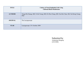
- 1. TITLE Culture of Nasal Epithelial Cells Using Chitosan-Based Membranes AUTHORS Tsung-Wei Huang, MD; Yi-Ho Young, MD; Po-Wen Cheng, MD; Yen-Hui Chan, MS; Tai-Horng Young, PhD JOURNAL The Laryngoscope YEAR Laryngoscope 119: October 2009 Submitted by Arunkumar Rengaraj (22141800)
- 2. Culture of Nasal Epithelial Cells Using Chitosan-Based Membranes Aim The aim of this study was to evaluate whether chitosan-based membranes can be used as scaffolds for growth and differentiation of nasal epithelial cells (NECs). Introduction • The nasal epithelium consists of ciliated epithelial cells, mucous blanket, and glands. • The nasal septum divides the nasal chamber into two cavities, aiding in the regulation of air flow through the nose. • Septal perforation, may cause inflammation, nasal crusting, bleeding, and whistling while breathing. • To date, various surgical repairs for the septal perforation are proposed • Chitosan- based membranes (scaffold) can be used for the culture of nasal epithelial cells (NECs). • It provides physical support and serves as a substrate to regulate cell growth, adhesion, and differentiation.
- 3. Materials and methods 2 % solution of chitosan chitosan + 1 M acetic acid spread on a glass plate Evaporated at 50C Mix 40 μg type I collagen chitosan-collagen membrane Tissues (Human inferior turbinates ) were treated with 0.5% pronase+1:1 mixture (DMEM) + Ham’s nutrient F12 centrifugation suspended in DMEM/F12 1.5-mL cell suspension was seeded on collagen coated Morphological Examination • The ciliary beat was observed under inverted microscope (Leica, DMI 6000, Solms, Germany) with 400 magnification. • Imaging of cilia movement was recorded at 240 frames per second with a high-speed complementary metal oxide semiconductor (CMOS) camera (CMC1300; VDS Vossku¨ hler GmbH, Osnabru¨ ck, Germany).
- 4. RESULTS • NECs were found to be successfully adhesive with collagen and chitosan-collagen membranes at day 3 after seeding. • Ciliated cells were first observed on collagen and chitosan- collagen membranes at day 7 • After confluence, which increased in number with active ciliary beating at day 21. • Confocal images demonstrated that NECs developed into a pseudo stratified polarized layer with apical cilia and basolateral nuclei on collagen and chitosan-collagen membranes resembling native respiratory epithelium, morphologically.