
Recommended
More Related Content
What's hot
What's hot (20)
Viewers also liked
Similar to What does endodontic microsurgery involve
Similar to What does endodontic microsurgery involve (20)
Recently uploaded
Recently uploaded (20)
What does endodontic microsurgery involve
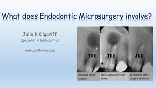
- 1. What does Endodontic Microsurgery involve? Julie K Kilgariff Specialist in Endodontics www.julie4endo.com Previous failed surgery Day surgical revision done 15 months after surgical revision JKK JKK JKK
- 2. Endodontic surgery is useful for treating infection at the end or sides of the root when the area cannot be accessed non-surgically or when a cyst is suspected. Less commonly, it is used to remove an unsalvageable root from a tooth with more than 1 root (so that half of the tooth – usually a molar – can be retained for chewing & aesthetics)
- 3. For example: This molar tooth originally had 2 roots. The further forward root was cracked and could not be saved. Rather than extract the whole tooth which would leave a sizable gap, the cracked root was divided from the rest of the tooth and removed. The remaining root was root canal treated and used to support a small bridge allowing the tooth to be used for chewing and completely filling the space left by the extracted root JKK JKK
- 4. For example: This large dark shadow associated with the end of the upper lateral incisor tooth was identified on x-ray after the patient had pain. There is a good quality root treatment already present. Owing to the size of the dark shadow a cyst was suspected and arrangements made to remove this surgically. Having done this, the removed tissue was sent to a laboratory (which confirmed that a cyst had been removed). At 1 year review, a large amount of new bone infill at the end of the root of the tooth can be seen. There is still more healing to occur (where the small dark area remains) JKK JKK
- 5. Traditionally (until 1990’s), endodontic surgery had poor success rates. Since the microscope was introduced into endodontics & microsurgical techniques developed, excellent success rates are achievable. Without using a microscope & microsurgical techniques, the success can be very low indeed, some studies report less than 30%
- 6. Generally, the microsurgical procedure involves: 1. The microscope is used 2. The tooth itself and neighbouring teeth are profoundly numbed 3. The gum is lifted to access the area of the root needed 4. Removal of infected soft and hard tissue +/- previous root filling materials 5. A tissue-friendly (biocompatible) material is placed into the root area to stimulate new bone formation around the tooth & ‘seal’ the root 6. The gum is placed back into position and held there with small stitches (sutures). 7. The sutures are removed 3-5 days later 8. Review
- 7. Small stitches (sutures) at 5 day removal JKK
- 8. If endodontic surgery is thought appropriate in your case, this will be discussed at your assessment. You will be given further information & leaflets depending on the type of surgery that it is.
- 9. If you have not found the answers to your questions regarding surgery here, please contact us via the email link Crack in tooth identified during microsurgery JKK