Eosinophils: Granulocytes in Inflammation
•Download as PPTX, PDF•
1 like•726 views

Eosinophils are granulocytic leukocytes that are involved in initiating and propagating many inflammatory responses, including those to parasitic infections and allergic diseases. They mature and are produced in the bone marrow, then migrate to mucosal tissues where they may play a role in preparing the uterus for pregnancy. Eosinophils contain orange-staining granules with cytotoxic proteins that can induce tissue damage, such as major basic protein, eosinophil cationic protein, eosinophil peroxidase, and eosinophil-derived neurotoxin.

Recommended
More Related Content
What's hot
What's hot (20)
Viewers also liked
Viewers also liked (19)
Similar to Eosinophils: Granulocytes in Inflammation
Similar to Eosinophils: Granulocytes in Inflammation (20)
Recently uploaded
Recently uploaded (20)
Eosinophils: Granulocytes in Inflammation
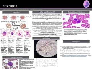
- 1. Eosinophils Anneka Pierzga & Geoffrey Latner | MLT 1042 | April 16, 2016 | College of Southern Maryland Maturation Hypersegmentation What is an Eosinophil? Eosinophilia Chediak-Higashi Syndrome • The Chediak-Higashi syndrome is a rare autosomal recessive condition associated with abnormally large leukocyte granules resulting from fusion of lysozymes. This disorder may affect granulocytes, leukocytes, and monocytes (UVA). • Phagocytosis is defective. Platelets lack dense granules and platelet function is abnormal (UVA). Bibliography • Eosinophilia occurs in association with hypersensitivity reactions, parasitic infestations, cancers (Hodgkin's disease, eosinophilic leukemia). (Blumenreich, 1990, p. 726) • Blood smear presents high percentage of eosinophils, as shown in the picture above. • A 100 cell differential count of 5 or more eosinophils is the basis for diagnosing eosinophilia. Pluripotent Stem Cell Myeloid Stem Cell CFU-GEMM CFU-Eo Myeloblast Eosinophilic Myelocyte Eosinophilic Metamyelocyte Eosinophilic Band Eosinophil Size: 12-18µm Nucleus: round to oval; may have 1 flattened side Nucleoli: not visible Chromatin: coarse; more condensed than promyelocyte Primary Granules: few to moderate Secondary Granules: variable number of refractile orange granules N/C Ratio: 2:1 to 1:1 “Dawn of Eosinophilia” Size: 10-15µm Nucleus: kidney bean shape; indentation < 50% width of nucleus Nucleoli: not visible Chromatin: coarse, clumped Primary Granules: few Secondary Granules: many orange, refractile N/C Ratio: 1.5:1 Size: 10-15µm Nucleus: constricted but no threadlike filament; indentation >50% nucleus Nucleoli: not visible Chromatin: coarse, clumped Primary Granules: few Secondary Granules: abundant orange, refractile N/C Ratio: cytoplasm predominates Size: 12-17µm Nucleus: 2-5 lobes connected by thin filaments Nucleoli: not visible Chromatin: coarse, clumped Primary Granules: rare Secondary Granules: abundant orange, refractile N/C Ratio: cytoplasm predominates IL-1, IL-6, IL-3 GM-CSF IL-3 GM-CSF, IL-3 GM-CSF, IL-5 GM-CSF, IL-5 GM-CSF, IL-5 Adapted from Harmening, 2009 Adapted from Carr & Rodk, 2013 Eosinophils are granulocytic leukocytes involved in the initiation and propagation of many inflammatory responses, including those of parasitic helminth infections and allergic diseases (Harmening 2009;Hogan and Rothenberg 2006). There is some evidence that they may act as APCs. They have been implicated in hypersensitivity reactions including allergic airway disease, asthma, eosinophilic gastritis and esophagitis, and Celiac disease (Hogan and Rothenberg 2006, Dyer, Foster et al. 2013) They are produced and mature in the bone marrow and are released into the blood, where they migrate to mucosal beds such as the skin, GI tract, vagina, and bronchial mucosa (Harmening 2009). Evidence suggests they may have a role in preparing the uterus for pregnancy and blastocyst implantation (Hogan and Rothenberg 2006). Eosinophils have orange-staining secondary granules which contain several cytotoxic proteins capable of inducing tissue damage and dysfunction: • Major Basic Protein – alters smooth muscle contraction responses and induces mast cell and basophil degranulation • Eosinophil Cationic Protein – shown to possess antiviral activity; induction of mast cell degranulation, stimulation of airway mucus secretion, suppression of T cell proliferation and IG synthesis by B cells • Eosinophil Peroxidase – catalyzes oxidation of several substances to produce highly oxidative substances that promote cell death • Eosinophil-derived Neurotoxin – shown to possess antiviral activity (Hogan and Rothenberg 2006) • Eosinophils typically have up to 4 nuclear lobes. • Hypersegmentation occurs when more than 3 eosinophils contain 5 lobes or 1 contains 6 or more lobes in a 100 cell differential (UVA). • This is sometimes called a “right shift” (UVA). • Hypersegmentation may accompany other disorders that affect maturation