
Recommended
More Related Content
What's hot
What's hot (20)
Similar to Medulla
Similar to Medulla (20)
More from anatomysrs
More from anatomysrs (20)
Recently uploaded
Recently uploaded (20)
Medulla
- 1. Medulla
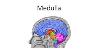
- 2. Location • Part of brainstem • Lower part of brainstem • Between pons and spinal cord • Part of Rhombencephalon • Hindbrain • Pons, medulla & cerebellum • Lies • Above spinal cord • Below Pons • Anterior to cerebellum
- 3. Extent • Superiorly • Pontomedullary sulcus • Inferiorly • Lower extent of pyramidal decussation • Through foramen magnum to the level of the atlas
- 4. External features • Ventral surface • Ventral median fissure • Pyramid • Olive • Nerves emerging • Hypoglossal (12th) • Sulcus between pyramid & olive • Glossopharyngeal (9th), Vagus (10th) & cranial part of accessory (11th) • Sulcus dorsolateral to olive
- 5. External features • Dorsal surface • Differ in • Caudal part (closed medulla) • Cranial part (open medulla)
- 6. Medulla - Closed • Cavity • Central canal • Dorsal median sulcus • Divides medulla into 2 halves • Fasciculus gracilis • On either side of dorsal median sulcus • Gracile tubercle • An elevation produced at upper part of fasciculus gracilis, • Site of gracile nucleus • Fasciculus cuneatus • On either side of fasciculus gracilis • Cuneate tubercle • An elevation produced at the upper part of fasciculus cuneatus • Site of cuneate nucleus Lateral to cuneate tubercle Tuber cinereum Spinal nucleus of V nerve is present
- 7. Medulla - open • Cavity • 4th ventricle • Caudal 1/3rd of floor of 4th ventricle • On either side of median sulcus • An inverted V-shaped sulcus divides the area into 3 parts • From medial to lateral • Hypoglossal triangle • Overlies hypoglossal nucleus • Vagal triangle • Overlies dorsal vagal nucleus • Vestibular area • Overlies vestibular nuclei
- 8. Internal structure • 2 important events happen by white matter • Pyramid decussation • Sensory decussation • By this action • Grey matter is subdivided
- 9. Pyramidal system • Primary system of voluntary movements • Upper motoneurons are located in cerebral cortex • Lower motoneurons can be found in • Motor nuclei of cranial nerves or in spinal ventral horn • Descending axons of upper motoneurons that terminate in • Motor nuclei of cranial nerves constitute • Corticonuclear Tract • Motor nuclei of Spinal cord constitute • Corticospinal tracts • Corticonuclear tract reaches the lower motoneurons of both sides (bilateral innervation) • While corticospinal fibres target the lower motoneurons of the opposite side only (crossed pathway)
- 10. Rubrospinal Tract • Origin • From red nucleus • Located in midbrain • Termination • Lateral column of spinal cord • Seen at • Lower midbrain • Pons • Medulla
- 11. Tectospinal Tract • Origin • Superior colliculus of midbrain • Termination • Anterior Column • Function • Motor function of the Skeletal muscles of the head and eyes in response to visual stimuli
- 12. Dorsal system • Medial lemniscus • Carry dorsal column modalities • From contralateral side • Can be seen • Upper medulla • Pons • Midbrain
- 13. Anterolateral system • Spinal lemniscus • Carry contralateral • Anteior spinothalamic • Crude touch • Lateral spinothalamic • Pain & temperature • Can be seen • Medulla • Pons • Midbrain
- 14. Spinocerebellar tracts • Carry impulses of a subconscious level • Fibres of spinocerebellar tracts form dorsal and ventral tracts • Carry information derived from muscle spindles, Golgi tendon organs and tactile receptors to the cerebellum
- 15. Dorsal (Direct) Spinocerebellar tract • Fibers originate from the cells of Clarke's column • At base of posterior horn • Axons ascend ipsilaterally to enter the cerebellum through inferior cerebellar peduncle
- 16. Ventral (Indirect) Spinocerebellar Tract • Fibers of ventral spinocerebellar tract decussate • Ascend on contralateral side of cord • Enter the cerebellum via • Superior cerebellar peduncle • Some axons then recross within the cerebellar white matter
- 17. Grey matter - At pyramidal (motor) decussation level • Central canal present • Anterior horn of grey matter • Cut off from remaining grey matter by • Fibres of pyramidal decussation • Becomes accessory nerve nucleus • Posterior grey horn shows • 2 projections • Nucleus gracilis – medially • Nucleus cuneatus – laterally
- 18. White matter - At pyramidal (motor) decussation level • Dorsal side • Fasciculus gracilis, fasciculus cuneatus & spinal tract of trigeminal • Lateral side • Lateral corticospinal tract • Anterior side • Pyramid & its decussation
- 19. At the level of sensory decussation • Fibres arising from nucleus gracilis and nucleus cuneatus • They cross the midline • Known as internal arcuate fibres • And turn upwards as medial lemniscus of the opposite side • Nucleus gracilis, cuneatus & nucleus of trigeminal nerve get detached from central grey matter
- 20. Grey matter - At sensory decussation level • Nuclei present in central grey matter • Hypoglossal • To tongue muscles • Dorsal nucleus of vagus • Gives parasympathetic fibres to • Heart, Lungs & abdominal organs • Nucleus tractus solitaries • Receives gustatory fibres from tongue • Through VII, IX & X
- 21. Grey matter - At sensory decussation level • Nuclei in dorsal side • Nucleus gracilis & Nucleus cuneatus • More prominent • Accessory cuneatus • Dorsolateral to cuneatus • Receives • Lateral fibres of fasciculus cuneatus • Sends fibres to • Posterior external arcuate fibers • Spinal nucleus of trigeminal • Nucleus ambiguus • Lies between • Nucleus of trigeminal & central grey matter • Supplies • Muscles of palate pharynx & larynx • Through IX, X & cranial accessory
- 22. Grey matter - At sensory decussation level • At ventral side • Inferior olivary nucleus
- 23. White matter - At sensory decussation level • Fasciculus gracilis & cuneatus terminates • Internal arcuate fibres • Medial longitudinal fasciculus • Connect the cranial nerve nuclei of • 3rd, 4th, 6th , 8th &spinal nucleus of XI • Spinocerebellar & lateral spinothalamic tracts • Pyramids
- 24. Grey matter – Inferior olivary level • In dorsal part • Central grey matter spread over floor of IV ventricle • Hypoglossal nucleus • Dorsal nucleus of vagus • Nucleus tractus solitaries • Vestibular nucleus (inferior & medial) • Spinal nucleus of trigeminal
- 25. Grey matter – Inferior olivary level • In middle part • Nucleus ambiguus • Lies in deep part of reticular formation • Gives fibres to • CN 9,10 & 11 • Olivary complex • Inferior olivary • Dorsal accessory olivary • Medial accessory olivary • In anterior part • Arcuate nucleus • Displaced pontine nucleui • Lies anteromedial part of pyramid
- 26. White matter – Inferior olivary level • On either side of midline • From dorsal to ventral • Medial longitudinal fasciculus • Tecto spinal tract • Medial leminiscus • Pyramidal tract • Lateral part • Lateral spinothalamic • Spino cerebellar • Posterolateral • Inferior cerebellar peduncle
- 27. Autonomic centres in medulla • Cardioinhibitory and cardiostimulatory centers • Affect the rate and force of cardiac contraction • Vasomotor centers • Affect smooth muscle fibers tone • Respiratory center
- 28. Other centres • Emesis • Deglutition • Coughing • Hiccupping • Sneezing
- 29. Blood supply • Ventrally by • Branches of vertebral and basilar arteries • Supply region next to midline • i.e. the part containing pyramid, medial lemniscus and hypoglossal nucleus • Laterally and dorsally by • Posterio-inferior cerebellar artery • Lateral and dorsal sides
- 30. Medial medullary syndrome • Due to loss of blood supply to ventral side • Structures affected • Pyramid • Medial lemniscus • Hypoglossal nucleus • Resulted in • Paralysis of tongue on same side • Hemiplegia (crossed pyramid damage) • Loss of touch and kinaesthetic sense on the opposite side ( medial lemniscus damage)
- 31. Lateral medullary syndrome • Loss of blood supply to lateral area
- 32. Lateral medullary syndrome • Loss of nucleus ambiguus • Paralyses of laryngeal, palatal and pharyngeal muscles on that side, causing dysphonia and dysphagia • Loss of uncrossed spinal tract of the trigeminal and of crossed spinal lemniscus • Loss of pain and temperature sensation on the same side of face and opposite side of the body
- 33. Lateral medullary syndrome • Involvement of vestibular nuclei • Causes vertigo and nystagmus with nausea and vomiting