2. OBJECTIVES
1. Describe external features of the brain stem
2. Identification of the tracts and nuclei in the brain stem.
3. Orientation of the cranial nerve nuclei in the brain stem.
4. Highlight the connections and significance of the reticular
formation.
3. INTRODUCTION
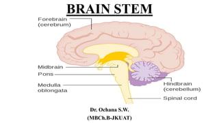
• The brainstem is made up of the medulla oblongata, the pons, and
the midbrain and occupies the posterior cranial fossa of the skull.
• It is stalk-like in shape and connects the narrow spinal cord with the
expanded forebrain.
• The brainstem has 4 broad functions:
It serves as a conduit for the ascending tracts and descending tracts
connecting the spinal cord to the different parts of the higher centers in
the forebrain.
It contains important reflex centers associated with the control of
vomiting, respiration and the cardiovascular system and with the
control of consciousness
It contains the important nuclei of cranial nerves III through XII.
It contains the reticular formation.
6. GROSS FEATURES
• The medulla oblongata is conical (bulb) in shape broad superiorly and
narrow inferiorly.
• It connects the pons with the spinal cord.
• On each side of its anteromedian fissure there is a swelling named
the pyramid.
• The pyramid tapers inferiorly where majority of corticospinal fibers
cross to the opposite side.
• Posterolateral to the pyramids are the olives which are oval elevations
produced by the inferior olivary nuclei.
7. • Rootlets of the hypoglossal nerve emerge between the pyramid and
olive on each side.
• Posterior to the olives are the inferior cerebellar peduncles.
• Rootlets of the glossopharyngeal, vagus and accessory nerves
emerge in the groove between the olives and inferior cerebellar
peduncles.
8. • The posterior surface of the rostral medulla forms the floor of the 4th
ventricle.
• The posterior surface of the caudal medulla possesses a posterior
median sulcus on each side of which there is an elongated swelling,
the gracile tubercle produced by gracile nucleus.
• Lateral to the gracile tubercle is a cuneate tubercle produced by
cuneate nucleus.
10. INTERNAL STRUCTURES OF THE
MEDULLA
• Internal features of medulla are illustrated in transverse sections
through 3 levels:
Caudal level, level of motor decussation.
Middle level, level of sensory decussation.
Superior level, level of open medulla.
11. Caudal medulla (Level of motor
decussation)
• At transition from the spinal cord to the medulla, the pattern of gray
and white matter undergoes considerable rearrangement.
• The ventral horn becomes much attenuated.
• The dorsal horn is replaced by the spinal trigeminal nucleus.
• Superficial to this nucleus is the spinal tract of the trigeminal nerve
descending to the spinal trigeminal nucleus.
• In the ventral aspect majority of pyramidal tract fibers decussate
(motor decussation) and pass laterally, dorsally and caudally to form
the lateral corticospinal tract.
12.
13. Middle medulla (Level of sensory
decussation)
• Gracile and cuneate nuclei appear beneath their respective tracts.
• Axons of gracile and cuneate nuclei course medially and ventrally as
internal arcuate fibers which decussate at this level forming sensory
decussation.
• The decussated fibers turn rostrally forming medial lemniscus (on
each side) which ascends in the brain stem to end in the thalamus.
• Posterior external arcuate fibers originate from accessory cuneate
nucleus (positioned lateral to cuneate nucleus) to reach the cerebellum
through the inferior cerebellar peduncle.
14.
15. Rostral (open) medulla
• Inferior olivary nucleus is shown within prominence of the olive.
• It is present dorsolateral to the pyramid.
• Lateral to the medial lemniscus is the spinothalamic tract.
• The dorsal surface of open medulla forms part of the floor of the 4th
ventricle.
• Immediately deep to this floor lie a number of cranial nerve nuclei
(hypoglossal nucleus, dorsal motor nucleus of the vagus nerve
(nucleus ambiguus), and inferior and medial vestibular nuclei).
• Ventromedial to the hypoglossal nucleus close to the midline is the
medial longitudinal bundle which links vestibular with ocular nuclei.
• Dorsolateral prominence of open medulla is the inferior cerebellar
peduncle.
16.
17. CLINICAL NOTES
• Tumors of the posterior cranial fossa produce increased intracranial
pressure (ICP).
• There is a tendency to downward herniation of the medulla and
cerebellar tonsils through the foramen magnum.
• This will produce headache, neck stiffness and paralysis of the lower 4
cranial nerves owing to traction.
• In this condition lumbar puncture is contraindicated.
19. GROSS FEATURES
• The pons (bridge) connects the medulla to the midbrain.
• It is located ventral to the cerebellum where it bridges to connect the
two cerebellar hemispheres.
• The anterior surface shows many transverse pontine fibers that
converge on each side to form the middle cerebellar peduncle.
• The trigeminal nerve fibers emerge from the anterolateral surface
between the pons and its peduncle.
20. • In the groove between the pons and the medulla emerge the abducent,
the facial and the vestibulocochlear nerves.
• The posterior surface of the pons forms the upper half of the floor of
the 4th ventricle.
• It is divided into two halves by a median sulcus.
• Lateral to this sulcus is the facial colliculus produced by fibers of the
facial nerve winding around nucleus of the abducent nerve.
• Lateral to the facial colliculus is the vestibular area produced by
underlying vestibular nuclei.
22. INTERNAL STRUCTURES OF THE
PONS
Inferior level, level of facial colliculus.
Middle level, level of trigeminal nuclei.
Superior level, level of 4 lemnisci.
• For purposes of description the pons is divided into:
A posterior part, the tegmentum
An anterior part, the basis pontis.
23. • Basis pontis is marked by transversely oriented pontocerebellar
fibers originating from scattered pontine nuclei and pass to the
contralateral cerebellar hemisphere through the middle cerebellar
peduncle.
• Corticospinal fibers appear dispersed between fascicles of transverse
pontine fibers.
• Tegmentum pontis contains reticular formation, lemnisci and
cranial nerve nuclei.
24. Caudal Pons (level of facial colliculus)
• The trapezoid body is located ventral to ascending lemnisci but dorsal
to basis pontis.
• Beneath the floor of the 4th ventricle lie a number of cranial nerve
nuclei.
• Abducent nucleus is medially located and close to the surface.
• Facial motor nucleus is located ventrolateral to abducent nucleus.
• Fibers of facial nerve wind around abducent nucleus before exiting
from the ventral aspect of the pons.
25. • This creates the facial colliculus seen in the floor of the 4th ventricle.
• Laterally in the floor of the 4th ventricle is the vestibular area deep to
which are the superior and lateral vestibular nuclei.
• The cochlear nuclei are located more laterally posterior and anterior
to the inferior cerebellar peduncle.
• Lateral lemniscus of the auditory pathway is seen in the tegmentum
of the caudal pons.
26.
27. Mid Pons (level of trigeminal nuclei)
• The four trigeminal nuclei reach their maximum extent at this level
adjacent to the origin of the trigeminal nerve which exits from the
ventral aspect between the pons and its peduncle.
Rostral Pons (level of four lemnisci)
• The superior cerebellar peduncles form the lateral walls of the 4th
ventricle.
• The thin superior medullary velum spans between the superior
cerebellar peduncles to form roof of the 4th ventricle.
• The 4 lemnisci are organized in order as medial, spinal, trigeminal and
lateral forming crescent of lemnisci.
29. Functional significances of the pons
Contains cranial nerve nuclei: V,VI VII, and VIII.
Pathway for ascending and descending tracts.
Pathway for hearing.
Contains pontine reticular formation and superior salivatory nucleus.
Contains part of respiratory centers.
31. GROSS FEATURES
• The midbrain connects the pons and cerebellum with the forebrain.
• It is traversed with the cerebral aqueduct which connects the 3rd and
4th ventricles.
• On the ventral surface there is a depressed area called the
interpeduncular fossa bounded on each side by the crus cerebri.
• Oculomotor nerve fibers emerge from a groove on the medial side of
the crus cerebri.
• On the dorsal aspect there are 4 colliculi (4 corpora quadrigemina);
two superior and two inferior colliculi.
32. • The superior colliculi are centers for visual reflexes.
• The inferior colliculi are centers for auditory reflexes.
• In the midline below the colliculi the trochlear nerves emerge and
then wind around the lateral aspects of the midbrain to enter the
cavernous sinuses.
34. INTERNAL STRUCTURES
• The cerebral aqueduct divides the midbrain into posterior part,
tectum, and anterior part, the cerebral peduncle.
• The cerebral peduncle is divided by the substantia nigra into an
anterior part, crus cerebri, and a posterior part, the tegmentum.
• In the periaqueductal gray matter is nucleus of locus ceruleus
(noradrenergic nucleus).
• Moreover there is the raphe nucleus (serotonergic neurons).
• The tegmentum is bounded by the crus cerebri (cerebral peduncle)
which consists of descending cortical efferents.
35. • Its middle half is composed of corticobulbar (medially) and
corticospinal (laterally) fibers.
• On each side of the pyramidal tract are the corticopontine fibers.
• The most ventral part of the tegmentum is occupied by the substantia
nigra composed of pigmented neurons that synthesize dopamine.
• These neurons project to the corpus striatum.
• The inner structures of the midbrain are studied in two levels:
Caudal level, level of inferior colliculi.
Rostral level, level of superior colliculi.
36. Caudal Midbrain
• The inferior colliculus is part of the hearing system.
• At this level the ascending auditory fibers in the lateral lemniscus end
in the inferior colliculus.
• Lemniscal crescent is obvious at this level.
• Ventral to the periaqueductal gray matter lays the trochlear nucleus.
• The superior cerebellar peduncles decussate in the midline of this
level.
37. • Periaqueductal gray matter contains several nuclei:
Nucleus of locus ceruleus (noradrenergic/dopaminergic). This
nucleus extends to the pons and is concerned with cognitive mapping,
affective behavior, thalamic and neocortical activities.
Raphe nucleus (serotonergic).
Dorsal tegmental nucleus (secretes endorphins and enkephalins).
Close to this area is the central tegmental tract containing the
reticulothalamic tract (reticular activating system).
38.
39. Rostral Midbrain
• The superior colliculus is part of the visual system.
• Its afferents are mainly corticotectal fibers originating from the visual
cortex and the frontal eye field.
• These inputs are concerned with controlling movements of the eyes
(following objects, accommodation and conjugate eye movements).
• Efferents from the superior colliculi decussate in the dorsal tegmental
decussation and descend as tectospinal tract.
40.
41. • A small number of fibers of the optic tract terminate in the pretectal
nucleus, just rostral to the superior colliculus.
• The pretectal nucleus is connected to Edinger Westphal
(parasympathetic) nucleus to control papillary light reflexes.
• Ventral to the periaqueductal gray matter are the oculomotor nuclei.
• The central portion of the tegmentum is occupied by the red nucleus.
• Efferents from the red nucleus decussate in the ventral tegmental
decussation and descend as rubrospinal tract.
42. Functional significances of the midbrain
It contains cranial nerve nuclei: III and IV.
The cerebral aqueduct passes through it.
It offers pathway for the corticofugal fibers and the lemnisci.
The substantia nigra is considered part of the basal ganglia.
It has the red nucleus which is one of the extrapyramidal system.
It contains midbrain reticular formation.