
Recommended
More Related Content
What's hot
What's hot (20)
Similar to Fowl pox virus
Similar to Fowl pox virus (20)
Recently uploaded
Recently uploaded (20)
Fowl pox virus
- 1. Assignment PATH-702 Ag no. 2020-ag-443 Submitted by: Ahmad Hassan Fowl POX Fowlpox is a common disease of poultry seen in many countries. Caused by the fowlpox virus, Avipoxvirus a DNA virus belonging to the family Poxviridae. The disease is characterized by production losses and cutaneous lesions, and mortality especially when the birds have more generalized forms of the disease. Fowlpox is transmitted mostly through mechanical transmission from the contaminated poultry house environment, and insects are also thought to assist transmission. Poxis a commonviral disease ofcommercial poultry as well as ofpet and wild birds. Fowlpox is an economically important disease of commercial poultry because it can cause a drop in egg production, slow growth, and unexpected mortality. Pox is a slow‐ spreading disease characterized by the development of discrete nodular proliferative lesions on the non‐ feathered parts of the body (cutaneous form) or fibro necrotic and proliferative lesions in the mucous membrane of the upper respiratory tract, mouth, and esophagus (diphtheritic form). Concurrent systemic infections also may occur. Economic Significance Mortality in flocks exhibiting the mild cutaneous form of the disease is usually low. However, it may become high with generalized infection, especially when lesions are primarily diphtheritic or when the disease is complicated by other infections or poor environmental conditions. Under those conditions the economic losses can be significant. Public Health Significance
- 2. Avian poxis not ofpublic health significance. It does notcause productive infection in mammalian species. Etiology Avianpox viruses infecting various avian species (canary, fowl, junco, mynah, pigeon, psittacine, quail, sparrow, starling, turkey, crow, peacock, penguin are recognized within the Avipoxvirus genus in the family Poxviridae. Morphology All avianpox viruses have a similar morphology. The mature virus (elementary body) is brick‐ shaped and measures about 330 × 280 × 200 nm. Incubation Period The incubation period of the naturally occurring disease varies from about 4–10 days in chickens, turkeys, and pigeons and is about 4 days in canaries. Clinical Signs The disease may occur in one of the two forms, cutaneous or diphtheritic, or both. In addition, a systemic form of infection with high mortality is usually seen in canaries. The cutaneous form of the disease is characterized by the appearance of nodular lesions on the comb, wattle, eyelids, and other non‐ feathered areas of the body. Cutaneous eye lesions interfere with the bird’s ability to find food and water. In the diphtheritic form (wet pox), cankers or diphtheritic yellowish lesions occur on the mucous membranes of the mouth, esophagus, or trachea with accompanying coryza‐ like mild or severe respiratory signs similar to those caused by infectious laryngotracheitis virus infection of the trachea. Lesions in the cornerof the mouth and on the tongue, throat, and upperpart of the trachea interfere with eating, drinking, and breathing. In pullets coming into lay and in older birds, the disease often runs a slow course accompanied by unthriftiness and reduced egg production. Morbidity and Mortality
- 3. The morbidityrate of poxin chickens and turkeysvaries from afew birdsbeing infected to involvementof the entire flock if a virulent virusis present and no preventive measures have been taken. Birdsaffected with the cutaneousform of the disease are more likely to recover than those with the diphtheritic form involving oral mucosa and the respiratory tract. Pathology Gross The characteristic lesion of the cutaneous form of pox in chickens is epithelial hyperplasia involving the epidermis and underlying feather follicles, with formation of nodules that first appear as small white foci and then rapidly increase in size and become yellow. In chickens infected intradermally, a few primary lesions appear by the fourth day. Papules are formed by the fifth or sixth day. This is followed by the vesicular stage, with formation of extensive thick lesions. Adjoining lesions may coalesce and becomerough and gray or dark brown. After about two weeks or sometimes sooner, lesions have areas of inflammation at the base and become hemorrhagic. Formation ofa scab, which may last foranother 1–2 weeks, ends with desquamation of the degenerated epithelial layer. If the scab is removed early in its development, there is a moist, seropurulent exudate underneath covering a hemorrhagic granulating surface. Attenuated vaccine viruses producelocalized lesions, which are mild in comparison to the severe ones due to pathogenic strains. The secondary lesions produced by pathogenic strains may persist for several weeks. In the diphtheritic form, slightly elevated, white opaque nodules or yellowish patches develop on the mucous membranes of mouth, esophagus, tongue, or upper trachea. Nodules rapidly increase in size and often coalesce to become a yellow, cheesy, necrotic, pseudo diphtheritic, or diphtheritic membrane. If the membranes are removed, they leave bleeding erosions. The inflammatory process may extend into sinuses, particularly the infraorbital sinus (resulting in swelling) and also into the pharynx and larynx (resulting in respiratory disturbances) and esophagus. It is not uncommon to find cutaneous as well as diphtheritic lesions in the same bird.
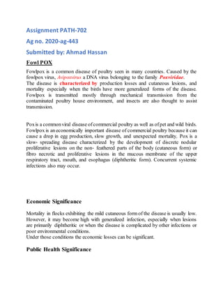
- 5. Cutaneous FWPV lesion on the eye and nostrils. Eosinophilic cytoplasmic inclusion bodies (arrows) are present in most of the infected
- 6. cells. Infected cells are enlarged, and some infected cells have lost their nuclei. Diagnosis Tentative diagnosis based onclinical signs and lesions, supported byhistopathology, is the method of choice to confirm the diagnosis in many laboratories. Hematology Parameters Microscopy Tissue sections from cutaneous ordiphtheritic lesions are processedbyconventional methods or by using a solution that fixes and dehydrates the tissues simultaneously followed by hematoxylin–eosin (H&E) staining for detection of cytoplasmic inclusions. Isolation and Identification of Virus Bird Inoculation Avianpox viruses can be transmitted to susceptible birds by applying a suspension of the lesion material from infected birds to their scarified comb or denuded feather
- 7. follicles of the thigh, or by the wing‐ web stick method. Fowlpox virus can be transmitted readily to susceptible chickens, with typical cutaneous lesions developing in 5–7 days. Serology Evidence for prior infection, or for ongoing infection in the flock, can be determined by serological methods. The older technique of gel precipitation has essentially been superseded by enzyme-linked immunosorbent assay (ELISA) as used in commercial flock monitoring kits Passive Hemagglutination A passive hemagglutination test detects antibodies in the serum of FWPV‐ infected chickens earlier than the immunodiffusion test. Although this test is very sensitive, its use has been limited because it requires sheep or horse red blood cells for sensitization with soluble pox virus antigens. Furthermore, differentiation of viruses is not possible because of cross‐ reacting antigens. Molecular Methods Restriction Endonuclease Analysis of Avianpox Virus DNA Restriction fragment length polymorphism (RFLP) can be used for comparing the genomes of avianpox viruses by examination of the relative mobilities of restriction endonuclease‐ generated fragments of their DNAs. The genetic profiles of FWPV strains are similar, with a high proportion of co‐ migrating fragments, although most strains could still be distinguished by the presence or absence of one or two DNA fragments. Control and Vaccination Fowlpox vaccine is commonly applied by the wing‐ web method to 4‐ week‐ old chickens and to pullets about 1–2 months before egg productionis expected to start. It is also used to revaccinate chickens held for the second year of egg production. Several live FWPV‐ vectored vaccines, for example, Newcastle disease–fowlpox vaccine for subcutaneous or wing‐ web stab immunization of 1‐ day‐ old chickens, is available commercially.