Conjunctival anatomy
•Download as PPTX, PDF•
1 like•258 views

Conjunctival anatomy,Blood supply ,nerve supply & Nerve Supply

Recommended
More Related Content
What's hot
What's hot (20)
Similar to Conjunctival anatomy
Similar to Conjunctival anatomy (20)
More from Md Afzal Mahfuzullah
More from Md Afzal Mahfuzullah (11)
Recently uploaded
Recently uploaded (20)
Conjunctival anatomy
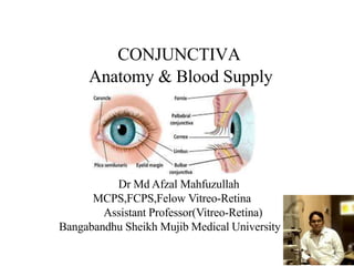
- 1. CONJUNCTIVA Anatomy & Blood Supply Dr Md Afzal Mahfuzullah MCPS,FCPS,Felow Vitreo-Retina Assistant Professor(Vitreo-Retina) Bangabandhu Sheikh Mujib Medical University
- 2. Introduction Conjoin = to join So the name conjuctiva has been given to this mucous membrane owing to the fact that it joins the eyeball to the lids It is a translucent mucous membrane which lines the posterior surface of the eyelids and anterior aspect of the eyeball
- 3. The normal conjuctiva is o Pink o Smooth o Thin o Transparent There are normally large deep blood vesells that run vertically
- 4. Functions of conjunctiva The conjunctiva helps lubricate the eye by producing mucus and tears, although a smaller volume of tears than the lacrimal gland It also contributes to immune surveillance and helps to prevent the entrance of microbes into the eye
- 5. Parts of the conjunctiva
- 6. Parts of the conjunctiva Conjunctiva Palpebral Marginal Tarsal Orbital Bulbar Scleral Limbal Fornix Superior Inferior Lateral Medial
- 7. Palpebral conjunctiva: o It is richly vascular, extremely thin and strongly bounded to the tarsal plate
- 8. oIt lines the lids and is subdivided into marginal tarsal orbital
- 9. Marginal- o Extends from the lid margin to about 2mm back of the lid upto the sulcus subtarsalis o Actually a transitional zone between skin and the conjunctiva proper o Lacrimal puncta open in the marginal zone
- 10. Tarsal- o Thin, transparent and highly vascular o Firmly adherent to the whole tarsal plate in the upper lid and only to half width of the tarsus in the lower lid o The tarsal glands are seen through it as yellow streaks
- 11. oOrbital- o It lies loose between the tarsal plate and the fornix o Orbital margin of the upper eyelid is loose and lies over the muller’s muscle
- 12. Bulbar conjunctiva- o It is transparent and lies loose over the underlying structures and thus can be moved easily o It is separated from the anterior sclera by episcleral tissue and o tenon’s capsule
- 13. o The average thickness is 33 microns o It is also known as ocular conjunctiva o It is further of two types Limbal Scleral
- 14. Limbal- o A 3mm ridge of bulbar conjunctiva around the cornea is called limbal conjunctiva o Strongly adherent to sclero-corneal junction Scleral- o Covers the eyeball above the anterior sclera and hence known as scleral conjunctiva o Thin, transparent & loosely attached to underlying sclera o Separated from the sclera by episcleral vessels and Tenon’s capsule
- 15. oConjunctival fornix: o It is thin, transparent , continuous circular cul-de- sac o It is broken only on the medial side by caruncle and the plica semilunaris o It joins the bulbar conjunctiva with the palpebral conjunctiva
- 16. Lateral Medial inferior It is further of four types Superior
- 17. oSuperior o Located at the level of superior orbital margin o Extends from slightly upper border of the tarsal plate to a distance about 10mm from the upper limbus o Here we can find the glands of Krause and o Mullers’s muscle in the subconjunctival tissue
- 18. oInferior fornix o Extends from slightly below the lower border of the lower tarsal plate to a distance about 8mm from the lower limbus o Located near the inferior orbital margin o Helps in maintaining the recess of the inferior fornix during movements of the lower lid
- 19. oLateral o Small in size like a cul de sac o Extends to just behind the equator of the eyeball o It is 14mm from the lateral limbus and about 5mm from the lateral canthus oMedial o It is a shallow cul de sac in which lie the caruncle and plica seminlunaris dipped in pool of tears called as tear lake
- 21. Structure of conjunctiva Histologically cornea consists of 3 layers Conjunctiva Epithelium Adenoid layer Fibrous layer
- 22. Epithelium a. The layers of epithelial cells in the conjunctiva vary from region to region and its different parts are oMarginal conjunctiva- oHave 5 layers non keratinised stratified squamous type of epithelium oSuperficial layer- squamous cell oIntermediate 3 layers- polyhedral cells oDeepest layer- goblet cells
- 23. oTarsal conjunctiva- oHas 2 layer epithelium in the upper eyelid oSuperficial layer- cylindrical cells oDeep layers- cubical cells oLower tarsal conjunctiva is made of 3-4 layers of cells like the cubical, polygonal, elongated wedge shaped and cone shaped cells
- 25. oFornix and bulbar conjunctiva o3 layered epithelium oSuperficial layer- cylindrical ells oMiddle layer- polyhedral cells oDeep layer- cuboidal cells
- 26. oLimbal conjunctiva o8-10 layers of stratified squamous epithelium oMost superficial 1-2 layers- squamous cells oIntermediate several layers- polygonal cells oBasal layer- cylindrical or cubicalcells
- 27. Cells Present In The Epithelium oGoblet cells- oPresent between the epithelial cells in all regions of conjunctiva oMelanocytes- oFound in conjunctiva at limbus, fornix, crancule and at the site of entry of anterior ciliary vessels
- 28. Cells Present In The Epithelium,Cont oLangerhans cells- oPresent in all parts of conjunctiva oConjunctival associated lymphoid tissue ( CALT)- oConsists of T and B lymphocytes
- 29. d. Mucosal associated lymphoid tissue(MALT) o MALT of the gut and bronchi are also found in the conjunctiva
- 30. Adenoid layer o Also called as lymphoid layer o Consists of fine connective tissue reticulum in the meshes of which lie the lymphocytes o Most developed in the fornices and ends at the subtarsal fold o Develops after 2-3 months of life The Adenoid Layer
- 31. oFibrous layer o Consists of a meshwork of collagenous and elastic fibres o Thicker than the adenoid layer o Except in the tarsal conjunctiva where it is very thin o This layer consist vessels and nerves of the conjunctiva o The adenoid layer and the fibrous layer are collectively called as substantia propia
- 33. Conjunctival glands Mucin secretory glans • Goblet cells • Crypts of henle • Glands of manz Acessory lacrimal glands • Glands of krause • Glands of wolfring
- 35. o Goblet cells o Round or oval in shape with an eccentric flat nucleus o Unicellular mucous cells located abundantly within the epithelium of all regions of conjunctiva
- 36. o These cells are formed from the deepest cells of the conjunctiva o Once discharging their content- the mucin they are destroyed o Density is more in children o than adults o More in the bulbar conjunctiva and inferior fornix
- 37. Henle’s glands o Not true glands but folds of mucous membrane present in the palpebral conjunctiva o These are tubular structures with lumina of 15-30 µm Glands of manz o Found in limbal conjunctiva in animals
- 38. Glands of krause o Microscopic glands that lie in the sub conjuctival tissue of the fornices o These are about 40-42 in the upper fornix and about 6-8 in the lower fornix Glands of wolfring o Also called as the glands of Ciaccio o These are microscopic glands present along the upper border of superior tarsus and lower border of inferior tarsus
- 39. Blood supply Arteries supplying the conjunctiva are derived from 3 sources. They are: 1. Marginal arcade of the eyelid 2. Peripheral arterial arcade of the eyelid 3. Anterior ciliary artery
- 40. Blood supply,Cont The palpebral conjunctiva and the fornices are supplied by branches from the marginal and peripheral arcades of the artery
- 41. Bulbar conjunctiva is supplied by posterior conjunctival arteries and anterior conjunctival arteries
- 42. Venous drainage The veins from conjunctiva drain into the venous plexus of eyelids which in turn drain into the superior and inferior ophthalmic veins.
- 43. Venous drainage,Cont A cicumcorneal zone of limbus drain into the anterior cilliary veins
- 44. Lymphatic drainage Lymphatics from the lateral side drain into the periauricular lymph nodes The lymphatics from the medial side drain the submandibular lymph nodes
- 45. Nerve supply A circumcorneal zone of the conjunctiva is supplied from the long ciliary nerves. Rest of the conjunctiva is supplied by the branches from the lacrimal, infratrochlear, supratrochlear, supraorbital and the frontal nevers