2. • Coined from latin word uva -
grape
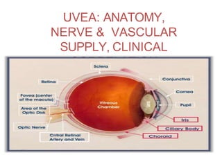
• Middle vascular coat of eyeball
• From anterior to posterior:
- Iris
- Ciliary body
- Choroid
INTRODUCTI
ON
3. Embryolog
y
NEUROECTODERM
Epithelial layers of the iris
Sphincter and dilator pupillae
muscles
Epithelial layers of the ciliary body
PERIOCULAR MESENCHYME
Blood vessels of iris, choroid,
ciliary body
Stroma of iris
Ciliary muscle and stroma of ciliary
body
4. Milestone
s
9th week of gestation Ciliary body begins to appear
12th week of gestation Sphincter pupillae appears
4th month Ciliary processes fully formed
5th month Iris and choroid are formed
6th month Dilator muscles begin to form
Sphincter muscle fully
differentiated
Postnatal period dilator muscles reach adult
proportion by 5 years
5. Greek Word iris - color
haloes/rainbows
Anterior most part ofuvea
Diameter -12 mm
Thickness -0.5 to 0.6 mm
3 to 4 mm aperture slightly nasally-
pupil
Attached to middle of anterior
surface of ciliary body
Thinnest at root and tears easily
away from its attachment to ciliary
body : Iridodialysis
IRI
S
6. POINTs TO BE NOTED
Pupillary margin
rests lightly on
anterior surface of
lens so when lens
is removed iris is
flat and often
tremulous:
IRIDODONESIS
CYCLODIALYSIS is
separation of ciliary
body from scleral spur,
creating a direct
connection betwn
anterior chamber and
suprachoroidal
space
increases aqueous
outflow and
predisposes the eye
to hypotony
9. Posterior
surface
- dark brown or black
- looks smooth
Under Magnification:
-schwalbe’s contraction folds:
radial furrows, commence 1mm from pupillary border
-schwalbe’s structural furrows:
start 1.5mm from pupillary border, narrow and deep to
start with but becomes wide and shallow as they
approach the ciliary margin
-circular furrows:
finer than radial furrows
cross the structural furrows at regular
interval more marked near the pupil
11. 1. Anterior limiting
membrane
• Condensed part of the stroma
• Consists of melanocytes and fibroblasts
• Deficient in areas of crypts, very thin at
contraction furrows
• Determines the color of iris
3 types of intercellular junctions present
- Gap junctions
- Intermediate junctions
- Discontinuous tight junctions
12. 2.
Stroma
• Main bulk of iris tissue
• Consists of loosely arranged collagenous
network with mucopolysaccharide ground
substance
contains
-The sphincter pupillae muscles
- Dilator pupillae muscles
-The vessels and nerves of iris
-Cellular elements: fibroblast, melanocytes,
clump cells and mast cells
13. Sphincter pupillae
muscles
- 0.7mm wide , 0.1-0.17mm thick
- Encircles pupillary margin
- lies in stroma deep to the surface
-Innervated by parasympathetic via short ciliary
nerve
Origin is from anterior epithelium,but actually
separated from this layer by a thin sheet of
collagen & dilator fibre processes, to which it is
firmly bound.
14. Dilator pupillae
muscle
- 60um long &7um
wide
- Filled with myofilaments
- Extend from iris root towards pupil
- When the muscle contract,it pulls the
pupillary margin towards the ciliary body,
dilating the pupil
- Innervated by sympathetics via long ciliary
nerve
18. 3. Anterior Epithelial
Layer
• Anterior continuation of pigment epithelium of
ciliary body
• Lacking of melanocytes
• Basal processes of the cells of this layer give
rise to dilator pupillae muscle
19. 4. Posterior pigmented epithelial
layer
• Anterior continuation of non pigmented
epithelium of ciliary body which in turn is the
continuation of sensory retina
21. Iris Nodules
- Accumulated deposits of epithelioid cells and
lymphocytes deposited onto the iris without tissue
destruction.
Two
types:
Koeppes nodule at pupillary
border
Busaccas nodules near
collarette
23. • Forward continuation of the choroid at ora
serrata
• In cut section, triangular in shape
CILIARY
BODY
24. • Anterior side of triangle- part of anterior
chamber angle
• In middle- attached to the iris
• Outer side of triangle- lies againstthe sclerawith
a suprachoroidal space in between
• Inner side of triangle divided into 2 parts:
1) Pars plicata-anterior
2) Pars plana-posterior
25. 1) Pars plicata / corona ciliaris
• Anterior part
• About 2 to 2.5 mm long
• Contain ciliary muscles
• Have finger like ciliary
processes
2) Pars plana / orbicularis
ciliaris
• Posterior smooth part
• 5mm wide temporally
• 3 mm wide nasally
26.
27. Microscopic structure
From without inwards, consists of five
layers:
1)Supraciliary lamina
2)Stroma of the ciliary body
3) Layer of pigmented epithelium
4) Layer of non-pigmented epithelium
28. 1) Supraciliary lamina
• Outermost condensed part of stroma
• Consists of pigmented collagen fibres
• Continuation of suprachoroidal lamina
• Anteriorly, continues with the anterior limiting membrane
of
iris
2) Stroma of the ciliary body
• Consists of collagenous connective tissue and fibroblast
• Embedded in it:
a.Ciliary muscle
b. Blood vessels
c.Nerves
d. Pigment cells & other cells
29. Ciliary
muscle
• Non striated muscle
• Occupies most of the outer part of the
ciliary body
Three main groups:
1) The longitudinal or meridional fibres
2) The oblique or radial fibres
3) The circular fibres
30. 1) Longitudinal or meridional fibres
-Most external and closest to the sclera
-Pass posteriorly into the stroma of ciliary
body
2) Oblique or radial fibres
-Radiate out from
the scleral spur
3) Circular fibres
-Occupy anterior and
inner portion of the
ciliary body
-Nearestto the lens
31. Main action of all parts of ciliary muscles is to
slacken the suspensory ligament of lens & thus
helps in Accommodation.
-Longitudinal muscle fibres form tendinous
attachment to the scleral spur: their contraction
increases aqueous flow by opening up the spaces
of trabecular meshwork
32. Contraction of the ciliary
muscle,especially
longitudinal and circular
fibres pulls the ciliary body
forward in
accommodation.
33. Vascular stroma
-Contains major arterial circle just in front of circular
fibres
- Arterial circle is formed by the anastomosis
between the long posterior ciliary arteries and
anterior ciliary arteries and send branches to iris
and ciliary body
34. 3) Layer of pigmented epithelium
• Forward continuation of RPE
• Anteriorly, continuous with anterior epithelium
of iris
4) Layer of non-pigmented epithelium
• Consists mainly of low columnar or cuboidal
cells
• Forward continuation of sensory retina which
stops at ora serrata.
• Continues anteriorly with posterior
pigmented epithelium of the iris
35. 5) Internal limiting membrane
• Lines the non-pigmented epithelium
• Forward continuation of internal limiting
membrane of the retina.
36. Nerve supply of the ciliary
body
• Sensory fibres run from the nasociliary branch of
the ophthalmic division of the trigeminal nerve, as
the long ciliary nerve
• These fibres enter the ciliary body and terminate
in iris, cornea and ciliary muscle
Sensory
Nerves
38. Ciliary
processes
• Whitish finger-like projections
from pars plicata part of the
ciliary body
• 70 to 80 in number
• Each process is about
2mm long and 0.5mm in
diameter
• Are the site of aqueous
40. a. The network of
capillaries
• Occupies the centre of each process
• Each capillary consists of a very thin
endothelium with fenestration
• Lined by basement m/m
• Mural cells or pericytes present
within basement
membrane
41. b) Stroma of the ciliary
process
• Very thin
• Separates capillary network from epithelial
layers
• Consists of ground substance:
mucopolysaccharide, proteins & solute of
plasma
• Few collagen connective tissue fibres
42. c) Two layers of
epithelium
• Their apical surfaces in apposition to each
other
Outer pigmented epithelium:
• Contains numerous melanin granules
Inner non-pigmented epithelium:
• Contain mitochondria, zonula occludentes &
lateral and surface interdigitations
• The tight junction between cells of this layer
form blood aqueous barrier
43.
44. • Posterior portion of the middle vascular coat
• Extremely vascular
• Extends from optic disc to ora serrata
• The inner surface: smooth, brown and lies in contact
with RPE
• The outer surface: rough and attached to sclera
• Posteriorly-0.22 mm thick
• Anteriorly-0.1 mm
CHOROI
D
46. 1) Suprachoroidal
lamina
• Thin membrane 10 to 34 μm
• Made of condensed collagen fibres,
melanocytes and fibroblasts
• Continues anteriorly with supraciliary lamina
• Space between this m/m and sclera:
suprachoroidal space (contain long & short
posterior ciliary arteries and nerves)
47. -Contains vessels, nerves, cells & connective
tissue
-Stromal cells include:
a.Melanocytes
b. Fibrocytes
c.Macrophages
d. Mast cells
e.Plasma cells
Main bulk is formed by vessels, arranged in
two layers:
a. Haller’s layer: outer layer of large vessels
2) Stroma of the
choroid
48. b
ore fenestrated
m medium &
large f sensory
retina
• Consists of a rich bed of
wide capillaries (18 to
50μm)
• Receives most of its blood
fro vessels of stroma
• Nourishes RPE & outer
layers o
• Density greatest at macula
3)
Choriocapillarie
s
• Choriocapillaries are divided into
non overlapping lobules or
hexagonal patches
49. • Innermost layer of
choroid
• Thin non cellular lamina
•Lies between
choriocapillaries
and pigment
epithelium of the
retina
4) Bruch’s
membrane
50. Comprises of five layers
a.Basal lamina of RPE
b. Inner collagen layer
c.Middle elastic layer
d. Outer collagen layer
e.Basal lamina of
choriocapillaries
51. Bruch’s membrane become thickened
with increasing age and produces
hyaline excrescence known as
Drusens
52. Uveal tract supplied by 3 sets of
arteries:
1) Short posterior ciliary arteries
2) Long posterior ciliary arteries
3) Anterior ciliary arteries
BLOOD SUPPLY OF THE
UVE
AL TRACT
53.
54. 1) Short posterior ciliary
arteries
Arise as two trunks from the ophthalmic
artery
Each trunk divides into 10 to 20
branches
Pierce the sclera around the optic nerve
Supply the choroid in segmental manner
55. 2) Long posterior ciliary
arteries
Arise as nasal and temporal branch from the ophthalmic
artery
Pierce the sclera obliquely on medial & lateral side of optic
nerve
Run forward in suprachoroidal space to reach ciliary
muscle, without giving any branch
Anastomose with each other & with the anterior ciliary arteries
to
form major arterial circle
And give branches which supply the ciliary body
56. 3) Anterior ciliary
arteries
Derived from muscular branch of ophthalmic artery
7 in number: 2 each from arteries of SR, IR & MR, 1 from
that
of LR
Pass anteriorly in the episclera
Give branches to sclera, limbus & conjunctiva
Ultimately pierce the sclera near the limbus to enter
ciliary muscle
57. Anastomose with two long posterior ciliary arteries to
form major arterial circle
Several Branches arise from major arterial circle & supply
ciliary process (one branch for each process)
Many branches from major arterial circle run radially
through iris towards pupillary margin
Anastomose with each other to form minor arterial
circle
58.
59. Venous
Drainage
Venae vorticosae
(vortexveins or posterior
ciliary veins)
• Four in number
(superior
temporal, inferior
temporal,
superior nasal
and inferior
nasal)
•Pierce sclera obliquely
60. • Two superior vortex veins open into superior
ophthalmic vein
• Two inferior vortex veins open into inferior
ophthalmic vein
61. Uveitis:
Inflammation of Uveal tissue
Classification:
1.Anterior uveitis:
Inflammation of Uveal tissue from iris upto pars
plicata of ciliary body
-Iritis: inflammation predominantly affect iris
-Iridocyclitis: iris and pars plicata part of ciliary
body are involved
-Anterior cyclitis: pars plicata part of ciliary
body is predominantly affected
62. 2.Intermediate
Uveitis:
Inflammation of pars plana and
peripheral part of retina and underlying
choroid
3.Posterior Uveitis:
Inflammation of choroid and retina
Hence the term choroiditis,
chorioretinitis, retinochoroiditis or
neurouveitis is used
4.Panuveitis:
66. Common clinical signs
a) Aqueous Cells:
-Presence of inflammatory
cells in the anterior
chamber
-The inflammatory response causes white blood
cells such as neutrophils, monocytes and
lymphocytes to leave the inflamed iris vessels to
reach aqueous humour.
67. Aqueous
Flare:
- Turbidity of the aqueous
humour caused by
increased protein level
- Blood aqueous barrier
breakdown results in protein (albumin) exudation in
the anterior chamber giving the normally clear,
colorless aqueous humour a milky appearance k/a
flare
68.
69. Sometimes strands
of this tissue are
left as Persistent
pupillary m/m
Towards end of gestation , the central iris
stroma (pupillary m/m) disappears forming
pupil.
-Represents remnants of vascular sheath of
lens
-Strands arise from and insert into iris
collarette.
70. Congenital Anomalies of Uveal
Tract
Aniridia
Partial or complete absence of iris
True aniridia : rare
A peripheral rim of iris present (although
hypoplastic) & this is called clinical aniridia
72. Corectopia
• Displacement of pupil
• Bilateral and symmetric
• A/w ectopia lentis, and the
lens and pupil are
commonly dislocated in
opposite directions
Polycoria
• More than one opening in
the iris
• Result of local hypoplasia
of iris stroma and
pigment epithelium
73. Coloboma
s
Colobomas represent an absence of tissue
may occur anywhere along optic fissure and
can affect the iris, choroid, macula & optic nerve
Generally Choroidal /Fetal Fissure Closes by 6th wk of
gestation Failure to fuse results typical COLOBOMAS
Because optic fissure closes first at equator of eye, &
then in a posterior and anterior direction, colobomas are
most frequently found at the two ends of the optic fissure,
i.e iris and optic nerve
74. Uveal
coloboma
- Coloboma : a condition where a portion of the
structure is missing
Typical coloboma:
Located inferonasally
in the region of
closure of embryonic
fissure
a. Complete coloboma:
Extends from pupil to optic
nerve Includes retina,
choroid, ciliary body, iris
b. Incomplete coloboma:
Involve the iris alone, or iris
and ciliary body, or iris,
ciliary body & partof choroid