5. PATHOGENESIS
• Invasion of microorganisms either from traumatized skin, or from a
distal site of infection into the lymphatic vessels that leads to
inflammation of the vessel.
6. PATHOGENESIS
Spesies of group A beta
hemolytic streptococci
Staphylococcus aureus
Pseudomonas spesies
Wucheria bancrofti
Invasion microorganism from
traumatic skin
Enter lymphatic channel
Subsequent infection ensuse
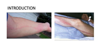
Manifesting as red streaks on
the skin
7. CLINICAL SYMPTOMS
• Local symptoms:
• Red macular linear streaks from site of infection toward the regional draining lymph
node
• Tenderness and warmth over affected skin
• May have lymph node involvement
• May have blistering of affected skin
• Systemic symptoms:
• Malaise
• Fever and chills
• Loss of appetite
• Headache
• Muscle aches
9. DIAGNOSIS
• LABORATORY
1. Complete blood count (CBC) may show leukocytosis.
Blood cultures
• IMAGING
1. Plain radiology unnecessary
• DIAGNOSTIC PROCEDUR
1. Aspirate and culture any pus.
2. Use sensitivity to guide antibiotic treatment.
10. THERAPHY
•Rest and elevation of the affect limb
•Symptomatic theraphy
•Etiology Specific antibiotic theraphy
•Surgical DebridementIncision and
drainage abscess areas
11. PROGNOSIS
• Without appropriate anti microbal theraphy cellulitis may extend
along the channel; necrosis and ulceration may occur.
• Anti microbal regimens are effective in more than 90% cases
• Morbidity and mortality is related to the underlyng infection
16. RISK FACTOR
•Congenital or inherited abnormalities
•Trauma,surgery, filariasis
•Recurrent skin infection
Non cancer
•Tumor causing obstruction of lymphatic channels or nodes
•Breast cancer
•Head and neck surgery
•Colorectal cancer
Cancer
•Radiation theraphy
•Lymph node biopsy or dissection
Cancer treatment
related
17. PATHOGENESIS
Non cancer
Cancer
Cancer related treatment
Inadequate lymphatic
outflow
Leads to lymphatic
hypertension and
decreased contraclity of
lymph
Lymphostatis,
accumulation of lymph,
interstisial fluid, protein
and glycosaminoglycans
Statis of lymph cause
accumulation of protein
and cellular metabolites
in the extracellular space
Water accumulation,
formation of edema and
increased interstitial
hydraulic pressure
24. THERAPHY
• The most common
treatments are a
combination of
manual compression
lymphatic massage,
compression garments
or bandaging.
25. PROGNOSIS
• There is no cure for lymphedema, and it is a progressive condition.
The outlook will depend to some extent on the severity of symptoms
Editor's Notes
Antibiotics (1)[A]:
Dicloxacillin:
Adults: 500 mg p.o. q6h
Children: 50 mg/kg/d divided into q.i.d. dosing
Nafcillin:
Adults: 2 g IV q4h
Children: 150 mg/kg/d divided into q.i.d. dosing
Cephalexin:
Adults: 500 mg p.o. q6h
Children: 50 mg/kg/d p.o. divided into q.i.d. dosing
Clindamycin (if penicillin or cephalosporin allergy):
Adults: 150–300 mg p.o. q6–8h or 600 mg IV q8h
Children: 8–20 mg/kg/d p.o. divided into t.i.d. or q.i.d. dosing; 20–40 mg/kg/d IV/IM divided into t.i.d. or q.i.d. dosing
Acetaminophen or ibuprofen for pain and fever
Second Line
Trimethoprim-sulfamethoxazole (TMP-SMZ) good for areas with high rates of methicillin-resistant S. aureus [MRSA]):
Adults: 160 mg TMP/800 SMZ mg p.o. q12h × 10–14 days
Children >2 months of age: 10–20 mg/kg/d p.o. or IM divided into t.i.d. or q.i.d. doses × 14 days