2. 1996). Immunohistochemistry revealed an increase in the
content of PGHS-2 in mononuclear trophoblast cells of
the placenta after day 140 of gestation. It was concluded
that PGHS-2 is the enzyme responsible for placental
prostaglandin production at term delivery. However, the
study of Gibb et al. (1996) did not elucidate the cellular
localization of PGHS-1 expression nor did it address the
factors that regulate enzyme formation.
Glucocorticoids regulate prostanoid production in many
tissues. PGHS activity increases after glucocorticoid treat-
ment in mouse myeloid leukaemia (MI) cells (Honma et al.,
1980), fetal rat lungs (Tsai et al., 1983) and in Swiss 3T3 mouse
fibroblast cells (Chandrabose et al., 1980). Human amnion
cells in primary culture have an increased capacity to convert
exogenous arachidonic acid into PGE2
in response to
dexamethasone treatment (Smieja et al., 1993). As labour
onset in ewes is preceded by activation of the fetal
hypothalamic–pituitary–adrenal axis (HPA axis), it is
possible that the prepartum increase in fetal cortisol
production acts to modulate intrauterine PGHS expression.
McLaren et al. (1996) demonstrated that the placentome is
the principal site of induced PGHS expression at the time
of glucocorticoid-induced labour in sheep, although the
distribution of the two forms of the enzyme within the
placentome was not addressed. The aim of the present
study was to characterize the cellular distribution of ir-
PGHS-1 and ir-PGHS-2 in ovine placental tissue using
immunohistochemical staining. The primary sites of
enzyme production were determined in placentomes after
glucocorticoid-induced and spontaneous labour and were
compared with those of ewes in late gestation but not in
labour.
Materials and Methods
Animals
Sixteen pregnant Border Leicester–Merino crossbred ewes
of known gestational age were used in this study. The animal
experiments were approved by the Monash University
Standing Committee on Ethics in animal experimentation.
Ewes underwent surgery between 118 and 125 days of
gestation to implant electromyogram electrodes. Electrodes
were implanted into the myometrium to enable labour onset
to be identified (Harding et al., 1982).
A paired treatment–control experimental paradigm
was used in the first part of the study to determine the effects
of glucocorticoid on PGHS expression. Fetal sheep were
injected using guided ultrasonography (via maternal
transabdominal injection) with either glucocorticoid
(betamethasone; Celestone Chronodose, Schering Plough
Pharmaceuticals, Baulkham Hills, NSW; 5.7 mg ml–1
in 1 ml
total volume; n = 5) or an equivalent volume of sterile
isotonic saline (control; n = 5) on day 131 of gestation.
Animals that received betamethasone were killed by i.v.
barbiturate injection when labour was established. Labour
onset occurred 56.6 Ϯ 0.8 h after glucocorticoid injection and
was determined from increased uterine electromyogram
activity and clinical factors such as swelling of the maternal
udder and vulva. Control ewes treated with saline were
killed at the same time as the experimental ewes to obtain
age-matched control tissues. The procedure of intrafetal
injection of glucocorticoid has been characterized and
validated (McLaren et al., 1996).
Six sheep were used for the second part of the study to
examine PGHS-1 and PGHS-2 expression at spontaneous-
onset labour. Ewes were allowed to progress to term and
were killed by i.v. barbiturate injection when increased
uterine contractile activity (consistent with labour onset) had
been observed for 6–8 h. The mean age to labour onset in
these ewes was 149 days.
Tissue collection
After the animals were killed, a post mortem examination
was conducted and tissues were collected in the shortest
possible period. Placentomes (sliced vertically along a radial
axis of symmetry) were placed in cold Bouin’s fixative
solution for 24 h. Tissues were washed five times in 70%
ethanol before dehydration and embedding. Tissues were
dehydrated using an automatic tissue processor (Histokinette,
Thomas Optical and Scientific Co. Pty Ltd). Tissues were
incubated sequentially in 70%, 90% and 100% ethanol for
12 h (2 ϫ 2 h washes), cleared of dehydrating agents by
washing in Histosol (2 ϫ 2 h washes) and embedded in
paraffin wax. Tissue sections (7 µm) were prepared by
standard techniques and placed on glass slides coated with
poly-L-lysine (0.01%; Sigma Chemical Company, St Louis,
MO).
Immunohistochemistry
Immunohistochemistry was performed using the DAKO
ENVISION™
System (DAKO Corporation, Carpinteria, CA).
After paraffin wax was removed from the tissue sections,
they were incubated with 0.03% (w/v) H2
O2
for 5 min to
quench endogenous peroxidase activity. Tissue sections were
incubated for 1 h at room temperature with PGHS-1 or
PGHS-2 antibody at 1:1000 dilution. PGHS-1 antiserum was
raised in rabbits against ram seminal vesicle PGHS (McLaren
et al., 1996). PGHS used for injection (99% purity) was
purchased from the Oxford Biochemicals Company (Oxford,
MI). The crossreactivity of the antibody with the PGHS-2
isozyme was estimated to be < 0.1%, as determined from
laser densitometry (McLaren et al., 1996). The polyclonal
PGHS-2 antiserum, raised in rabbits against murine PGHS-2,
was purchased from the Cayman Chemical Company (Ann
Arbor, MI). There was no detectable crossreactivity of the
PGHS-2 antibody with PGHS-1 protein at amounts up to
2.5 mg (as determined from western blot analysis; McLaren
et al., 1996). After washing with Tris-buffered saline (TBS)
(pH 7.5), the sections were incubated with peroxidase-
labelled polymer conjugated to goat anti-rabbit IgG second
antibody for 30 min. Slides were rinsed once and the
substrate–chromogen solution was added for 10 min.
Specific immunostaining was identified using diamino-
benzidine. The sections were counterstained with Harris’
34 W. J. McLaren et al.
3. haematoxylin, dehydrated and mounted with DPX moun-
tant. Immunoreactivity in tissue sections was examined
using a Leitz Wetzlar Dialux 20 microscope.
Controls
Tissues from animals of each group were processed
simultaneously to allow direct comparison between staining
runs. The following negative controls (derived from the
same specimen) were included in every staining run to
monitor daily variations in the immunohistochemical
staining procedure and to verify the performance of the
reagents: (i) the PGHS-1 or PGHS-2 primary antibody
was substituted either by antibody dilution buffer or by non-
immune rabbit serum (1:1000 dilution); (ii) the peroxidase-
labelled secondary link antibody (goat anti-rabbit immuno-
globulins) was substituted with TBS (pH 7.5) wash buffer;
and (iii) the slide section was only incubated with TBS
(pH 7.5) diluent before the addition of the substrate–
chromogen solution.
If any of the control specimens demonstrated residual
background staining, the results of the staining run were
considered to be invalid.
Pre-absorption controls were also performed to verify that
the staining observed in tissue sections was due to binding of
the primary antibody to its target antigen. PGHS-2 antibody
(protein concentration: 33 µg µl–1
) was pre-absorbed with an
excess (4000 times greater protein concentration) of PGHS-2
antigen (protein concentration: 1.04 µg µl–1
) for 18 h at 4ЊC.
Similarly, the PGHS-1 antibody (protein concentration: 83 µg
µl–1
) was pre-absorbed with an excess (4000 times greater
protein concentration) of PGHS-1 antigen (protein concen-
tration: 1 µg µl–1
) for 18 h at 4ЊC.
After addition of precipitating solution (0.05 mol phos-
phate buffer l–1
, 0.25% BSA, 5% polyethylene glycol 6000,
0.125% (v/v) normal rabbit serum and 0.5% (v/v) goat anti-
rabbit IgGs), the mixtures were centrifuged at 1200 g for
30 min at 4ЊC. The supernatant was incubated with the tissue
section. The tissue sections used for the PGHS-1 and PGHS-2
pre-absorption control incubations were derived from a
betamethasone-injected animal in labour previously shown
to demonstrate positive staining for both isozymes. A serial
section of the same specimen was run concurrently with
every pre-absorbed antibody control tissue to act as a
positive control.
Quantitation of immunohistochemical staining
Quantitative analysis of positive staining for ir-PGHS-1
and ir-PGHS-2 in placental tissue sections was performed
using image analysis software (Zeiss KS 400, Version 3.0).
Five placentomes collected from the different animals within
each treatment group (saline injection and betamethasone
administration) and six placentomes collected from the
animals in the spontaneous labour group were examined for
immunoreactivity. The percentage area of positive staining
was calculated from five fields of view (ϫ 20 magnification)
for each tissue section examined. Two sections per placen-
tome were assessed for the degree of positive staining. The
results are expressed as the mean percentage area of positive
staining for PGHS-1 and PGHS-2.
Statistical analysis
Statistical analysis of the data was performed using a
commercially available statistical program (GraphPad Prism
Version 2.01). Data were first analysed by a univariate
homogeneity of variance test (Bartlett’s Box F test). If
significance was found for a particular parameter (that is the
raw data were non-homogeneous), the test was repeated
using log10
transformation and square root transformation.
The transform most closely attaining homogeneity was then
used for all subsequent statistical analyses. Significant
interactions occurring between two or more factors were
identified using multifactorial ANOVA. The post hoc test of
least significant difference (LSD) was used subsequent to
the ANOVA to identify significant differences between pairs
of mean values. A probability level of 5% (P < 0.05) was
specified as significant. The data are presented graphically in
an untransformed state. Values are expressed as mean Ϯ SEM.
Results
PGHS-1 immunoreactivity
The location and the percentage area of tissue expressing
PGHS-1 immunoreactivity in sheep placentomes obtained
after intrafetal saline injection, glucocorticoid-induced
labour and spontaneous parturition are shown (Figs 1
and 2, respectively). In all groups studied, PGHS-1 im-
munostaining was localized to mononuclear cells of the
trophoblastic epithelium. The binucleate cells were clearly
immunonegative. The endothelial cells of fetal vessels in the
chorionic villi demonstrated weak immunostaining (Fig. 1b,
arrow). Moreover, the fibroblasts of the fetal stroma were
weakly immunopositive for PGHS-1. Both the fetal and
maternal mesenchymal core demonstrated diffuse staining
for PGHS-1. There was no significant effect of spontaneous
parturition or treatment (saline injection or glucocorticoid-
induced labour) on the location and the percentage area of
positive staining for PGHS-1 in placental tissue sections.
When control incubations were performed with primary
PGHS-1 antibody that had been pre-absorbed with purified
PGHS-1 isolated from ram seminal vesicles, no staining was
observed (Fig. 3a). Similarly, no staining for ir-PGHS-1 was
observed when the primary antibody was substituted in the
staining procedure with normal non-immune rabbit serum
(Fig. 3b).
PGHS-2 immunoreactivity
PGHS-2 was present in placental cotyledon tissue sections
on day 133 of gestation, as demonstrated in the saline-
injected control animals (Fig. 4a). Staining was confined to
the trophoblastic mononuclear epithelial cells. Binucleate
PGHS-1 and PGHS-2 immunoreactivity in ovine placenta 35
4. cells were consistently immunonegative. In contrast to the
PGHS-1 isozyme, the endothelial cells of fetal vessels in the
chorionic villi and the fibroblasts of the fetal stroma did not
contain ir-PGHS-2. Furthermore, both the fetal and maternal
stroma were immunonegative for PGHS-2. The location of
staining for PGHS-2 did not alter in response to glucocorticoid-
induced (Fig. 4b) or spontaneous labour (Fig. 4c); however,
there was a significant increase in the percentage area of
positive staining for PGHS-2 at labour onset (Fig. 2; P < 0.05).
The amount of positive staining for PGHS-2 in placental
tissue collected after glucocorticoid-induced labour and
spontaneous parturition was significantly higher than that
after intrafetal saline injection.
When control incubations were performed with primary
PGHS-2 antibody that had been pre-absorbed overnight with
purified PGHS-2 isolated from sheep placenta (70% purity),
no staining was observed in cells that previously demon-
strated positive staining for the PGHS-2 isozyme (Fig. 3c).
Moreover, no positive immunoreactivity was observed in
tissue sections in which the primary antibody was substituted
with non-immune rabbit serum (Fig. 3d).
Discussion
This study has demonstrated the presence and localization of
ir-PGHS-1 and ir-PGHS-2 in sheep placental tissue after
glucocorticoid-induced labour and spontaneous parturition.
Animals administered an intrafetal injection of isotonic
saline on day 131 of gestation acted as non-labour controls.
In all groups studied, the PGHS-2 isozyme was localized
exclusively to trophoblastic epithelial cells; no detectable
36 W. J. McLaren et al.
(a)
(b)
(c)
Fig. 1. Patterns of immunostaining for immunoreactive prosta-
glandin G/H synthase 1 (ir-PGHS-1) in placental tissue collected
from sheep after (a) intrafetal saline injection, (b) glucocorticoid-
induced labour and (c) spontaneous parturition. PGHS-1
immunoreactivity was localized to mononuclear cells of the
trophoblastic epithelium and the fibroblasts of the fetal stroma.
Binucleate cells were clearly immunonegative. Maternal placental
tissue demonstrated weak staining for the PGHS-1 isozyme.
Endothelial cells of the fetal vessels demonstrated weak
immunoreactivity for PGHS-1 (arrow in left-hand corner of (b)).
BNC: binucleate cell; F: fetal stroma; M: maternal placenta; T:
trophoblastic epithelium. Scale bar represents 50 µm.
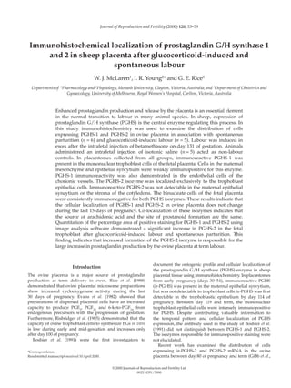
30
25
20
15
10
5
0
PGHS-1 PGHS-2
a
a a a
b
b
Areaofpositivestaining(%)
Fig. 2. Computer-assisted quantitation of positive staining for
immunoreactive prostaglandin G/H synthase 1 and 2 (ir-PGHS-1
and ir-PGHS-2) in placental tissue collected from sheep after
intrafetal saline injection (ᮀ), glucocorticoid-induced labour ()
and spontaneous parturition (). Bars with different letters are
significantly different (P 0.05).
5. staining was observed in the intervening maternal epi-
thelium or stroma of the cotyledons. Similarly, ir-PGHS-1
was present primarily in the trophoblast cells, although cells
in the maternal mesenchyme and epithelium were weakly
immunopositive for this enzyme. There were no cell-specific
alterations in PGHS-1 or PGHS-2 immunoreactivity in sheep
placentomes during the last 10–15 days of pregnancy.
Furthermore, localization of enzyme staining was not
influenced by labour-onset, whether spontaneous or
glucocorticoid-induced.
The placenta has been identified as the major site of
prostaglandin production in the uterus of pregnant sheep.
Risbridger et al. (1985) demonstrated that prostaglandin
synthesis by dispersed ovine cotyledonary cells is low
during early and mid-gestation but increases markedly from
day 110–120 of gestation to term. In that study, exogenous
administration of arachidonic acid to incubations of
cotyledonary cells did not significantly increase prosta-
glandin synthesis. This finding is consistent with the
suggestion that PGHS activity in placental trophoblast cells
increases only after day 110 of gestation. The results of the
present study indicate that the fetal trophoblast cells are the
primary site of prostaglandin formation during the last
10–15 days of pregnancy.
In the past, studies of placental endocrine function in vitro
have relied on the use of heterogeneous placental explants
(Hoffmann et al. 1979; Matt and MacDonald, 1984) or cell
suspensions (Branchaud et al., 1983; Shemesh et al., 1984a,b)
to elucidate possible sites of hormone formation. Although
these studies have contributed to our knowledge of placental
endocrinology, the contribution of individual types of cell
was not elucidated. In an attempt to delineate the principal
sites of prostaglandin output in sheep placenta, Mitchell and
Flint (1978) manually separated the fetal cotyledon from the
maternal caruncle. The prostaglandin synthesizing capacity
of the two sides of the placentome was then investigated
using a dispersed cell preparation. Since the fetal cotyledon
had a greater prostaglandin output than the maternal uterine
epithelium, it was suggested that PGHS activity was
primarily localized to the fetal side of the placentome.
However, in that study, no anatomical evidence was pre-
sented to support the completeness of separation of fetal and
PGHS-1 and PGHS-2 immunoreactivity in ovine placenta 37
(a) (b)
(c) (d)
Fig. 3. Control tissue sections for the prostaglandin G/H synthase 1 and 2 (PGHS-1 and PGHS-2) staining procedures. When the
primary PGHS-1 antibody was pre-absorbed with purified immunoreactive PGHS-1 (ir-PGHS-1) isolated from ram seminal vesicles
(a), no staining was observed. Similarly, when the primary PGHS-2 antibody was pre-absorbed with ir-PGHS-2 isolated from sheep
placenta (c), no positive staining was observed. When the primary antibodies were substituted in the immunohistochemical staining
procedure with non-immune rabbit serum, no staining was observed (b,d). The tissue used for the control staining procedures was
derived from an animal in labour after intrafetal injection of betamethasone. This tissue was previously shown to demonstrate
positive staining for the PGHS-1 and PGHS-2 isozymes. BNC: binucleate cell; F: fetal stroma; M: maternal placenta; T: trophoblastic
epithelium. Scale bars represent 50 µm.
6. maternal components. Given the high degree of
interdigitation of the chorionic villi with the crypts of the
uterine mucosa, complete separation of the ovine cotyledon
into fetal and maternal components is not technically
feasible. Thus, the results are potentially confounded by the
possibility of a non-homogeneous cell preparation.
Evans et al. (1982) measured PGE2
, PGF2α
and 6-keto-PGF1α
output by sheep cotyledons at different stages of pregnancy
using an in vitro cell culture system. The capacity of
dispersed placental cells to synthesize these prostaglandins
was higher on days 130 and 145 than on days 50 and 100 of
gestation. This finding is consistent with the suggestion of
increased PGHS expression; however, the localization of
enhanced PGHS production could not be characterized as no
histological assessment was made of the types of cell in the
preparation. Risbridger et al. (1985) used a similar dispersion
protocol and suggested that cells of the fetal trophoblast
were the major site of prostaglandin production during
pregnancy and parturition. Although these investigators
demonstrated that enzymatic digestion of the placental
tissue yielded binuclear and mononuclear cells, the
dispersion technique excluded syncytial cells from the final
preparation. Thus, the contribution of maternal epithelial
cells to prostaglandin output was not demonstrated.
The data presented in the present study clearly
demonstrate that PGHS-1 and PGHS-2 enzyme formation
predominantly localizes to the mononuclear cells of the
fetal trophoblast. Other studies have confirmed that the
subcellular locations of PGHS-1 and PGHS-2 are also the
same. PGHS-1 was first shown to be localized to the
endoplas-mic reticulum and nuclear membrane of kidney
tissue sections using immunofluorescence (Smith and
Wilkin, 1977; Smith and Bell, 1978). This result was later
confirmed by immunoelectron microscopy of cultured
mouse fibroblasts (Rollins and Smith, 1980). Regier et al.
(1993) demonstrated that PGHS-2 is associated with the
endoplasmic reticulum and nuclear envelope of mouse 3T3
fibroblast cells.
Unlike the mononuclear trophoblast cells of the fetal
syncytium, binucleate cells of the trophoblast were shown to
be immunonegative. This result is consistent with the
findings of Boshier et al. (1991) and Gibb et al. (1996) and
indicates that these cells are not important to placental
prostaglandin formation. Rather, binucleate cells have two
alternative functions that are important to the normal
progression of pregnancy: (i) to form the fetomaternal
syncytium essential for successful implantation and
subsequent placental growth; and (ii) to produce and secrete
protein and steroid hormones. During the last two-thirds of
pregnancy, placental lactogens are measurable in the
maternal and fetal circulations of sheep (Chan et al., 1978;
Martel and Lacroix, 1978), cattle (Wallace et al., 1985) and
goats (Currie et al., 1990). The binucleate cells are the sole
source of these hormones. Furthermore, binucleate cells of
sheep and cows are capable of considerable progesterone
production from endogenous sources (Reimers et al., 1985;
Ullman and Reimers, 1989; Wango et al., 1991).
Quantitation of immunoreactive staining in placental
tissue sections using image analysis software demonstrated
a significant increase in PGHS-2 in association with
glucocorticoid-induced and spontaneous-onset labour. This
result strongly supports previous reports of increased
PGHS-2 protein concentrations in sheep placenta after
glucocorticoid-induced labour using western blot analysis
38 W. J. McLaren et al.
(a)
(b)
(c)
Fig. 4. Patterns of immunostaining for immunoreactive prosta-
glandin G/H synthase 2 (ir-PGHS-2) in placental tissue collected
from sheep after (a) intrafetal saline injection, (b) glucocorticoid-
induced labour and (c) spontaneous labour. PGHS-2 immuno-
reactivity was localized to mononuclear cells of the trophoblastic
epithelium. Binucleate cells and fibroblasts of the fetal stroma were
immunonegative. Maternal placental tissue did not express the
PGHS-2 isozyme. BNC: binucleate cell; F: fetal stroma; M:
maternal placenta; T: trophoblastic epithelium. Scale bar represents
50 µm.
7. (McLaren et al., 1996). In saline-injected control animals,
expression of the PGHS-2 isozyme was low. PGHS-2 staining
patterns in placental tissue sections from animals in labour
reflect higher enzyme production rates by uninucleate
trophoblast cells rather than increased expression by a more
diverse group of cells.
In summary, co-localization of PGHS-1 and PGHS-2
implies that the source of arachidonic acid, the site of
prostanoid formation and the mechanism of product
transport from the inside to the outside of the cell are the
same for these two isozymes. The primary difference
between PGHS-1 and PGHS-2 lies in the differential
regulation of enzyme expression. PGHS-1 isozyme
formation is constitutive. Conversely, PGHS-2 expression is
induced by a variety of stimulatory factors including
glucocorticoid-induced enzymes and cytokines (Liggins and
Thorburn, 1994). Regulation of PGHS-2 formation is critical
to myometrial activation that results in birth.
The work described in this manuscript was supported by a
Project Grant from the National Health and Medical Research
Council of Australia (G. E. Rice). G. E. Rice is in receipt of an NH
MRC Principal Research Fellowship. The authors wish to thank Carl
Zeiss Pty Ltd for providing the imaging software for quantitative
analysis of the data.
References
Boshier DP, Jacobs RA, Han VKM, Smith W, Riley C and Challis JRG (1991)
Immunohistochemical localisation of prostaglandin H synthase in the sheep
placenta from early pregnancy to term Biology of Reproduction 45 322–327
Branchaud CP, Goodyer CG and Lipowski LS (1983) Progesterone and
estrogen production by placental monolayer cultures: effect of dehydro-
epiandrosterone and luteinizing hormone-releasing hormone Journal of
Clinical Endocrinology and Metabolism 56 761–766
Chan JS, Robertson HA and Friesen HG (1978) Maternal and fetal
concentrations of ovine placental lactogen measured by radioimmunoassay
Endocrinology 102 1606–1613
Chandrabrose KA, Bonser RW and Cuatrecasas P (1980) Role of extracellular
arachidonate in regulation of prostaglandin biosynthesis in cultured 3T3
fibroblasts Advances in Prostaglandin and Thromboxane Research 6 249–253
Currie WB, Card CE, Michel FJ and Ignotz G (1990) Purification, partial
characterisation and development of a specific RIA for goat placental
lactogen Journal of Reproduction and Fertility 90 25–36
Evans CA, Kennedy TG and Challis JRG (1982) Gestational changes in
prostanoid concentrations in intrauterine tissues and fetal fluids from
pregnant sheep and the relation to prostanoid output in vitro. Biology of
Reproduction 27 1–11
Gibb W, Matthews SG and Challis JRG (1996) Localisation and
developmental changes in prostaglandin H synthase (PGHS) and PGHS
messenger ribonucleic acid in ovine placenta throughout gestation Biology
of Reproduction 54 654–659
Harding R, Poore ER, Bailey A, Thorburn GD, Jansen CAM and Nathanielsz
PW (1982) Electromyographic activity of the non-pregnant and pregnant
sheep uterus American Journal of Obstetrics and Gynecology 142 448–457
Hoffmann B, Wagner WC, Hixon JE and Bahr J (1979) Observations
concerning the functional status of the corpus luteum and the placenta
around parturition in the cow Animal Reproduction Science 2 253–266
Honma Y, Kasukabe T, Hozumi M and Koshihara Y (1980) Regulation of
prostaglandin synthesis during differentiation of cultured mouse myeloid
leukemia cells Journal of Cell Physiology 104 349–357
Liggins GC and Thorburn GD (1994) Initiation of parturition. In Marshall’s
Physiology of Reproduction Vol. 4 pp 863–1002 Ed. GE Lamming. Chapman
and Hall, London
McLaren WJ, Young IR, Wong MH and Rice GE (1996) Expression of
prostaglandin G/H synthase-1 and -2 in ovine amnion and placenta
following glucocorticoid-induced labour onset Journal of Endocrinology 151
125–135
Martal J and Lacroix M-C (1978) Production of chorionic somato-
mammotropin (oCS), fetal growth and growth of the placenta and corpus
luteum in ewes treated with 2-bromo-alpha-ergocryptine Endocrinology 103
193–199
Matt DW and MacDonald GJ (1984) In vitro progesterone and testosterone
production by the rat placenta during pregnancy Endocrinology 115 741–747
Mitchell MD and Flint APF (1978) Prostaglandin production by intra-uterine
tissues from preparturient sheep: use of a superfusion technique Journal of
Endocrinology 76 111–121
Regier MK, DeWitt DL, Schindler MS and Smith WL (1993) Subcellular
localisation of prostaglandin endoperoxide synthase-2 in murine 3T3 cells
Archives of Biochemistry and Biophysics 301 439–444
Reimers TJ, Ullman MB and Hansel W (1985) Progesterone and prostanoid
production by bovine binucleate trophoblastic cells Biology of Reproduction
33 1227–1236
Rice GE, Wong MH and Thorburn GD (1988) Gestational changes in
prostaglandin synthase activity in ovine cotyledonary microsomes Journal of
Endocrinology 118 265–270
Risbridger GP, Leach Harper CM, Wong MH and Thorburn GD (1985)
Gestational changes in prostaglandin production by ovine fetal trophoblast
cells Placenta 6 117–126
Rollins TE and Smith WL (1980) Subcellular localisation of prostaglandin-
forming cyclooxygenase in Swiss mouse 3T3 fibroblasts by electron
microscopic immunocytochemistry Journal of Biological Chemistry 255
4872–4875
Shemesh M, Hansel W and Strauss JF, III (1984a) Modulation of bovine
placental prostaglandin synthesis by an endogenous inhibitor Endocrinology
115 1401– 1405
Shemesh M, Hansel W, Strauss JF, III, Rafaeli A, Lavi S and Mileguir F
(1984b) Control of prostanoid synthesis in bovine trophoblast and
placentome Animal Reproduction Science 7 177–194
Smieja Z, Zakar T and Olson DM (1993) Stimulation of cultured amnion cell
prostaglandin endoperoxide H synthase activity by glucocorticoids and
phorbol ester American Journal of Obstetrics and Gynecology 169 653–661
Smith WL and Bell TG (1978) Immunohistochemical localisation of the
prostaglandin-forming cyclooxygenase in renal cortex American Journal of
Physiology 235 F451–F457
Smith WL and Wilkin GP (1977) Immunohistochemistry of prostaglandin
endoperoxide-forming cyclooxygenases: the detection of the cyclo-
oxygenases in rat, rabbit and guinea-pig kidneys by immunofluorescence
Prostaglandins 13 873–892
Tsai MY, Josephson MW, Handschin B and Brown DM (1983) The effect of
prenatal dexamethasone on fetal rat lung prostaglandin synthesis
Prostaglandins, Leukotrienes and Medicine 11 171–177
Ullman MB and Reimers TJ (1989) Progesterone production by binucleate
trophoblastic cells of cows Journal of Reproduction and Fertility Supplement 37
173–179
Wallace CR, Collier RJ, Bott DJ, Byatt JC and Bremel RD (1985) Bovine
placental lactogen concentrations in maternal and fetal fluids Journal of
Animal Science 61 Supplement 1 Abstract 377
Wango E, Heap RB and Wooding FBP (1991) Progesterone and 5β-
pregnanediol production by isolated fetal placental binucleate cells from
sheep and goats Journal of Endocrinology 129 283–289
PGHS-1 and PGHS-2 immunoreactivity in ovine placenta 39