In The Cretaceous, Insects Already Possessed A Range Of Defensive Mechanisms
Naimi_Waheeda Masters Thesis
1. Localization of Chitin Synthase in Drosophila Melanogaster
The author of this item has granted worldwide open access to this work.
APA Citation: Naimi, W.(2016). Localization of Chitin Synthase in Drosophila Melanogaster. Retrieved
from http://libra.virginia.edu/catalog/libra-oa:11539
Accessed: May 18, 2016
Permanent URL: http://libra.virginia.edu/catalog/libra-oa:11539
Keywords: Arthropod Exoskeleton Cuticle Chitin Chitin Synthase kkv CRISPR/Cas9
Terms: This article was downloaded from the University of Virginia’s Libra institutional
repository, and is made available under the terms and conditions applicable as set forth
at http://libra.virginia.edu/terms
(Article begins on next page)
4. Naimi 3
1. Abstract
Proper cell-cell adhesion and communication are essential during
development. Both are heavily maintained and regulated by the content present in
the extracellular matrix (ECM), which composes the tough exoskeleton called the
cuticle. An enzyme called Chitin synthase (CS) provides the exoskeleton with much
of its strength and stability through the production of chitin. Chitin, a polymer of N-
acetyl-ß-D-glucosamine, is an important element in the exoskeleton of invertebrates
and functions much like cellulose in plants and keratin in vertebrates, that is, to
provide hardness, strength, and protection against the external environment. The
underlying component for both chitin and cuticle formation is CS, which is found
across several different species. It is now known that nearly 5-6 different copies of
CS in yeast and fungi have been condensed into only two copies in insects. During
insect development, one of the two copies is involved in formation of the gut lining
while the other is involved in epidermal tissue development, helping to produce the
tracheal lining as well as the exoskeleton. The gene that encodes for the chitin
synthase involved in the epidermal tissue in D. melanogaster is called krotzkof
verkehrt (kkv); we are specifically interested in its involvement in the formation of
the exoskeleton. kkv is an important gene during development as it is involved in
production of chitin by CS, which is then used to synthesize the cuticle found in the
ECM. Mutant kkv results in detachment of the cuticle from the apical end of the
cellular body, which then dilates and results in a lethal, curved, short embryo with a
scrambled head that is unable to hatch. Not only does kkv need to function properly
but CS must also be localized to the appropriate region in order to synthesize chitin.
There are currently two hypotheses as to how that may occur: (1) CS rests on the
apical membrane, and secrets chitin into the ECM, which is then guided to the
proper location, and a cuticle is formed or (2) CS is carried around in vesicles
termed chitosomes, which localize kkv to the right region while chitin synthesis is
initiated inside. Upon proper localization, chitin is finally released. Recent
discoveries in CRISPR/Cas9 have been used to facilitate understanding of this
predicament.
5. Naimi 4
Keywords: Arthropod, Exoskeleton, Cuticle, Chitin, Chitin Synthase, kkv,
CRISPR/Cas9
2. Introduction to the Arthropod Exoskeleton
The word arthropod comes from the Greek words “arthro” and “podos”
meaning “jointed legs” and rightfully describes a diverse group of invertebrate
animals with an external skeleton, segmented body, and joined appendages. Much of
the reason for their survival is due to the exoskeleton, which is composed of a tough
element called the cuticle. The arthropod cuticle provides the organism with a
number of properties ranging from stabilization of body and appendage shape,
protection from predators, infection, and dehydration (10). In particular, the
exoskeletons of insects are comprised of lightweight material that also provides the
organism with fast locomotive skills both on land and in the air (76). The life cycle of
insects is separated into two defined stages: larval and adult. Molting of the initial
rigid, external skeleton does not hinder insect growth as the organism
metamorphoses from a larva to an adult causing the cuticle to detach from the
epithelial surface, shed, and be replaced with another (10). This allows insects to
inhabit multiple ecological niches and specialize in different roles for each
developmental stage: larval for feeding and adults for reproduction (76).
3. Cuticle Formation during Development
The insect cuticle as described by Neville (76) is a multi-laminate structure
that is secreted by a single layer of epithelium in a variable time sequence allowing
for the formation of several layers throughout the cuticle. Collectively, the insect
cuticle forms the apical extracellular matrix (aECM) and is composed of lipids,
waxes, glycosylated and unglycosylated proteins, and most importantly a
polysaccharide called chitin (62). Despite the large amount of diversity among
arthropods, the chitin containing cuticle is one element that has remained fairly
conserved throughout. Chitin is not only found in the exoskeleton, but also in the
internal head skeleton, foregut, hindgut, trachea, and mouthparts (27). Aspects of
the cuticle vary within the organism’s anatomical framework and even among
6. Naimi 5
developmental stages. For example, the larval cuticle is usually soft and tender
whereas the thoracic cuticle is stiff and dense (70).
The integument is a monolayer of epidermal cells that produce and secrete
cuticular components. During embryogenesis, these cells undergo differentiation
where a change in the shape of the cells produces an overall layer that displays
strong cell-cell interactions and can withstand many sources of tension and
pressure (70). Recent research on the ECM has made clear that this structure is not
solely involved in maintaining organ shape but it also contributes to other aspects of
cellular behavior and genetic programming.
Overall, the cuticle is composed of three layers. Much has been written on the
nomenclature of these different regions however in the following thesis, the cuticle
will be divided up into the envelope, epicuticle, and procuticle. Before these layers
are formed, the plasma membrane of the integument epithelial cells forms
protrusions termed apical undulae (70) similar to microvilli that are stabilized by
microtubules and run perpendicular to the horizontal laminae (Fig. 1A). Topology
and correct localization of these undulae are thought to be essential for proper
chitin microfibril orientation (Fig. 1B).
3.1 Cuticle Layers
3.1.1 Envelope
Figure 1 – Apical Undulae: (A) Model of apical undulae formed at the surface of epidermal cells. Microtubules
help stabilize the longitudinal protrusions of the undulae, but the underlying interactions of the cytoskeleton
with microtubule are unknown. (B) Zoomed-in model of the apical undulae showing D. melanogaster larval
cuticle production. Plaques at the surface represent clusters of CS secreting chitin into the aECM perpendicular
to the undulae (und) underneath the envelope (env) and epicuticle (epi) (10).
(A) (B)
7. Naimi 6
The outermost cuticle layer (Fig. 2), which faces the environment is
comprised primarily of neutral lipids, wax esters, quinones, and long chain alcohols
that give it a hydrophobic nature. This thereby provides the organism with a means
of protection against dehydration and also acts as a pheromone in certain insects
(10). The envelope can be further divided into the inner epicuticle and outer
envelope or cuticulin, which is the major component of the envelope and composed
of lipids and sclerotin (12). In D. melanogaster, the outer envelope is deposited in
fragments at the tips of protrusions made by epithelial cells. These fragments fuse to
form a single layer and are thickened by the addition of extra layers during cuticle
differentiation (70, 12). A large discussion still remains as to how envelope
components are transported across the plasma membrane of epithelial cells and
through the several layers underlying the envelope. It has been suggested that pore
canals that is, tubes that traverse throughout the entire cuticle from the apical
epithelium to the tip of the cuticle, are responsible for transporting the material via
an unknown mechanism (10)
Figure 2 – Model of Cuticle Layers and Laminae Sheet Rotation: (A) The envelope is laid down in fragments
that fuse together at the plasma membrane surface and proteins needed for the epicuticle are secreted
through the valleys between crests. Both of these layers are thin relative to the procuticle. The final layer
contains microfibrils of chitin fibers that form sheets of laminae. These sheets rotate with respect to one
another as they are stacked up (17, 26). Epi, epicuticle; pro, procuticle; env, envelope; tri, Trichomes. (B) Zoom
in on the procuticle where chitin polymers are lined up anti-parallel to form chitin microfibrils. These
microfibrils are depicted here running parallel to form sheets of laminae. Tagging endogenous kkv with GFP
(in green) would allow us to visualize this organization (12). (C) Model showing laminae arranged in a helical
stack. Tagging kkv with GFP would allow us to visualize these microfibrils indicated in green (70).
8. Naimi 7
3.1.2 Epicuticle
Not much is known about the epicuticle, which is composed of unidentified
small proteins with low structure complexity (10). Unlike the envelope, the
epicuticle is not formed in any sequential manner. Epicuticle material is deposited
into the valleys between epithelial cell protrusions and slowly thickens during
cuticle formation (12, 70)
3.1.3 Procuticle
Unlike the other two layers of the cuticle, the procuticle is the largest and
harbors the polysaccharide chitin, whose microfibrils contain a specific
organizational scheme (10). Chitin fibrils, arranged in an antiparallel manner,
associate to form microfibrils, which are subsequently arranged parallel to one
another (Fig 2B). These parallel microfibrils form a 2D sheet called laminae. These
laminae form a helicoid pattern by which each new sheet is rotated by some degree
from the previous sheet (Fig. 2C). This was originally discovered by Bouligand in
1965 in crustaceans and later confirmed in insects by Luke and Neville in 1969 (10).
The orientation of laminae differs from organism to organism. The overall
architecture of this layer is also stabilized through chitin-protein interactions.
Resilin, one of the chitin-binding proteins found in this region, provides the cuticle
with high elasticity (12).
3.2 Laying Down the Drosophila melanogaster Cuticle
While it was originally thought that each layer of the cuticle is temporally
separated, recent work by Moussian (70) has shown otherwise. Through a series of
images taken via light and fluorescence microscopy and transmission and scanning
electron microscopy, Moussian demonstrated that the previously thought sequential
layers were in fact not so temporally separated. The envelope precursor was seen in
fragments at stage 15 of development at the tip of epithelial cell protrusions. These
gaps fused and another layer was added during stage 17. Both the epicuticle and
procuticle components are secreted during envelope development in stage 16. The
9. Naimi 8
chitin filaments required for the procuticle is secreted mostly during the latter half
of stage 17. In contrast, for adult cuticle there is a clear temporal separation in
deposition, although the earlier deposited layers appear to be modified at later
times. In comparison to exoskeleton, the chitin filaments found in the trachea are
first seen at stage 15 and cover the entirety of the tracheal lumen. It isn’t until stage
17 when chitin degradation occurs and the lumen of the trachea is cleared allowing
for air to fill the space (70). Overall development thus involves establishment of the
first three layers, thickening of the cuticle, and finally the formation of a helical
structure by chitin laminae in the procuticle. .
4. Chitin background
Chitin, one of the components that constitute the procuticle layer, is the
second most abundant polymer after cellulose. It is a linear polymer composed of ß-
(1à4)-linked N-acetyl-D-glucosamine (GlcNAc monomers) where the reaction is
catalyzed by an enzyme called chitin synthase (CS). Chitin is made up of alternating
residues linked in ß-(1-4)-glycosidic bonds (7). Much research was implemented
towards understanding the stereochemistry of the overall reaction. It was thought
that in order to accommodate for the 180º turn between consecutive monomers,
two GlcNAc residues were added during each catalytic cycle (1). Yeager’s lab proved
the presence of two active sites using dimeric inhibitors to prove greater overall
inhibition over monomeric inhibitors (21). This provided a more in-depth look into
the overall stereochemistry of the reaction where two GlcNAc monomers are used
per catalytic cycle. GlcNAc monomers are essential sugars involved in various
reactions, however one of their most important roles is contribution to the function
and architecture of the ECM (18). The Leloir pathway (7) is used to convert a
trehalose sugar into the most active form of GlcNAc, UDP-N-acetylglucosamine,
where CS completes the final conversion step. This pathway is highly conserved in
both arthropods and fungi (6).
Chitin exists in three different crystalline modifications called α, β, and γ
chitin (7). The most prevalent form, α-chitin, is found in arthropod cuticles and
contains chains in anti-parallel orientation. The anti-parallel orientation allows for
10. Naimi 9
tight packing into chitin microfibrils, maximizing the number of hydrogen bonds
and simultaneously minimizing room for any water. This is one of factors behind the
strength and stability of arthropod cuticles. β chitin chains are arranged in a parallel
orientation whereas γ-chitin chains contain two parallel strands with one that is
anti-parallel. β and γ chains are more commonly found in cocoons. They lack the
tightness and stability provided by α chains and therefore have an increased
number of hydrogen bonds with water. This property gives them a more flexible and
soft chitinous structure that is also found in the peritrophic membrane in the gut
lining (8). This difference in chitin chains also results in different arrangement of
chitin microfibrils later on. Whereas cuticle microfibrils are arranged in a helicoidal
formation, peritrophic matrices and even those found in the trachea are structured
as a random network of chitin fibrils and are very rarely found in an organized
manner (7).
5. Chitin Synthase
Chitin synthase (CS) is the enzyme required for converting UDP-GlcNAc into
chitin (8). CS is part of the glycotransferase family, which contains a group of
enzymes that catalyze the transfer of sugar from donor to acceptor while forming a
glycosidic bond. The overall reaction requires the presence of a divalent metal
cation like Mg2+ or Mn2+ (7-8) Although much research has been done on fungal CS,
especially yeast, the first CS sequence found in arthropods was identified by
Tellam’s lab in 2000 (1). He used degenerate primers with similarities to fungal
chitin synthases to sequence the enzyme from Lucilia cuprina and further tested its
presence with fungal CS inhibitors. From there, Tellam was able to repeat similar
procedures with C. elegans, D. melanogaster, and arachnids. In situ localization of CS
mRNA in 3rd instar larvae resulted in stained layers of epidermal cells underneath
the procuticle.
From the sequence analysis, it was found that chitin synthases are relatively
large proteins with 15-18 transmembrane segments. The enzyme can be split up
into three domains: a, b, and c.
11. Naimi 10
5.1 Domains A, B, and C
5.1.1 Domain A
Domain A, at the N-terminal, substantially varies in length when compared
across different species. It also varies in terms of number of transmembrane
segments which regulate whether this region is found intra- or extracellularly (7).
Research has shown that a deletion of up to 389 base pairs in yeast CS1 and a 221
base pair deletion in yeast CS2 does not affect the enzymatic activity of either
enzyme (75). From this, one may conclude that this region is rather insignificant. An
alternative interpretation is that domain A has specific functions that have rapidly
evolved leading to many segments.
5.1.2 Domain B
Domain B is known as the catalytic domain with hydrophobic properties and
no transmembrane segments. Because UDP-GlcNAc is located in the cytosol, it is
assumed that this domain faces the interior of the cell (7). Initial sequence analysis
by Tellam’s lab showed a conserved sequence among all four organisms; the
sequence, QRRRW, is thought be a product-binding site. Point mutations in this
region were implemented in yeast and resulted in decreased overall CS activity but
had no effect on the Km values for the substrate (8). A second conserved sequence
was also found in the following domain: (S/T)WGT(R/K). Originally it was thought
to be required for catalysis as any mutations resulted in a loss of activity, however
because this region is located extracellularly in yeast, that idea was quickly
abandoned. Although further experimentation is necessary, this second conserved
sequence has been hypothesized to be part of the translocation process that moves
chitin polymers into the extracellular matrix. (8). Other homologous sequences
include Walker A and B motifs (Walker et al, 1982) and a GEDRxx(T/S) motif at the
acceptor binding site (27).
5.1.3 Domain C
Domain C contains multiple transmembrane segments, which are conserved
among C. elegans, D. melanogaster, and arachnids but not in yeast. Among
12. Naimi 11
arthropods, the domain is fairly conserved with respect to location and spacing
between transmembrane segments (27). One important feature of this domain
involves five transmembrane segments that are located immediately after the
catalytic domain with two further downstream near the C-terminal. These segments
are thought to be involved in translocation of polymerized chitin chains.
5.2 CS Class A vs. Class B
While probing segments of digested DNA with a segment from the catalytic
domain of L. cuprina (LcCS-1), Tellam’s lab (1) came across a new CS, which they
termed LcCS-2. Further analysis using the TBLASTN computer program and a
similar probe revealed that this was also the case in D. melanogaster (DmCS-2) and
C. elegans (CeCS-2). Sequence analysis just on the catalytic domain region
demonstrated a near 72% similarity between LcCS-1 and DmCS-1 genes and 98%
similarity between DmCS-1 and DmCS-2. Cross species sequence analysis showed
similar results.
Based on this information, researchers have grouped CS into two classes CS-
A and CS-B. With the exception of a few arthropods, most insects have these two CS
genes. In Drosophila, both of these genes are located on chromosome 3 and are
thought to have evolved from a common ancestor via gene duplication (7). Class B is
the more ancient form and expressed in the gut epithelial cells producing the
peritrophic matrix. Much of the difference between class A and B can be seen at the
c-terminal in Domain C (Fig 3). This domain contains a total of seven
transmembrane segments. Class A genes are predicted to have a coiled coil region
after the fifth transmembrane segment (27). This coiled coil is thought to face the
extracellular matrix and might be involved in protein-protein interactions, vesicle
fusion, or oligomerization (7, 49). Class B enzyme lacks this region.
Class A has two mutually exclusive exons that result in two mRNA splice
variants (Fig. 3A). Both exons code for 59 amino acids and result in an additional
site for N-linked glycosylation. This variation is located in Domain C, c-terminal to
the five transmembrane segments. This change may result in different interactions
with cytosolic or extracellular proteins, which can then regulate chitin synthesis,
14. Naimi 13
5.4 Krotzkopf Verkehrt
In D. melanogaster, the gene that encodes for the CS expressed in the trachea
and epidermis is called krotzkopf verkehrt (3); it is also a vital component of proper
exoskeletal cuticle formation (11). It was found through a mutant screen in 1984 by
Nüsslein-Volhard and Wieschaus (2) who strived to characterize mutant alleles on
the third chromosome involved in the larval cuticle. kkv mutants displayed a
crumbled head skeleton, narrower denticle bands across the abdomen, and some
embryos were inverted in the egg case. Further research found they contained a
distinct “blimp” phenotype, whereby the cuticle of mutant embryos detaches from
the body and dilates (3) (Fig. 4B). This phenotype is wider than the wildtype and
indicates a loss of cuticle integrity (11). This became especially clear when these
embryos were mechanically devitellinized. The denticle belt was stretched laterally
and although the number of belts had not changed, the level of chitin within each
belt had decreased. These embryos were also less pigmented (3, 11). Because kkv is
also expressed in the trachea, that structure was no longer visible in the mutant
embryos. The word krotzkopf means, “scrambled head” in German and rightfully
describes the deformed and non-pigmented embryos. A scrambled head is not ideal
for hatching and many times one will find that certain hyperactive embryos will
have inverted in the egg case in an attempt to hatch (11).
Further research on mutant embryo cuticles found it to be altered. The
cuticle had variable thickness, particularly in the epicuticle and procuticle (Fig 5).
The epicuticle was broadened, penetrating into the procuticle, which contained free
procuticular chitin-binding proteins instead of a protein-chitin laminae. The overall
Figure 4 – Mutant kkv: Darkfield microscopy of cuticle preparations of (A) Wildtype kkv and (B) mutant kkv.
Mutants show the characteristic blimp phenotype where the cuticle is detached from the apical surface losing
their normal body morphology. Kkv mutants are seen with a scrambled head and larger body (11).
15. Naimi 14
Figure 5 – Mutant Cuticle Model: Wildtype cuticle (left) shows laminae (yellow) stabilizing upper levels via
association with the adhesion zone below. Mutant CS leads to a mutated cuticle where the layers are not
separated into distinct regions. The epicuticle protrudes into the lower procuticle and appears larger than in
the wt. The cuticle is not attached to the epidermal layers below, causing the blimp phenotype. Chitin laminae
are required for proper cuticle formation. Env, envelope; epi, epicuticle; pro, procuticle; adh, adhesion zone;
epid, epidermis (11).
adhesion between the epithelial cells was also non-existent (11). The epidermis and
cuticle depend on one another to form the exoskeleton and are required to stabilize
body morphology.
5.4.1 Other Blimp Phenotype Genes
Through a collaborative effect, several labs during the 1980’s were able to
come across three genes that also produced the blimp phenotype of kkv. These
include knickkopf (knk), grainy head (grh), and retroactive (rtv) (2). Another gene,
zeppelin (zep) was added later (3). These five genes are vital for proper cuticle
integrity. In terms of viability, zep mutants are the most hyperactive and therefore
are able to hatch but die at roughly the same stage as knk and rtv mutants. Kkv and
grh cause more severe damage to the head skeleton, denticle belt, and result in
lower hyperactivity.
In situ hybridization of knk showed low levels of mRNA throughout all stages
of development (3). Knk is thought to interact with the epidermis prior to cuticle
formation along with zep, as both were found to be interacting with mutations in
Drosophila E-cadherins encoded by shotgun (shg). They are needed for shg to form
proper epithelial cell adhesion and subsequently the cuticle it secretes. Once it was
sequenced, the knk gene was found to encode for an extracellular protein anchored
to the plasma membrane via a GPI moiety with no enzyme domain (14). Much like
knk, rtv is also an extracellular membrane-anchored protein and is thought to
16. Naimi 15
coordinate binding with chitin via six aromatic residues. Rtv mutants show the
standard blimp phenotype and mutant cuticle organization. It is thought to function
in lamellar procuticle organization (14) via two possible methods: anchoring chitin
chains to the plasma membrane, or binding other blimp phenotype proteins such as
knk to organized chitin chains (63). Both knk and rtv are structural proteins
assisting more with chitin filament assembly downstream of kkv and less with the
overall chitin synthesis process.
Grh is a transcription factor that belongs to the GATA family of transcription
factors. It is responsible for activation of several genes during development, one of
them being dopa-decarboxylase, which is ultimately needed to produce the quinones
required for proper crosslinking of cuticular proteins. While grh might increase
expression of kkv, grh mutant cells have been shown to display normal kkv activity.
5.4.2 Genes Interacting with kkv
Recent work done by Moussian’s lab has found two genes, expansion (exp)
and rebuff (reb), which are required for kkv function. Without either gene, chitin
deposition does not occur. Overexpression of both in chitin-devoid regions resulted
in chitin deposition. Exp and reb are hypothesized to participate in chitin polymer
translocation, microfibril formation, or in the direct or indirect posttranscriptional
modification of kkv (13).
5.5 Forms of Regulation
CS regulation occurs at all stages of development. Tissue specific expression
of LcCS-1 mRNA was measured by RT-PCR and showed expression of CS in 1st, 2nd,
and 3rd instar larvae, pupae, adults, and eggs (1). Any mutations would be
detrimental to the overall growth of the organism. Several forms of regulation are in
place to prevent such issues.
a. Hormonal control
Insect molting and metamorphosis are controlled by ecdysterone, a steroid
hormone that acts primarily on gene transcription. It exhibits a regulatory
role over CS-A and CS-B transcript levels (4, 8). Experiments done with
17. Naimi 16
Drosophila CS have shown that transcripts of either gene are not detected
prior to and during late larval ecdysone pulses. Once the pulse ceases
however, both genes are upregulated (4).
b. Transcriptional/post transcriptional control
kkv has five potential binding sites for the transcription factor grh, whose
exact role in chitin synthesis is currently unknown. Post-transcriptional
regulation includes phosphorylation, dephosphorylation, and N-
glycosylation, which have been found to regulate the localization, activity,
and stabilization of certain CS (8). CS activity can also be controlled via
regulation of components in the Leloir pathway. In the pathway, the rate-
limiting step is undergone by the glutamine-fructose-6-phosphate
aminotransferase (GFAT). In Drosophila two GFAT genes, Gfat1 and Gfat2,
have been recognized (Adams et al 2000; Graack et al 2001). Gfat1 is
inhibited by UDP-GlcNAc via a feedback mechanism and stimulated by
protein kinase A (PKA). This in turn controls levels of UDP-GlcNAc available
to be converted into chitin by CS.
c. Chitinases
Insect chitinases belong to a family of glycohydrolases responsible for the
catalysis of glycoside hydrolysis.
d. Zymogenic behavior
In some yeast and insects, it has been suggested that certain CS activity is
regulated by trypsin and other proteases categorizing them as zymogens.
Trypsin experiments have been done in vivo and appear to increase overall
activity; to date however, no endogenous protease has been identified to
cleave CS zymogens in insects (27).
e. Environmental factors
In certain mosquitos, it has been shown that CS regulation in the peritrophic
matrix is dependent on a blood meal (8).
5.6 Localization
18. Naimi 17
One of the many unknowns about kkv and
CS in general involves attempting to
understand the mechanism by which CS
localizes itself. Attempts to purify CS from
yeast have allowed for the discovery of
chitosomes (5), or vesicles that contain CS on
their plasma membrane. Although they have
been recorded in yeast, evidence for their
involvement in insects has not yet been found.
Chitin synthesizing enzymes are thought to
cluster at the tip of microvilli formed by
epidermal cells. Even with that assumption, it
is still not known whether CS is an integrated
membrane protein or if it resides in vesicles
that cluster near the apical membrane. One
model based on the idea of chitosomes
suggests that chitin is secreted into the lumen
of specialized vesicles, which then fuse with
the plasma membrane thus allowing for the
secretion of chitin into the ECM (Fig 6A). This
model however has a few shortcomings that
should be considered. The vesicles are
relatively small in size and may not provide
adequate space on the membrane for a large enzyme nor may they contain the
necessary space inside for chitin. Another aspect to consider is that CS is activate as
it makes its way from the ER to the plasma membrane, thus leaving room for excess
chitin production. If the catalytic domain of CS is inside the chitosome, a mechanism
should exist for UDP-GlcNAc transport into the vesicle. The second model suggests
intracellular vesicles that merely act as exocytotic conveyors transport CS from the
Figure 6 – CS Localization: (A) Vesicles loaded
with CS are transported from the golgi to the
plasma membrane at the apical surface, fuse
and are then activated via unknown
mechanism (proteolytic cleavage,
oligomerization, etc.). (B) CS vesicles called
chitosomes carry the activated enzyme
through the cytosol, producing chitin fibrils
into the lumen of the vesicles. Once the
chitosome docks and fuses with the plasma
membrane, the chitin fibrils are released into
the aECM (7).
19. Naimi 18
ER to the plasma membrane, where it could be activated by proteases or other
proteins to form chitin (1, 7, 8) (Fig. 6B).
6.7 Fungal CS
The fungal genome contains somewhere between 2-20 genes per species,
which have been categorized into five or seven classes (6). The most well studied
species, yeast, contains three types of CS (22, 32, 71), whose activity is spatially and
temporary dependent on the cell cycle (36). Domain A is quite variable among the
different species with most lacking transmembrane segments. Classes V, VI, VII
enzymes however contain a myosin motor domain (MMD) (Weiss et al 2006). With
the exception of a number of transmembrane segments in domain C, domain B and C
follow similar sequence schemes to those found in arthropods. Zymogenicity is
variable but has been shown in crude extracts. When attempting to purify CS from
Mucor rouxii, Ruiz-Herrera discovered the presence of chitosomes, vesicles that
harbor CS on its membrane. These vesicles had a lower buoyant density than other
exocytotic vesicles and were capable of producing chitin fibrils when substrate and
activator were added to the extract (Bracker et al 1976). They also did not contain
any yeast plasma membrane markers such as ß-1,3-Glucan synthetase (5).
6. Chitin applications and usages
Chitin polymers are the second most abundant polymers following cellulose.
Investigation of these polymers would allow one to manipulate certain properties
for other uses outside its natural involvement with the arthropod cuticle. Recent
interest in chitosan, a deacetylated chitin derivative, has led to its usage in tissue
engineering. Chitosan, a linear polysaccharide, is the second most abundant natural
biopolymer commonly found in crustacean shell and fungal cell walls. It has variable
solubility properties depending on the pH of the solution it is incorporated into.
Tissue engineering advances in cartilage, bones, skin (55, 56) and even drug
delivery systems (57) have made it clear that the material used has to be
bioabsorbable, with certain porosity and degradable properties that do not hinder
growth or normal physiology of the tissue within which it is placed. Chitosan, when
20. Naimi 19
conjugated with other chemicals, helps facilitate those properties within these
scaffolds. In one example, chitosan was combined with hyaluronan, a polysaccharide
found in the ECM, to form a lightweight matrix for chondrocytes, cells found in
cartilage. The combination of both chitosan and hyaluronan helped to increase not
only the level of chondrocyte adhesion and proliferation but allowed for synthesis of
collagen as well (56). The ability of chitosan to bind certain anionic molecules such
as DNA and several growth factors also opens up the opportunity for further uses
outside of tissue engineering, including developmental research (55).
Understanding the mechanism via which chitin is synthesized and deposited
opens doors for ways to inhibit its production. These would prove extremely useful
when developing insecticides for both agrarian and domestic purposes. Certain
insecticides, which have been used substantially in research to understand chitin
metabolism do not inhibit CS itself but other properties related to cuticle
development. Nikkomycine Z, a potent competitive inhibitor of fungal chitin
synthase, was tested on insects and found to be a growth regulator (1) without
evidence of having direct effects on CS itself. Lufenuron, another fungal insecticide
has also been used on insects and thought to affect chitin polymerization. While
these and other insecticides may do the trick, affecting other reactions or properties
related to the framework of the cuticle, they may have targets in vertebrates.
Targeting CS itself would reduce such risks. This would also be extremely important
when developing drugs for certain fungal infections that affect human lives (42).
7. CRISPR/Cas9 Overview
Targeted genome editing has become a powerful tool for biological research
with great potential for therapeutic discoveries against genetic disorders. Precise
editing in the past has been limited to certain organisms such as yeast and mice;
even in those cases, complications arose with off-target effects, limited efficiencies,
and high costs. An important first step to genome editing is creating DNA double-
stranded breaks (DBS). These breaks can then be repaired via two mechanisms: (1)
non-homologous end joining (NHEJ) and (2) homology directed repair (HDR). NHEJ
is an easy method for inducing small insertions and deletions that cause changes in
21. Naimi 20
the reading frame. HDR meanwhile takes advantage of a donor template to repair
the damage. That template can be engineered in the lab to implement specific
changes to the genome such as insertions or deletions of specific nucleotides, tags,
and even resistance markers to name a few. In the past, these DSB have been
induced via two methods: zinc finger nucleases (ZFN) and transcription activator-
like effector nucleases (TALENs). Both are chimeric proteins made up of a nuclease,
Fok1, which is guided by a programmable DNA-binding domain (61). Fok1 must
dimerize to be active thereby requiring two ZFNs/TALENs to produce a DSB. While
these techniques have been successful with modifications, they do present a
significant number of drawbacks. Two ZFN or TALEN designs per modification
require time and effort; sometimes, assembly of the separate parts alters the
interaction between them (61). A new method called CRISPR/Cas9 has found a
method by which to circumvent such issues.
The CRISPR system is the adaptive immune system used by bacteria whereby
sequences from invading DNA are incorporated into CRISPR repeat sequences.
When regions are transcribed, CRISPR RNAs (crRNAs) also known as protospacer
sequences, are formed harboring both foreign bacterial DNA and parts of the
CRISPR repeat. crRNAs bind to transactivating CRISPR RNAs (tracrRNA) and form a
complex with the Cas9 nuclease. This complex can then cut foreign DNA if they are
adjacent to a protospacer adjacent motif (PAM) (60). When it comes to
implementing this in research, a cas9 nuclease and 20 nucleotide guide RNA (gRNA)
target cut sites via RNA-DNA complementary base pairing. The targets must be 5’ of
a PAM sequence, which vary depending on the type of organism one is working
with. This allows gRNAs to be of the general form, 5’ 20 nucleotides –PAM 3’, with a
section of core RNA encoded by the gRNA vector.
This method has a wide variety of uses from bacteria to cell cultures to entire
animals. Regions in the genome can be edited ranging from small to large insertions,
deletions and replacements, to being modified with activation/inhibition domains,
effector domains to produce conditional alleles, or fluorescent tags for a more
precise understanding of localization and interactions during development. Delivery
22. Naimi 21
of separate components has also been simplified down to merely injections instead
of using viruses or electroporation like in the past (60).
7.1 Drawbacks
7.1.1 gRNA efficiency
gRNAs needed for this series of reactions are rather simple to make. They are
made up of a 5’ 20 nucleotide homology region upstream of a PAM sequence. Being
that the specificity of the Cas9 is based on the PAM sequence, there is a great chance
of off target cuts that must be repaired to prevent unknown mutations. Several
websites have been set up to screen regions of interest for potential gRNA
sequences and provide efficiency levels for each. The present issue concerns the
discrepancy between sites and the fact that most do not contain distinctions
between mismatches in their PAM sequences vs distal sequences in their algorithms
(59). Data from each site should be compared to select the best gRNA. Also when
choosing gRNAs, it has been found that those beginning with two guanine residues
before the complementary sequence yield a better on-target to off-target ratio (60).
The gRNA promoter should also be considered; recent work done in Port’s lab
demonstrated the importance of this requirement. CRISPR work done in Drosophila
recommends using RNA polymerase III-dependent promoters from the U6 snRNA
genes, which include U6:1, U6:2, and U6:3 (67). gRNA constructs were created using
the three different promoters. Flies expressing each gRNA-y gene were then crossed
to transgenic flies expressing act-cas9. The results showed that those gRNAs under
U6:1 and U6:3 promoters developed cuticles that were phenotypically more yellow
than those under U6:2 control.
7.1.2 Cas9 Nuclease Specificity
Modulating Cas9 activity will help reduce off target cuts and increase overall
efficiency of a given experiment. It has been suggested that paired nucleases on
adjacent strands in combination with two gRNAs provide greater specificity. This
method also allows for equal levels of HDR vs NHEJ induction instead of one or the
other (65).
23. Naimi 22
The PAM sequence is a key requirement for the Cas9 target sights. Although a
specific Cas9 recognizes specific PAM sequences, there are cases where regions of
non-canonical PAM sites get cleaved (58). However if the Cas9 is modified, where its
nuclease function is only partially active, that issue can be overcome. This would
allow for nicks to occur in the genome instead of DSB and induce HDR instead of
NHEJ (59).
Catalytically dead Cas9 have also been fused with Fok1. Two gRNAs and
chimeric Cas9 nucleases would be required for this process. Because Fok1 needs to
dimerize, it wouldn’t be constitutively active; this combines the honing in aspect of
CRISPR/Cas9 with a more target specific nuclease. One would have to consider the
effect of the size of fok1 on the overall transduction efficiency of the plasmid DNA
(69).
8. Methods
8.1 Fly care
All flies used were grown on standard fly food at 25ºC. OregonR (OreR)
wildtype flies, and y!; CyO/Gla and w hs-flp; TM3/TM6 balancers were from the
Adler lab stock collection. The various chromosomes were originally obtained from
the Bloomington Drosophila Stock Center. act-Cas9, and vas-Cas9 stocks were
obtained from the Bloomington Drosophila Stock Center. Nos-cas9 was obtained
from BestGene and kkv PB from the Exelixis Collection at Harvard Medical School.
8.2 Yeast Overexpression via Gateway Cloning
We overexpressed transcript D of kkv (Flybase ID: FBtr0301398) in yeast
cells using the gateway system (Fig. 7). This procedure takes advantage of two site-
specific recombination events through the use of attP sites. Using Accuprime pFx
DNA Polymerase (Catalog number: 123444-024), we flanked transcript D with
forward and reverse primers.
Forward: attB1; translation start codon; 18-20 gene specific nucleotides
GGGG ACAAGTTTGTACAAAAAAGCAGGCT TC ATG TTCAGTTTAGCGAAGACAACGAACCCGAAA
Reverse: attB2; translation stop codon; 18-20 gene specific nucleotides (reverse complement)
GGGG ACCACTTTGTACAAGAAAGCTGGGT C TTA CTGTTTGATGCTTCTATTTATTGTTTTAAA
24. Naimi 23
Gateway® BP Clonase® II Enzyme mix and Gateway® LR Clonase® II
Enzyme mix were used (procedures included). Entry vector pDONR 221
(invitrogen) was used during the BP reaction and the destination vector
pAG426GPD-ccdB, a gift from Susan Lindquist (Addgene plasmid # 14156), was
used during the LR reaction. The destination vector was than transformed into yeast
cells, KKY1035 and KKY1037 (provided by Keith Kosminski at the University of
Virginia) and transformant yeast colonies were picked from URA(-) plates. The
pYES2 Yeast Expression Vector manual (Cat. no. V825–20) was used to express our
protein in yeast cells.
8.3 kkv Antibodies and Western Blots
Two kkv antibodies T01812 and G3453 (both anti-rabbit) were made using SDIX
and LLC services. We isolated newly formed OreR pupa (wt) from their vials and placed
them in 25ºC incubators for 48hrs. These pupae were removed from the pupal case and
separated into three e-tubes with two containing five flies and one with two flies. 250μl of
SDS sample buffer was added to each tube and the content was ground up. 25μl of 2-
mercaptoethanol was added to each mixture and the tubes were heated accordingly for
10 min:
Tube A of 5: 90ºC
Tube B of 5: 60ºC
Tube C of 2: 90ºC
Figure 7: Overview of Gateway System (19)
25. Naimi 24
The samples were spun down at 14000 rpm for 5min and the supernatant transferred to
fresh e-tubes and used for western blot. All antibodies were diluted 1:1000. The
secondary antibody was goat anti-rabbit (purchased through Li-Core
https://www.licor.com/bio/products/reagents/secondary_antibodies/).
8.4 Injections
Rainbow Transgenic Flies Inc. provided all injection services
(http://www.rainbowgene.com/default.html).
8.5 Vectors
8.5.1 pUAST-attB GAL4 construct
kkv transcript D was cloned from a cDNA clone in
lab using the following primers*. The pUASTattb kkvR
and pUASTattb GFPF primers were designed to create an
overhang at the end of kkv and beginning of GFP, which
could subsequently be annealed together. pUASTattb
kkvF and pUASTattb GFPR primers were designed to
allow for insertion of the fragment kkv-GFP between EagI
and XhoI restriction sites in the vector, pUAST-attB
(Bischof et al, 2007) (Fig 8).
pUASTattb kkvF: 5’ ATCG CGGCCG CTGTTTGATGCTTCTATTTA
pUASTattb kkvR: 5’ reverse complement CCTTGCTCACCATTTCAGTTTAGCGA
pUASTattb GFPF: 5’ TCGCTAAACTGAAATGGTGAGCAAGG
pUASTattb GFPR: 5’ reverse complement GTCA CTCGAG TTACTTGTACAGCTCGTCCA
*Green designating GFP sequences; bold represent restriction sites;
underlined are regions of overlap
Ligation was done using the Gibson Assembly Cloning Kit (New England BioLabs
catalog#: E5510S). Although the protocol does not specify, 5μl of each flanked gene
was mixed with 10μl of Gibson Assembly Master Mix and left overnight on ice at 4ºC
before proceeding with the protocol provided. This construct underwent a standard
phiC31-attP40 injection on the second chromosome.
26. Naimi 25
8.5.2 pHD-DsRed HDR construct
For the homology directed repair
construct, pHD-DsRed (ADDGENE plasmid
#51434) was used. For this construct, three
separate fragments, kkva, GFP, and kkvb were
ligated together using Gibson Assembly. kkva
and GFP regions of overlap were then annealed
together and inserted between the restriction
sites SacII and NdeI. kkvb was inserted
between PstI and XhoI (Fig. 9). The following
primers* were used. All annealing and ligation
was done via Gibson assembly. kkva and kkvb were cloned from the genomic clone
BCR34M23 (created by the Berkeley Drosophila Genome Project) and GFP was
cloned from pUASTattB-GFP.
kkva 5’ CCGCGG TAGCATGCTGTGGAGATCGT
5’ reverse complement CCTTGCTCACCATGCTAAGCATAATG
GFP 5’ CATTATGCTTAGCATGGTGAGCAAGG
5’ reverse complement CATATG TTA CTTGTACAGCTCGTCCATGC
kkvb 5’ CTGCAG ATGTACTATACTATCATTTG
5’ reverse complement CTCGAG GCACAGTTCGCTGTGGGGTC
* Green designating GFP sequences; bold represent restriction sites; underlined are regions
of overlap
8.5.3 pCFD4 Double gRNAs
The pCFD4 vector (addgene 49411) (Port et al,
2014) combines together two promoters, U6:1 and U6:3,
adjacent to the gRNA core sequence allowing for the
expression of two gRNAs at one time (Fig. 10). Forward and
reverse primers* were provided in the supplementary figure
3D. gRNA primers were purchased from
http://www.operon.com/ and the vector pCFD4 was used as
the template during PCR. The primers contain a 3’ homology
to the vector backbone to allow for PCR amplification, which can then be inserted
28. Naimi 27
constructs to target regions around the kkv PB
transposable mutation with one gRNA inside a kkv
intron and the other outside of kkv near its 5’ start.
https://chopchop.rc.fas.harvard.edu/ was used to find
the most efficient gRNA, made through
http://www.operon.com/, and annealed together
using the following protocol. 20μl of each primer was
mixed together, placed on a 90ºC heat block for 2min,
and then cooled down to room temperature. Annealed primers were inserted into a
BbsI-digested pCFD3 backbone via Gibson Assembly. The primers* are listed below.
These gRNAs will be co-injected with the ssODN repair template into nos-Cas9;kkv PB
and yw vas-Cas9;kkvPB flies.
gRNA-Out 5’ forward: GAAGAC ATG GTTCGGATGTAGCTTATCCTTGG
5’ reverse complement: GAAGAC TTA CCAAGGATAAGCTACATCCGAAC
gRNA-In 5’ forward: GAAGAC ATG GATAAACAGCCTGAACCATTAGG
5’ reverse complement : GAAGAC TTA CCTAATGGTTCAGGCTGTTTATC
* Bold regions represent BbsI restriction sites followed by either a start or stop
codon and then the underlined gRNA sequence
8.5.6 pattB Repair construct
This construct consists of annealing the region between gRNA-Out and
gRNA-In with a GFP tag and inserting it into the EagI and XhoI digested backbone of
pattB. The region between the two gRNAs was cloned from the genomic clone
BCR34M23 and GFP was cloned from pUASTattB-GFP (Fig 12A). The following
primers* were used and ligated via Gibson Assembly. These flies will undergo a
phiC31-attP40 injection on the 2nd chromosome.
GFP repair 5’ Forward: ATCG CGGCCG ATGGTGAGCAAGGGCGAGGA
5’ Reverse complement: CATCCGAACCCAC CTTGTACAGCTCG
kkv repair 5’ Forward: CGAGCTGTACAAG GTGGGTTCGGATG
5’ Reverse complement: GTCA CTCGAG CCATCCTAATGGTTCAGGCT
* Bold regions represent restriction sties; green represents GFP; underlined
regions are areas of overlap.
29. Naimi 28
A second kkv pattB construct without GFP was also constructed using the
following primers inserting kkv between EagI and XhoI (in bold) (Fig. 12B).
5’ Forward ATCG CGGCCG GTGGGTTCGGATGTAGCTTA
5’ Reverse GTCA CTCGAG CCATCCTAATGGTTCAGGCT
9. Predicted Results and Discussion
9.1 Yeast overexpression
We cloned kkv transcript D into a destination vector via Gateway Cloning.
This procedure avoids the use of restriction enzymes and thereby bypasses
problems related to excess cutting or inversions of the template DNA. The
procedure consists of two recombination reactions, the first being called BP, which
inserts the fragment into an entry vector. These vectors are quite useful as they
allow for the movement of a sequence of interest into a destination or expression
vector via a second recombination reaction called LR. A wide variety of destination
vectors exist with different fluorescent tags, resistance markers, etc.
Once we had transformed our yeast vector (verified via DNA sequencing), we
induced expression using both liquid induction medium and plates. We had hoped
to observe some morphological change within the yeast cells, however that did not
occur. From what we have discussed regarding CS activity, this could be caused by
an array of things. It is possible that the enzyme needed to be phosphorylated or
required the presence of other transcription factors and proteins. CS also doesn’t
function properly without the presence of a divalent cation Mg+2 or Mn+2.
30. Naimi 29
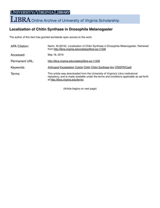
Figure 13 – Western Blot Results: (A) Antibody G3454 tested on wt OreR pupae incubated at 25ºC for
48hrs (B) Antibody T01812 tested on wt OreR pupae incubated at 25ºC for 48hrs (C) Antibody
T01812 test on transformed yeast cells and wt yeast cells
To check for the expression of kkv, we performed a western using two kkv
antibodies, G3453 and T01812. Before attempting to do so, we confirmed their
activity on first instar pupa. Post 2-mercaptoethanol addition, the mixture was
heated up to either 90ºC or 60ºC due to the unknown but potential polymerization
property of CS, which may affect the speed with which it travels on a gel. The
molecular weight of transcript D is around 182kDA but because the enzyme was
cleaved, we did not see one concise band but rather several bands (Fig 13A, 13B).
Cleavage might have occurred in areas where the protein passes through the plasma
membrane, with the heaviest band around 100kDA being the catalytic domain.
Temperature did not have any effect on polymerization of the protein. These
antibodies were then tested on our transformed yeast cells, and we were able to
show the presence of kkv. The antibodies used were specific for kkv and therefore
should not have stained any endogenous yeast CS; indeed, no bands were seen for
wt KKY1035 or KKY1037 (Fig 13C).
9.2 Overexpression of kkv in Drosophila
31. Naimi 30
Continuing with the idea of overexpression, we wanted to attempt to do one
of two things within Drosophila: overexpress kkv or express kkv in certain patterns.
To do so, we cloned kkv from a cDNA clone and GFP from a pUAST-attB-GFP vector
and flanked each with overlapping regions. These two separate pieces were ligated
together using Gibson Assembly into the pUAST-attB vector. This vector is
particularly useful in our case because it has an upstream activation sequence that is
driven by a yeast transcription activator called GAL4 (Fig. 14A). This will allow us to
express the gene and fluorescent tag in controlled manners. Because CS is so vital
for survival, it is very difficult to turn this gene on and off. We could for example use
vestigial-GAL4 for expression all over the wing or patched-GAL4 to express kkv-GFP
in a stripe along the wing (Fig. 14B). We could then try to induce knockouts in that
same region and see if the phenotype can be rescued. We do not expect to see much
change in cell morphology, however there could be an excess production of chitin
polymers. In the thoracic region, it has been previously shown that excess chitin is
deposited early on during development that disappears at later stages. We expect to
see less of these polymers disappearing (Adler et al, 2015). Future antibody staining
of this region for other proteins would allow us to decipher certain protein-protein
interactions such as those between CS and potential chitin-organizing proteins.
Presently, this construct has been sent out for injection into a pC31 fly line.
An attempt to create this construct using Gateway Cloning did not work
properly. Donor221 was used as the entry vector and PTWG was used as the
destination vector from the Drosophila Genomic Resource Center
(https://dgrc.bio.indiana.edu/product/View?product=1076). The PTWG vector is
useful in the sense that it already had a GFP tag and the overall construct was made
using just two recombination reactions. The construct was sent in for a p-element
injection twice, and the G0 generation was crossed to a whshep; TM3/TM6 balancer.
This however did not produce any transformants. While the first group of injected
flies was kept at 25ºC, the second was moved to the 21ºC incubator. This was done
in case kkv expression was temporally sensitive; the slower growth rate of the 21ºC
incubator would allow us to observe this. While no transformants were recorded,
32. Naimi 31
Figure 14 – Gal4/UAS: (A) Gal4 is a regulatory gene found in yeast. When produced, it acts as a
transcription activator that binds an upstream activation sequence (UAS), which drives the production of
genes that may follow. Our overexpression vector will help to tag endogenous kkv. (B) Different types of
transcription activators can be used to express a gene. Patched GAL4 will express kkv-GFP in a stripe along
the wing. Knockouts (blue circles) can be induced to see if the wt phenotype can be rescued.
there was a greater level of fertility in injected females when crossed with a male
balancer than with female balancers crossed with injected males. This lack of
transformation could be due to enhancer trapping of the insert leading to
expression lethality.
9.3 Tagging Endogenous kkv
9.3.1 HDR and 2gRNAs
We are attempting to tag kkv by taking advantage of a cell’s DNA repair
mechanism that is, homology directed repair. By injecting our HDR and pCFD4 gRNA
constructs together into nos-Cas9 attp40 flies, our gRNA will produce two cuts near
the c-terminal (Fig. 15). Once these cuts have been produced, the HDR construct we
provide will repair regions adjacent to both cuts and tag kkv with a GFP tag. The
pHD-DsRed vector has a DsRed marker that will be expressed in the eyes and will
allow us to identify transformants. We should also be able to detect the GFP under a
fluorescent dissection scope, as CS is such a vital enzyme in the exoskeleton.
Currently, these constructs have been sent out for co-injection into a nos-Cas9
attP40 line.
Tagging kkv would allow us to understand several aspects of CS activity
particularly localization. There are thought to be two manners in which CS can
33. Naimi 32
Figure 15 – HDR and 2gRNA: 2 gRNAs will be used to target regions in Domain A of kkv to induce double
strand breaks. A HDR template with homology arms to kkv and a GFP tag will be provided to repair these
regions while tagging our enzyme.
localize at the apical membrane. One model, based on the idea of chitosomes,
suggests that chitin is secreted into the lumen of specialized vesicles, which then
fuse with the plasma membrane secreting the chitin content out into the ECM. The
second model suggests intracellular vesicles that merely act as exocytotic conveyors
to transport CS from the ER to the plasma membrane, where it could potentially be
activated by proteases or other proteins to form chitin. Based on the literature
discussed, the latter model holds great potential. With chitosome-like vesicles, one
would need to consider how a large enzyme like CS could fit into a small vesicle,
how the size of the vesicle might limit the level of chitin production, manners for
transporting UDP-GlcNAc into the lumen of the vesicle where the catalytic domain
resides, and finally what the advantages are of having CS constitutively active in the
cytosol. Normally large amounts of chitosomes are seen in EM studies done in Fungi
and presently, no evidence of such vesicles has been found in EM studies done in D.
melanogaster.
34. Naimi 33
This construct could also allow us to understand the rotational configuration
of the cuticle (Fig. 2B, 2C). When the chitin polymer is secreted into the ECM, it
arranges in a pattern to form sheets called laminae. These sheets stack up on each
other with a small rotation in each layer allowing for the formation of a helicoidal
structure. An endogenous GFP tag could potentially allow us to visualize this
stacking effect. One attempt to co-inject a pCFD4 with two gRNAs along with the
HDR construct has been made, however, we did not obtain any transformants. Both
constructs were checked again via sequencing and PCR to confirm that they were
appropriately made. There is also the issue with efficiency of a given gRNA.
Although there are a great variety of websites available to provide potential gRNA
sequences, there is very little overlap between them. While a particular gRNA may
have a high efficiency score on one site, the same gRNA may prove insufficient when
cross-checked with a second source. This proved to be true with our first set of
gRNAs. We checked the efficiency of these gRNAs in lab by injecting them separately
into a P{nos-phiC31int.NLS}X, P{CaryP}attP40 fly line (on the 2nd chromosome). The
G0 transformant siblings with vermillion eyes, a marker present on the vector, were
crossed to each other in order to increase progeny count. F1 virgin females were
then crossed with yv; CyO/Gla balancer males and virgin female progeny with y!;
gRNA/CyO was collected. These flies had wt red eyes with curly wings. This stock
was then crossed to one of three Cas9 nucleases, act-Cas9, nos-Cas9, and vas-Cas9. If
the gRNA worked as expected, larvae should be seen but no flies; we did however
see progeny. Female progeny were observed under the scope and equal ratios of
curly to straight winged flies were seen. Those with straight wings showed wt
phenotypes however, due to the low efficiency values of these gRNAs, 1-2 flies with
bent wing hinges were seen per vial. CS does produce chitin in that particular region
so it is possible that the gRNAs were cutting the CS expressed there. It is also
possible that the wing was damaged in the fly vial. These results suggest that the
gRNAs were not efficient. Further imaging of these wings must be done to
understand other possible phenotype changes. Moving forward, the regions we
targeted with our HDR were reevaluated for other gRNA targets using both
http://crispr.mit.edu/ and https://chopchop.rc.fas.harvard.edu/; two new gRNAs
35. Naimi 34
with the highest efficiency scores from both sites were used to make a second
pCFD4 construct described in the method section above.
Besides gRNA efficiency levels, there is a chance that our gRNA construct is
targeting our HDR construct instead of the genomic kkv since both the gRNA and
HDR construct are injected together. This issue might be resolved if the amount of
gRNA and Cas9 nuclease injected is reduced or if several base pairs in the homology
arms are mutated. Although slightly rare, there is the potential for polymorphisms
to exist in the region being targeted. One could resolve this issue through PCR and
sequencing of several injected flies.
9.3.2 ssODN Repair Template and gRNA constructs
Another way to endogenously tag kkv is through the use of an attP site. attP
sites are regions for site specific recombination reactions. One can insert this site
through the use of an ssODN repair template. It works exactly like the HDR and
gRNA constructs but instead of inserting a tag, we are inserting an attP site for
future purposes and the gRNAs are in separate pCFD3 vectors instead of one. The
pCFD3 constructs are slightly easier to make and contain the optimal U6:3 promoter.
The ssODN construct and two gRNAs will be co-injected into a nos-Cas9;kkvPB and
yw vas-cas9;kkvPB fly line developed in our own lab. For screening purposes, we
choose to target the attP to a region that also contains a transposable element
insertion named kkv PB marked with white+ near the 5’ end (Fig. 16A). Flies with this
mutation are viable. The oligonucleotide provided as a repair template contains an
attP site and four point mutations to the homology arm. We expect insertion of the
oligonucleotide will result in deletion of the transposon, hence all transformants
with the attP site insert should show a loss of eye color (Fig. 16B). In the future, we
could use the attP site to insert any desired DNA that contains an attB site. A
separate repair construct using the pattB vector will be made encompassing the
region between the two gRNAs used in the kkv PB injection along with a GFP tag. This
construct will rescue the wt phenotype for kkv and tag the gene at the same time
(Fig. 16C).
36. Naimi 35
Figure 16 – kkvPB ssODN gRNA and Repair Template – (A) kkvPB is a non-lethal transposable mutation near
the 5’ end of kkv. It is targeted to provide a method of screening for insertion of an attP site. Two gRNAs
incorporated into separate pCFD3 vectors will target regions surrounding this mutation creating two
double stranded breaks. The repair template is an oligo that contains homology arms for the break, and
the attP docking site. (B) The oligo will repair the cut sites while incorporating an attP-docking site in
between. This attP site will allow for site specific recombination reactions whereby the attP site can be
replaced with a specific marker, mutations, etc. (C) One such recombination reaction includes replacing
the attP site with kkv incorporated into an pattB vector. (D) This would result in replacement of attB with
the wt kkv.
10. Conclusion
Arthropods are a very abundant and diverse species. A common element
among them however is a tough exoskeleton that helps to protect the organism from
external harm, dehydration, etc. The underlying element of the exoskeleton is the
cuticle, made up of a polymer called chitin produced by chitin synthase.
Understanding the mechanism via which CS functions and localizes will open doors
to (1) analyzing the fundamental aspect of the cuticle itself and (2) means of
controlling its activity. The latter will be especially important because CS is also
found in the cell walls of fungi, a common parasite to livestock, agriculture and