
Recommended
Recommended
More Related Content
What's hot
What's hot (20)
Similar to General anatomy of the vertebral artery
Similar to General anatomy of the vertebral artery (20)
More from spine spine
Recently uploaded
Recently uploaded (20)
General anatomy of the vertebral artery
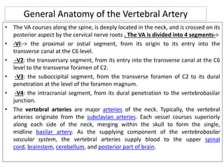
- 1. General Anatomy of the Vertebral Artery • The VA courses along the spine, is deeply located in the neck, and is crossed on its posterior aspect by the cervical nerve roots . The VA is divided into 4 segments-> • -VI:-> the proximal or ostial segment, from its origin to its entry into the transverse canal at the C6 level. • -V2: the transversary segment, from its entry into the transverse canal at the C6 level to the transverse foramen of C2. • -V3: the suboccipital segment, from the transverse foramen of C2 to its dural penetration at the level of the foramen magnum. • -V4: the intracranial segment, from its dural penetration to the vertebrobasilar junction. • The vertebral arteries are major arteries of the neck. Typically, the vertebral arteries originate from the subclavian arteries. Each vessel courses superiorly along each side of the neck, merging within the skull to form the single, midline basilar artery. As the supplying component of the vertebrobasilar vascular system, the vertebral arteries supply blood to the upper spinal cord, brainstem, cerebellum, and posterior part of brain.
- 4. • The V1 segment courses vertically from its origin on the posterior aspect of the subclavian artery to the transverse foramen men of C6. It is accompanied by 2 veins anteriorly and posteriorly . • The V2 segment courses vertically from one transverse foramen to the next. It is surrounded by the perivertebral venous plexus and enclosed in a periosteal sheath that is continuous with the periosteum of the foramina of the transverse processes . At its entrance into the transverse canal at C6, the periosteal sheath adheres somewhat to the VA, forming a proximal fibrous ring. Otherwise, the VA is free inside this periosteal sheath.
- 5. • The V3 segment has a complex course . First, it runs horizontally from the C3 to the C2 transverse foramen. Unlike the other transverse processes, which are perpendicular to the vertebral bodies, the C2 transverse process is oblique inferiorly and laterally. Moreover, the C2 transverse process is longer than the other transverse processes. Therefore, the VA must turn laterally and course almost horizontally to reach the C2 transverse process. Then it courses vertically" from C2 to C1 .
- 6. • At the C1 transverse foramen it again changes direction, coursing horizontally in the groove of the posterior arch of the atlas . The end of this groove is often clearly indicated by an increase in the height of the posterior arch of the atlas. At the end of this groove, the VA turns obliquely and vertically and medially toward the dura. It pierces the dura mater, invaginating the dura and the periosteal sheath 3 to 4 mm, making a double furrow around the VA. At this level the VA is also adherent to the periosteal sheath forming a distal fibrous ring. Otherwise, the VA is free inside the periosteal sheath, still surrounded by the venous plexus
- 8. • Branches of the VA ->Embryologically, the VA is formed by the junction of metameric segments. Therefore, each segment gives rise to similar branches: one radiculomeningeal branch and one muscular branch . • The muscular branches are connected with the muscular branches of the ascending cervical artery, the deep cervical artery, and the external carotid artery, forming a vascular network that may become enlarged in some cases. When the VA is occluded proximally, this network allows the VA to refill with blood distally to compensate for the decrease in vertebrobasilar flow . This vascular network always exists even when not visible on angiography.
- 9. • Another important branch is the anterior meningeal artery from the radicular branch of the third interspace (C2-C3) . • The last interesting branch is the posterior meningeal artery, which originates from the VA just after its dural penetration or occasionally from the occipital artery.
- 10. • Anomalies and variations ->In 40% of subjects, one VA is larger than the contralateral VA. The former is called dominant, the latter is called minor. • The most important variation in the course of the VA concerns level of entry into the transverse canal. Instead of entering the transverse foramen of C6, the VA may enter at C5, C4, or even C3. • Occasionally, the VA is duplicated at its origin, with two trunks that join into one VA at the entrance of the transverse canal. • Branches-> • The most common variation is an extracranial origin of the PICA, which may be present in 20% of the population. PICA may arise from the VA above C1, piercing the dura just beside the VA, or between C1 and C2, or even lower in the neck. • Another variation involves the intradural course of the VA from C2 rostrally . In this variation, VA is composed of an atretic component coursing normally along the spine between C2 and C1 and a major component piercing the dura between C1 and C2 and then coursing intradurally.
- 11. • Dynamic changes The VA is free inside the periosteal sheath along the V2 and V3 segments. Therefore, during movements of the head and neck, the VA may be stretched or compressed. This mechanism especially affects the V3 segment during head rotation . During this movement, the atlas follows the movements of the head, rotating around the odontoid process. As a result, the 2 parts of the contralateral V3 segment are stretched and become parallel to the posterior arch of atlas. This effect is of particular importance when positioning patients for surgery.
- 12. Vertebral artery injuries in cervical spine surgery • Injury to the vertebral artery is a potentially devastating complication of cervical spine surgery. While the overall incidence rate in the cervical spine is 1.4%. • patients are at the greatest risk when undergoing posterior instrumented upper cervical spine surgery (4-8% incidence) In contrast, the risk of injury is only 0.3-0.5% for anterior subaxial cervical spine procedures. • The clinical sequelae of an iatrogenic vertebral artery injury can vary widely depending on anatomic variations, circulation dominance, and the presence of collateral blood flow. • Patients can have minimal, but noticeable, sequelae, and serious complications such as lateral medullary (Wallenberg) syndrome, quadraparesis, and even mortality have been reported.
- 13. • AVOIDANCE • Anterior cervical surgery-> Vertebral artery injuries in anterior subaxial cervical spine surgery are rare and most occur during the decompression. • An important mode of prevention involves evaluation of a tortuous vertebral artery on preoperative advanced imaging. Assessment of spinal canal width can also aid in the avoidance of the vertebral artery. • Since the vertebral artery is located between 0.8 and 1.6 mm from the lateral tip of the uncinate process, avoiding an aggressive removal of the uncinate joint is prudent when pathology is appropriate. • Finally, one potential pitfall from the use of an operating microscope is the potential for an asymmetric decompression. An oblique or asymmetrical decompression during a corpectomy may put the vertebral artery at risk, especially if a tortuous path is missed.The keys to preventing this complication include symmetric and stable patient placement on a head holder and positioning of the microscope perpendicular to and over the center of the wound
- 14. • Posterior cervical spine-> Instrumented posterior surgery of the upper cervical spine places the vertebral artery at the highest risk for injury. • Avoidance measures start with preoperative evaluation of the vessel course on advanced imaging, preferably a CT scan. In 18-23% of patients, at least one side will not accommodate a transarticular (Magerl) screw, and in up to 6%, a transarticular screw cannot be placed in either side. • Vertebral artery injuries during posterior exposure can also be avoided. When exposing the posterior ring of the atlas, lateral dissection should be limited to a point 15 mm lateral to the midline. Furthermore, since the vertebral artery typically runs along the superior aspect of C1 posterior ring, dissection. along the inferior aspect is safest. No dissection should occur on the superior edge of C1 more than 8 mm lateral to the midline. • all patients who sustained a vertebral artery injury after the placement of transarticular screws had a poorly reduced C1/C2 joint when screw placement occurred. The authors argued that with an anatomically aligned C1/C2 joint and use of biplanar fluoroscopy, the safety of placement of a transarticular screw increases significantly.
- 15. • During placement of a C1 lateral mass screw, avoidance of a vertebral artery injury hinges upon use of an appropriate starting point, which is the junction of lateral mass and inferior aspect of posterior arch ,The screw is then angled 10-15 degrees medially to avoid the vertebral artery, which courses laterally. • Similarly, a properly placed C2 pedicle screw has minimal risk to the vertebral artery. The C2 pedicle screw is placed utilizing a starting point slightly superior and medial to the center of the lateral mass and aimed 10 degrees medially and 15 degrees cephalad . Placing subaxial cervical spine lateral mass screws can also put the vertebral artery at risk. Techniques such as the Magerl, Roy-Camille, and Anderson all describe a laterally directed screw (10- 25 degrees) from the midpoint of the lateral mass to avoid the vertebral artery in the foramen transversarium .
- 16. • ACTION-> Once a vertebral artery injury has occurred, the surgeon must always keep the treatment goals in mind in the appropriate order: • (1) Achieve control of the hemorrhage. • (2) Prevent acute central nervous system ischemia. • (3) Prevent postoperative complications such as embolism and pseudoaneurysm • Control of vertebral artery bleeding can be achieved in three different ways: Primary repair, bypass surgery, or sacrifice. • Primary repair->when available, remains the best option. After vascular surgery consultation is obtained and aggressive intravenous access for fluid resuscitation has been communicated to the anesthesia team, the first step should be to ensure that the head is in a neutral position, as cervical extension and axial rotation can lead to occlusion of the contralateral vertebral artery. Digital pressure can often be used to obtain hemostasis, followed by large pieces of hemostatic agents such as thrombin soaked gel foam and cottonoids. It is important to use only large items that cannot accidentally embolize. After proper exposure, a temporary aneurysm clip or vessel loop is placed proximal and distal to the injury site. The injury can then be directly repaired with 7-0 or 8-0 Prolene (Ethicon Sumerville, NJ).[7,10] Prior to completion of the repair, the temporary clips should be removed to prevent air embolism or the propagation of other emboli.
- 17. • If direct repair is not possible, the remaining options are to bypass the injury site or vessel sacrifice. Ligation of the vessel should only occur if there is good retrograde flow, as this may be a sign of considerable contralateral/collateral flow. To evaluate this, place an aneurysm clip proximal to the injury on the vertebral artery and look for significant back flow from the cephalad end. • The decision to sacrifice the vessel should be a last resort, as the severe neurologic complication rate can be as high as 43%. If the vessel cannot be repaired, and there is poor retrograde flow, bypass is indicated. Alternatively, the vascular surgeon may choose to perform traditional bypass surgery. Tamponade alone is not effective in achieving hemostasis, as multiple reports exist of complications after use of this method. • Postoperatively the patient should be started on an antiplatelet medication after 6 hours and should be evaluated with conventional angiography, as often times there may be significant artifact with an MR angiogram.
- 18. • Symptomsvariable in presentation and time of onset • vertebrobasilar insufficiency manifests with – dizziness – vertigo – nausea – diplopia – blindness – ataxia – bilateral weakness – oropharyngeal dysfunction