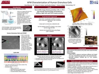
AFM Characterization of Human Granulosa Cells
Vera Marsova 18’1, Brian D. Cohen1 and Sudhir Khetan1, 1Union College, Schenectady, NY
Conclusions
Experimental Approach
References and Acknowledgements
• Whited, A., & Park, P. (2013). Atomic force microscopy: A multifaceted tool
to study membrane proteins and theirinteractions with ligands. Biochimica
Et Biophysica Acta, 1838, 56-68.
• Frederix, P., Bosshart, P., & Engel, A. (2009). Atomic Force Microscopy of
Biological Membranes. Biophysical Journal, 96(2), 329-338.
• Li, G., & Xi, N. (2007). Nanotechnology and Membrane Receptors:
Focused on Angiotensin II Receptors. Med Clin North Am., 91(5), 929-936.
• (2006, March 23). Lipid Rafts and Cell Function. The Keystone
Symposium. Lecture conducted in Steamboat Springs, CO
• Union College Summer Research Fund
Goals
Successfully image human granulosa cells as a first
step towards characterizing cell membrane
stiffness, lipid rafts and FSH receptor proteins
Atomic Force Microscopy (AFM)
Human Granulosa Cells and hFSHR
•AFM is an imaging technique that
is able to produce real time
topographic maps on the
nanometer scale
•Concept: cantilever movement
which adjusts to the relative
position on the sample is recorded
through laser deflection on the
photodiode detector. The image
resolution is determined by the
magnitude of the probe (tip radius
42 nm)
• hGrC1 cell line was
chosen as the most
physiologically relevant
one in terms of studying
the human follicle
stimulating hormone
receptor (hFSHR)
• hFSHR is a G-protein-
coupled receptor (GPCR)
activated by FSH, located
in granulosa cells in
ovaries and responsible
for follicular development
stimulation in females
Introduction
Deflection trace and retrace as a collection of surface height
measurements as the probe rasters over the surface
• The artifacts can be seen when the probe goes over the highest points
• The graphs indicate precision of the scan
Grow hGrC1 at optimized conditions on poly-D-
lysine coated petri dishes/cover slips compatible
with AFM
Use the adapted protocol to engage on the surface
and live image the cells
Interpret and analyze AFM data using ARgyle
Light™ Software
2-step wash to remove any unattached cells
•Most optimal AFM protocol and probe were experimentally
determined
•Future studies:
• utilize AFM to measure membrane stiffness and height
as a way of identifying lipid rafts on the cell membrane
• generate antibody modified tips for FSH receptor
visualization
From Whited, A., & Park, P. (2013 Biochimica Et
Biophysica Acta, 1838, 56-68.
•AFM Advantages: simple sample preparation,
can be operated in liquid, less destructive than
other biological imaging methods.
TR400PB probe from Asylum Research Probe Store
Lipid rafts
From Prof. W. Stutts of University of Florida
• Lipid raft microdomains are the native context of the
hFSHR on the cell membrane.
• Membrane rafts:
o Small (10–200 nm)
o Heterogeneous and highly dynamic
o Sterol- and sphingolipid-enriched
o Compartmentalize cellular processes
Results
Compiled image of deflection retrace overlaid on
height retrace data of an hGrC1 cell in a petri dish
Compiled image of deflection
retrace overlaid on height retrace
data of an hGrC1 cell on a clear
cover slip in an open fluid cell
assembly
Open fluid cell assembly from Asylum
Research
TR400PB probe from Asylum Research Probe Store BL-AC40TS probe from Asylum Research Probe Store
The least damaging probe was determined both experimentally and on the
basis of low spring constant (0.02
𝑁
𝑚
) and frequency (10 kHz).
✓ ✘From Prof. W. Stutts of University of Florida

veramarsova_hGRC1_AFM_poster

  • 1.
    AFM Characterization ofHuman Granulosa Cells Vera Marsova 18’1, Brian D. Cohen1 and Sudhir Khetan1, 1Union College, Schenectady, NY Conclusions Experimental Approach References and Acknowledgements • Whited, A., & Park, P. (2013). Atomic force microscopy: A multifaceted tool to study membrane proteins and theirinteractions with ligands. Biochimica Et Biophysica Acta, 1838, 56-68. • Frederix, P., Bosshart, P., & Engel, A. (2009). Atomic Force Microscopy of Biological Membranes. Biophysical Journal, 96(2), 329-338. • Li, G., & Xi, N. (2007). Nanotechnology and Membrane Receptors: Focused on Angiotensin II Receptors. Med Clin North Am., 91(5), 929-936. • (2006, March 23). Lipid Rafts and Cell Function. The Keystone Symposium. Lecture conducted in Steamboat Springs, CO • Union College Summer Research Fund Goals Successfully image human granulosa cells as a first step towards characterizing cell membrane stiffness, lipid rafts and FSH receptor proteins Atomic Force Microscopy (AFM) Human Granulosa Cells and hFSHR •AFM is an imaging technique that is able to produce real time topographic maps on the nanometer scale •Concept: cantilever movement which adjusts to the relative position on the sample is recorded through laser deflection on the photodiode detector. The image resolution is determined by the magnitude of the probe (tip radius 42 nm) • hGrC1 cell line was chosen as the most physiologically relevant one in terms of studying the human follicle stimulating hormone receptor (hFSHR) • hFSHR is a G-protein- coupled receptor (GPCR) activated by FSH, located in granulosa cells in ovaries and responsible for follicular development stimulation in females Introduction Deflection trace and retrace as a collection of surface height measurements as the probe rasters over the surface • The artifacts can be seen when the probe goes over the highest points • The graphs indicate precision of the scan Grow hGrC1 at optimized conditions on poly-D- lysine coated petri dishes/cover slips compatible with AFM Use the adapted protocol to engage on the surface and live image the cells Interpret and analyze AFM data using ARgyle Light™ Software 2-step wash to remove any unattached cells •Most optimal AFM protocol and probe were experimentally determined •Future studies: • utilize AFM to measure membrane stiffness and height as a way of identifying lipid rafts on the cell membrane • generate antibody modified tips for FSH receptor visualization From Whited, A., & Park, P. (2013 Biochimica Et Biophysica Acta, 1838, 56-68. •AFM Advantages: simple sample preparation, can be operated in liquid, less destructive than other biological imaging methods. TR400PB probe from Asylum Research Probe Store Lipid rafts From Prof. W. Stutts of University of Florida • Lipid raft microdomains are the native context of the hFSHR on the cell membrane. • Membrane rafts: o Small (10–200 nm) o Heterogeneous and highly dynamic o Sterol- and sphingolipid-enriched o Compartmentalize cellular processes Results Compiled image of deflection retrace overlaid on height retrace data of an hGrC1 cell in a petri dish Compiled image of deflection retrace overlaid on height retrace data of an hGrC1 cell on a clear cover slip in an open fluid cell assembly Open fluid cell assembly from Asylum Research TR400PB probe from Asylum Research Probe Store BL-AC40TS probe from Asylum Research Probe Store The least damaging probe was determined both experimentally and on the basis of low spring constant (0.02 𝑁 𝑚 ) and frequency (10 kHz). ✓ ✘From Prof. W. Stutts of University of Florida