
Recommended
More Related Content
What's hot
What's hot (20)
Similar to Presentation on Pityriasis Versicolor .pptx
Similar to Presentation on Pityriasis Versicolor .pptx (20)
Recently uploaded
Recently uploaded (20)
Presentation on Pityriasis Versicolor .pptx
- 2. Outline Introduction Epidemiology Etiology Predisposing factors Pathogenesis Clinical features Investigations Treatment Differential diagnosis References
- 3. Introduction Pityriasis Versicolor AKA Tinea versicolor is a common, benign, superficial cutaneous fungal infection usually characterized by hypo-pigmented or hyper-pigmented macules and patches on the chest and the back. Pityriasis versicolor is not contagious and usually does not cause any health issues and post people recover fully following treatment. In patients with a predisposition, Pityriasis versicolor may chronically recur.
- 4. Epidemiology Tinea versicolor occurs worldwide, with prevalence reported to be as high as 50% in the humid, hot environment. Race Although the alteration in skin pigmentation is more apparent in darker-skinned individuals, the incidence of tinea versicolor appears to be the same in all races. Sex Several studies have addressed the frequency of tinea versicolor based on sex, and no dominance of either sex is apparent. Age It is most common in persons aged 15-24 years, when the sebaceous glands are more active. The occurrence of tinea versicolor before puberty or after age 65 years is uncommon.
- 5. Etiology Malassezia furfur (Pityrosporon ovale). M. furfur is a member of normal flora of the skin found in 18% of infants and 90-100% of adults. Most cases of tinea versicolor occur in healthy individuals with no immunologic deficiencies. Nevertheless, several factors predispose some people to develop this condition. These factors include genetic predisposition; warm, humid environments; excessive sweating; immunosuppression; malnutrition; application of oily preparations or excessive oily skin; corticosteroid usage; hormonal imbalance Predisposing factors Human peptide cathelicidin LL-37 plays a role in skin defense against this organism.
- 7. Pathogenesis
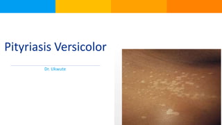
- 8. Clinical Features Most individuals with tinea versicolor report cosmetically disturbing, abnormal pigmentation. The involved skin regions are usually the trunk, the back, the abdomen, and the proximal extremities. The face, the scalp, and the genitalia are less commonly involved. In patients with fair skin, the color of each lesion varies from almost white to reddish- brown or fawn colored. In darker skin types, involved areas can have varying degrees of either hypo- pigmentation or hyper-pigmentation. A fine, dustlike scale covers the lesions. Occasionally, a tinea versicolor patient also reports mild pruritus. In most instances, the condition is asymptomatic.
- 9. The clinical presentation of tinea versicolor is distinctive, and the diagnosis is often made without any laboratory documentation. The ultraviolet black light (Wood lamp) can be used to demonstrate the coppery-orange fluorescence of tinea versicolor. However, in some cases, the lesions appear darker than the unaffected skin under the Wood lamp, but they do not fluoresce. The diagnosis is usually confirmed by potassium hydroxide (KOH) examination, which demonstrates the characteristic short, cigar-butt hyphae that are present in the diseased state. The KOH finding of spores with short mycelium has been referred to as the spaghetti and meatballs or the bacon and eggs sign of tinea versicolor Investigations
- 10. Counsel Patient: Patients should be informed that tinea versicolor is caused by a fungus that is normally present on the skin surface and is therefore not considered contagious. The condition does not leave any permanent scar or pigmentary changes, and any skin color alterations resolve within 1-2 months after treatment has been initiated. Recurrence is common, and prophylactic therapy may help reduce the high rate of recurrence Treatment Treatment depends on factors such as severity, climate, thickness and area of infection. It involves a combination of counselling, lifestyle change and pharmacotherapy
- 11. Pharmacotherapy: Topical agents- Selenium sulphate Sodium thiosulphate solution Azole like ketoconazole, itraconazole Allylamine antifungals like Terbinafine Life style change: Avoid prolonged exposure to ultraviolet rays Avoid going out in hot and humid temperature Daily bath to avoid excessive accumulation of oil and dirt on the skin Use prescribed medications appropriately. Systemic Agents- ketoconazole 200mg OD for 7-10 days Fluconazole single oral dose of 400mg Itraconazole 200-400mg OD for 7days
- 12. Differential Diagnoses Erythrasma Guttate Psoriasis Pityriasis Alba Seborrheic Dermatitis Tinea Corporis Vitiligo
- 13. References Medscape Common Skin Diseases in Africa: An illustrated guide (Colette van Hees & Ben Naafs) Slideshare