2. Introduction
The kidneys represent the primary organs of homeostasis
in the regulation of both volume and composition of
body fluids and the excretion of metabolic waste
products in urine.
The kidneys are large, bean-shaped organs which lie on
the dorsal side of the visceral cavity.
They are protected by a tough fibrous coat called the
renal capsule.
2
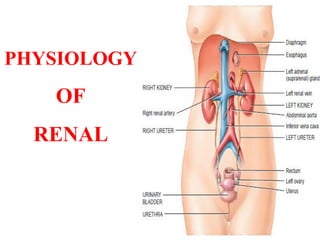
3. Components of renal system
The renal system composed of
• Kidneys: Formation of urine
• Ureters: Transport urine from the kidneys to the bladder
• Urinary bladder: Provides a temporary storage
reservoir for urine
• Urethra: Transports urine from the bladder to out of the
body
3
4. Kidneys
On a longitudinal section of
kidney there are two distinct
regions, cortex & medulla.
The outer cortex surrounds
darker triangular structures
called pyramids which
form the medulla.
The inner part of the
kidneys, the renal pelvis
collects the urine from the
calyces draining it into
the ureter.
4
5. Nephron
The basic functional unit of the kidneys.
Each kidney is made up of approximately 1 million
nephrons.
The nephron consists of the following components :
Glomerulus & Bowman’s capsule
Proximal convoluted tubule
Loop of Henle
Distal convoluted tubule and
Collecting duct
5
6. Nephron…
The glomerulus and
Bowman’s capsule are
situated in the cortex of the
kidney and form the renal
corpuscle.
The capsule continues with
the proximal convoluted
tubule, the long U-shaped
tubule called loop of Henle,
the distal convoluted tubule
that connects to collecting
tubule which eventually
merge to join the ureter.
6
7.
8.
9. Functions of the Urinary System
1. Excretion of nitrogenous metabolic waste products:
- urea, uric acid and creatinin.
2. Regulate
• ABP by controlling blood volume and RAAS
• RBC formation by producing EPO
• Electrolytes concentration (Na+, K+, Ca2+, PO43-)
• ECF and blood volume
• Acid-base balance
• Osmolality of the body fluid (300 mosm/l ) maintain
the proper balance between water and salts
3. Endocrine function: Renin, EPO, Calcitriol
4. Filter 180 liters of blood daily to eliminate: toxins,
metabolic wastes and excess ions
5. Drug metabolism & detoxification of certain chemicals
9
10. Functions of the Urinary System…
6. Filtration of the blood
Occurs in the glomerulus of the kidney nephron
Contributes to homeostasis by removing toxins or
waste
7. Reabsorption of vital nutrients, ions & water
Occurs in most parts of the kidney nephron
Contributes to homeostasis by conserving important
materials
10
11. Functions of the Urinary System…
8. Secretion of excess materials
• Assists filtration in removing material from the blood
• Contributes to homeostasis by preventing a build-up
of certain materials in the body such as drugs,waste,etc.
9. Activation of Vitamin D
• Vitamin D made in the skin is converted to Vitamin D3
by the kidney.
• Active Vitamin D (D3) assists homeostasis by
increasing calcium absorption from the digestive tract
and reabsorption from renal tubules.
11
12. Functions of the Urinary System…
10. Release of Erythropoietin (EPO) by the kidney
EPO stimulates new RBC production
New RBC’s assist homeostasis by insuring adequate
Oxygen & Carbon dioxide transport
11. Release of Renin by the kidney
Renin stimulates the formation of a powerful
vasoconstrictor called Angiotensin II
Angiotensin II assists homeostasis by causing
vasoconstriction which increases blood pressure
12
13. Functions of the Urinary System…
12. Release of Prostaglandins
Prostaglandins dilate kidney blood vessels
Dilated blood vessels contribute to homeostasis by
maintaining blood flow in the kidneys
13. Secretion of H+1 & reabsorption of HCO3-1
Eliminates excess hydrogen ions & conserves buffer
material such as bicarbonate contributes to
homeostasis by controlling acid/base conditions in
body fluids
13
14. Structures of Kidney
Capsule
The outer membrane that encloses, supports and
protects the kidney
Cortex
The outer layer of the kidney that contains most of
the nephron, main site for filtration, reabsorption &
secretion
Medulla
Inner core of the kidney that contains the pyramids,
columns, papillae, calyces, pelvis and parts of the
nephron not located in the cortex.
14
15. Structures of Kidney…
Renal Pyramids
Triangular shaped units in the medulla that house the
loops of Henle and collecting ducts of the nephron.
Site for the counter-current system that concentrates
salt and conserves water and urea
Renal Column
A passageway located between the renal pyramids
found in the medulla and used as a space for blood
vessels
Nephron
The physiological unit of the kidney used for
filtration of blood and reabsorption and secretion of
materials 15
16. Structures of Kidney…
Renal Papilla
Tip of the renal pyramid that releases urine into a
calyx
Calyx
A collecting sac surrounding the renal papilla that
transports urine from the papilla to the renal pelvis
Renal Pelvis
Collects urine from all of the calyces in the kidney
Ureter
Transports urine from the renal pelvis to the bladder
16
19. 19
Blood supply to kidneys
Renal blood flow/RBF
The amount of blood flow to kidney per minute.
Arterial flow into and venous flow out of the kidneys follow
similar paths.
20.
21. 1. RBF = 1200 ml/min, or 21%
of the CO. 94% to the cortex
2. Two capillary beds:
Glomerulus and peritubular
capillaries
3. High hydrostatic pressure in
glomerular capillary (about 60
mmHg) and low hydrostatic
pressure in peritubular
capillaries (about 13 mmHg)
4. It is unique that glomerular
capillaries are found b/n 2
arterioles.
Characteristics of the RBF
21
22. Nerve supply to the kidneys
Kidneys receive sympathetic nerve supply from the
last thoracic & upper 2 lumbar segments of the spinal
cord which relay in the paravertibral and mesentric
ganglia.
Sympathetic stimulation results in
Constriction of arteries & arterioles →↓RBF
(α-AR effect)
↑Na reabsorption in renal tubules (α-AR
effect)
↑Renin secretion by JG-cells (β-AR effect)
Dilation of efferent arterioles (β-AR effect)
Parasympathetic supply from vagus nerve function
is not clear so far 22
23. Mechanisms of urine formation
The mechanism by which
nephrons clear the plasma of
unwanted substances is:
It filters the plasma through
the fenestrated glomerular
membrane into renal tubules
As the filtrate flows through
the tubules, reabsorption
of needed substances &
Secretion of unwanted
substances into the renal
tubules
23
24. GFR 125 ml/min, 180L/day, about 1% is excreted
Process of urine formation
24
25. Mechanism of formation of concentrated urine
When there is a shortage of H2O in
the body
↓ECF volume, ↑Osmolality
Stimulates osmoreceptors in the HT
↑ADH secretion
↑ H2O reabsorption in the DT &
CD
↑Excretion of solutes
Concentrated (1200 mosm/l), in
small volume of urine is produced
ADH
25
26.
27. Mechanism of formation of diluted urine
When there is excess H2O in
the body
↑ECF vlume, ↓Osmolality
↑Aldosterone secretion
↓ADH secretion
↑NaCl reabsorption in the DT &
CD
↑H2O excretion
Diluted urine (50-100 mosm/l)
28.
29. Glomerular Filtration
It is the filtration of fluid through the glomerular
capillaries.
The kidneys filter the body’s entire plasma volume 60
times each day. The filtrate contains:
All plasma components (except protein); water,
nutrients, and essential ions to become urine
Plasma proteins are not filtered and are used to
maintain oncotic pressure of the blood.
The glomerulus is more efficient than other capillary
beds b/c 1. Its filtration membrane is significantly more
permeable
2. Glomerular blood pressure is higher ;it has
a higher net filtration pressure.
The urine contains metabolic wastes & unwanted
substances.
29
30.
31. Glomerular membrane
Made up of 3 layers
1. Endothelial layer
2. Basement membrane
3. Epithelial cell (podocytes)
Thickness: 1 µm
Fenestrated, highly
permeable
Allows the passage
of all components of
plasma except
plasma proteins
and blood cells.
31
32. Glomerular Filtration Rate (GFR)
The amount of fluid filtered per minute in all
nephrons of both kidneys.
GFR = 125 ml/min, or 180 L/day
Filtration fraction (FF): the fraction of RPF (renal
plasma flow) that becomes glomerular filtrate.
RBF = 1200 ml/min RPF = 55% of RBF, 650 ml/min
FF = GFR/RPF, 125/650 = 19%
32
33. Glomerular Filtration Rate (GFR)
Filtration Pressure (FP): the net pressure forcing fluid to
be filtered through the glomerular membrane.
Determined by
1.Glomerular capillary pressure (60 mm Hg)
2.Glomerular capillary colloid osmotic pressure (32mm Hg)
3.Capsular hydrostatic pressure (18 mm Hg)
FP = GCP – (GCCOP + CHP) = 60 – (32 + 18) = 10 mm Hg
36. Factors affecting GFR
1. Filtration pressure
2. Permeability of the glomerular capillary membrane
3. Diameter of afferent arterioles: dilation increases GFR
Caffeine & diuretics dilate AA & increase GFR.
Sympathetic stimulation constricts AA and decreases
GFR.
4. Diameter of efferent arterioles: dilation decreases GFR
↓RBF→↓GFR →↑Renin →↑Ang-II →EA
constriction→ ↑GFR
5. Concentration of plasma proteins:
↑Proteins → ↑PCOM →↓GFR
6. Renal blood flow: ↑RBF → ↑GFR
7. Arterial blood pressure: ↑ABP (limits) → ↑GFR
36
37. GFR regulation : Adjusting blood flow
GFR is regulated by three mechanisms
1. Renal Autoregulation
2. Neural regulation
3. Hormonal regulation
All three mechanism adjust; renal blood pressure and
resulting blood flow
37
38. Autoregulation of GFR
When the GFR is increased
Tubular fluid will pass with minimum reabsorption
of the required substances.
When the GFR is decreased
Tubular fluid will pass with maximum reabsorption
of unwanted substances.
Therefore, the glomerular filtrate must flow into the
tubular system at an appropriate rate to:
Allow unwanted substances to pass into the urine
Reabsorb nutritionally important substances
38
39. There are two autoregulation mechanisms of GFR
1. Afferent arteriole vasodilator feedback mechanism
↓GFR→Tubular fluid flows slowly→↑Na+ & Cl-
reabsorption →Detected by the macula densa
secret PG-E1 & E2 →Dilation of AA → ↑GFR
2. Efferent arteriole vasoconstrictor Feedback mech.
↓GFR →Tubular fluid flows slowly →↑Na+ & Cl-
reabsorption →Detected by the macula densa
secret PG-E1 & E2 →Stimulate JG-cells to secret
renin → ↑Ang-II → vasoconstriction of EA →
↑GFR
Autoregulation of GFR …
39
41. Neural regulation of GFR
Sympathetic nerve fibers innervate afferent and
efferent arteriole
Sympathetic stimulation is low but can increase
during hemorrhage and exercise
Sympathetic stimulation constricts AA and ↓GFR
Vasoconstriction occurs as a result which
Conserves blood volume (hemorrhage) and
Permits greater blood flow to other body parts
(exercise)
41
42. Hormonal regulation of GFR
Several hormones contribute to GFR regulation
1. Angiotensin II
Produced by renin (released by JG-cells) is a
potent vasoconstrictor. ↓ GFR
2. Atrial Natriuretic Peptide (ANP)
Released by atria when stretched, ↑ GFR by
increasing capillary surface area available for
filtration
3. NO
A potent vasodilator, ↑ GFR
4. Endothelin
A potent vasoconstrictor, ↓ GFR
5. Prostaglandin E2
A potent vasodilator on AA, ↑ GFR 42
43. Parts of the Nephron
1. Proximal convoluted tubules
15 mm long & 55 µm in diameter, lined with
granulated cuboidal epithelial cells
2. Loop of Henle
U-shaped tubules lie b/n PCT and DCT
Has descending and ascending limbs with 2 segments:
thin & thick segments
3. Distal convoluted tubules
5 mm long, 35 µm in diameter, joined to collecting
ducts entering the renal pyramids in the medulla
4. Collecting ducts
43
44. 1. proximal tubules
Reabsorption of nutrients
Reabsorption of Na+ (70-75%)
Almost total reabsorption of K+. Fluid in the Loop
of Henle is free of K+. K+ is secreted in the DT
Passive reabsorption of Cl-, HCO3
-
Obligatory reabsorption of H2O (70-75%) along
with Na, K, Cl, HCO3
- independent of ADH
Reabsorption of urea
Secretion of H+, NH4, creatinin, sulphate and drug
metabolites
44
Tubular reabsorption and secretions
45. Tubular reabsorption and secretion (cont’d)
2. Loop of Henle
Descending limb
Passive reabsorption of H2O
Ascending limb
Active reabsorption of Na,Cl
Impermeable to H2O
3. Distal tubules (diluting segment)
Active reabsorption of Na,Cl
Impermeable to H2O & urea
Late DT is permeable to H2O
ADH dependently
4. Collecting ducts
Reabsorption of Na, Ca and H2O
hormone dependently
45
46. Descending limb of Loop of Henle
Permeable to water, but impermeable
to Na+, K+, Cl-,urea
Water flows out down the osmotic
gradient
Osmolality of tubular fluid increases
progressively up to 1200 mosm/L
Thick segment of the ascending limb
of Loop of Henle
Impermeable to water & urea, but
active reabsorption ofelectrolytes
(Na+, Cl- and K+)
The osmolaltiy of tubular fluid is
progressively decreases up to 150
mosm/l 46
47.
48. Tubular reabsorption of H2O and electrolyte
Reabsorption of H2O in the DT and CD is dependent
on the presence of ADH
Reabsorption of Na+ in the DT and CD is dependent
on aldosterone
Reabsorption of Ca2+ in the DT and CD is dependent
on the presence of PTH and calcitriol
48
49.
50. Tubular transport maximum (Tm)
The maximum amount of substance (mg) transported
(reabsorbed/secreted) by tubules per minutes.
TmG (Tmax of glucose =350 mg/min)
The maximum amount of glucose in mg that can be
reabsorbed by the renal tubules per minute.
It means glucose that is filtered in the glomerulus is
reabsorbed.
Determination of TmG is used as a renal function test
b/c it measures the reabsorptive power of the kidneys.
50
51. Tubular load (TLoad) of substances
The rate of a particular substance filtered through the
glomeruli into the tubules per minute
It equals GFR times the concentration of the substance in
the filtrate.
TLoad of a subs (freely filtered) = Conc. In the filtrate X GFR
TLoad of Glucose = 100 mg/dl X 125 ml/min = 125 mg/min
TLoad of Na+ = 142 meq/1000 ml X 125 ml/min=18 meq/min
TLoad of Cl- = 13 meq/min
TLoad of Urea = 33 mg/min
51
52. Renal plasma clearance
A measure of the volume of plasma that is completely
cleared of a given substance per minute.
It is the ratio of the renal excretion rate of the substance
to its concentration in plasma.
It can be calculated using the following formula:
Cx = Ux V
Px
Where:
Cx = Clearance of the subs (ml/min)
Ux = Concent. of the subs. In urine (mg/ml)
V = Volume of UO (ml/min)
Px = Concent. of the subs in plasma (mg/ml) 52
53. 1. Stimulation of stretch
receptors by large volume of
urine (200-400 ml)
2. Sensory impulse transmitted
to the spinal cord through
PNS
3. Motor impulse stimulates
smooth muscle lining
bladder & Relax internal
urethral sphincter (IUS)
4. Stretch receptors also send
impulse to higher centers
(Pons, HT and cerebral
cortex)
5. Motor impulse from higher
centers promote readiness to
urinate
6. Identify places for urination
7. Relax external urethral
sphincter
Micturition reflex
53