The Urinary System Explained
•Download as PPTX, PDF•
0 likes•81 views

The urinary system consists of the kidneys, ureters, bladder, and urethra. The kidneys filter waste from the blood to produce urine. Each kidney contains around 1 million nephrons, the functional units of the kidney. Nephrons filter blood in the glomerulus and remove waste while reabsorbing useful substances. Hormones regulate urine concentration and volume. Urine is stored in the bladder and expelled through the urethra in a process called micturition.

Recommended
More Related Content
Similar to The Urinary System Explained
Similar to The Urinary System Explained (20)
More from Jagruti Marathe
More from Jagruti Marathe (20)
Recently uploaded
Recently uploaded (20)
The Urinary System Explained
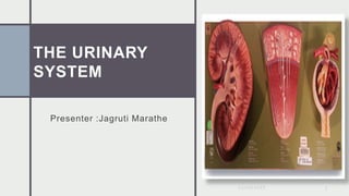
- 1. THE URINARY SYSTEM Presenter :Jagruti Marathe 20/06/2022 1
- 2. The urinary system is composed of : a pair of kidneys a pair of ureters a bladder and a urethra. The urinary system 20/06/2022 2
- 3. Introduction Thousands of metabolic processes in myriad body cells produce hundreds of waste products. The urinary system removes them by filtering and cleansing the blood as it passes through the kidneys. 20/06/2022 3
- 4. Introduction Another vital function is the regulation of the volume, acidity, salinity, concentration, and chemical composition of blood, lymph, and other body fluids. Under hormonal control, the kidneys continually monitor what they release into the urine to maintain a healthy chemical balance. 20/06/2022 4
- 5. THE KIDNEYS The kidneys sit at the back of the abdominal wall and at the start of the urinary system.
- 6. Content • These organs are constantly at work: • Nephrons, tiny structures in the renal pyramids, filter gallons of blood each day. • The kidneys reabsorb vital substances, remove unwanted ones, and return the filtered blood back to the body. • As if they weren’t busy enough, the kidneys also create urine to remove all the waste. The urinary system 20/06/2022 6
- 7. Content • Kidney cross section This cutaway shows the kidney’s main layers, the cortex and the medulla, which form segments known as renal pyramids. • The renal artery and vein circulate huge amounts of blood – about 2 1/2 pints/min at rest, which is up to one- quarter of the heart’s total output. The urinary system 20/06/2022 7 Kidney cross section
- 8. Content • The kidneys are located behind the peritoneum, and so are called retroperitoneal organs. • They sit in the back of the abdomen between the levels of the T12 and L03 vertebrae. • The right kidney is slightly lower than the left kidney to accommodate the liver. • Both kidneys are bean shaped and about the size of an adult fist. The urinary system 20/06/2022 8
- 9. Content • Blood enters the kidneys through renal arteries. • These arteries branch into tiny capillaries that interact with urinary structures inside the kidneys (namely the nephrons). • Here the blood is filtered. Waste is removed and vital substances are reabsorbed back into the bloodstream. • The filtered blood leaves through the renal veins. • All the blood in the body moves in and out of the kidneys hundreds of times each day—that’s about 200 quarts of liquid to be filtered every 24 hours. The urinary system 20/06/2022 9
- 10. The Nephron • Each kidney consists of about 1 million basic functional units called nephrons where blood filtering and urine formation occur . • Each nephron is composed of 10 parts – • afferent arteriole → glomerulus →bowman's capsule → efferent arteriole → proximal convoluted tubule (PCT) → descending limb of loop of Henle → loop of Henle ascending limb of loop of Henle → distal convoluted tubule(DCT) → collecting duct (not part of the nephron). The urinary system 20/06/2022 10
- 11. The Nephron • molecules in the blood that will be transformed to become part of urine travel through the above structures , while molecules that will be retained and reabsorbed back to the blood will come out of the bowman's capsule , and go into efferent arteriole and the peritubular capillaries . The urinary system 20/06/2022 11
- 12. Urine Formation • The kidneys filter unwanted substances from the blood and produce urine to excrete them. • There are 3 main steps of urine formation: Glomerular filtration, Reabsorption Contraction Secretion. • These processes ensure that only waste and excess water are removed from the body. The urinary system 20/06/2022 12
- 13. Content • Each nephron has a glomerulus, the site of blood filtration. • The glomerulus is a network of capillaries surrounded by a cuplike structure, the glomerular capsule (or Bowman’s capsule). • As blood flows through the glomerulus, blood pressure pushes water and solutes from the capillaries into the capsule through a filtration membrane. • This glomerular filtration begins the urine formation process. The urinary system 20/06/2022 13
- 14. Glomerulus Filtration: • small molecules in blood plasma are forced from the glomerulus to bowman's capsule , through the pores in the capillary walls of glomerulus. • any molecules smaller than the plasma proteins will be filtered across – e.g. water , glucose , amino acids , fatty acids , vitamins , minarets , electrolytes , antibodies , enzymes , hormones , drugs, and nitrogenous wastes . The urinary system 20/06/2022 14
- 15. Functions of nephron components • Renal capsule: • Glomerulus's : filtration of H2O and dissolved substances from the plasma . • Glomerular capsule : receives the glomerular filtrate. • proximal convoluted tubule: • Reabsorption of glucose, amino acids, creatine, lactic acid, citric, uric, and ascorbic acids; phosphate , sulfate , calcium , K , and Na by active transport . • Reabsorption of proteins by pinocytosis . Reabsorption of H2O by osmosis. Reabsorption of Cl - and other negatively charged ions by electrochemical attraction. • Active secretion of substances such as penicillin, and hydrogen ions. The urinary system 20/06/2022 15
- 17. The urinary system 20/06/2022 17 Descending limb of nephron loop : • Reabsorption of H2O by osmosis Ascending limb of nephron loop : • Reabsorption of Na, K and Cl- by active transport . Distal convoluted tubule : • Reabsorption of Na by active Transport . • Reabsorption of H2O by osmosis . • Active secretion of hydrogen ions . • Secretion of K both actively and by electorchemical attraction (passives). Collecting duct : • Reabsorption of H2O by osmosis
- 18. Juxtaglomerular Apparatus • The JG apparatus is located at the point of contact between the distal convoluted tubule and the afferent and efferent arterioles . • In its convolutions , the DCT comes into very close contact with the afferent arterioles . • At this point the cells in the afferent arteriols are more numerous, forming a cuff , and are called JG cells these are mechanoreceptors that detect changes in Blood pressure in the afferent arterioles , and secrete renin. The urinary system 20/06/2022 18
- 19. Juxtaglomerular Apparatus • The distal convoluted tubule cells contacting these JG cells are called macula densa (chemo or osmoreceptors) that respond to changes in the solute concentration of the filtrate , in the tubule . The urinary system 20/06/2022 19
- 20. • The distal convoluted tubule cells contacting these JG cells are called macula densa (chemo or osmoreceptors) that respond to changes in the solute concentration of the filtrate , in the tubule . The urinary system 20/06/2022 20
- 21. Glomerular Filtration a. urine formation begins when waste and water and dissolved materials are filtered out of the glomerular capillary . Urinary excretion = glomcrular filtration + Tubular secretion – tubular reabsorption The urinary system 20/06/2022 21
- 22. Glomerular Filtration b. the glomerular capillaries are much more permeable than the capillaries in other tissues . • Filtration pressure = Forces favoring filtration (Glomerular capillary hydrostatic pressure & capsular osmotic pressure) – forces opposing filtration (capsular hydrostatic pressure & Glomerular capillary osmotic pressure). • Thus, filtration pressure is the net force acting to move material out of glomerulus and into the glomerular capsule . The urinary system 20/06/2022 22
- 23. Regulation of GFR • Neural regulation where sympathetic nerves , upon the activation of chloride ion levels , can cause the constriction or relaxation of the afferent arteriole , resulting in a change of GFR. • Renal autoregulation where the juxtaglomerular apparatus (JGA) (formed by the afferent arteriole and DCT) secretes vasoconstriction substances to either afferent arteriole, in response to GFR changes and NaCl levels. The urinary system 20/06/2022 23
- 24. Regulation of GFR • Hormonal regulation involves the JGA secreting a hormone called renin which activates an inactive hormone from the liver called anigotensinogen , resulting in an active hormone (angiotenesin I) which will be converted to angiotensin II by the angiotensin converting enzyme (ACE) [released from the lungs]. Angiotensin II causes constriction of afferent arteriole & release of Aldosterone from adrenal cortex which leads to salt & water retension. The urinary system 20/06/2022 24
- 25. GFR • The rate of filtration varies with filtration pressure. Filtration pressure changes with the diameters of the afferent and efferent arterioles • constriction of afferent arterioles due to sympathetic stimulation decreases glomerular filtration rate. • As the osmotic pressure in the glomerulus increases, filtration decreases • As the hydrostatic pressure in a glomerular capsule increases, filtration decreases. • The kidney produce 125 ml of glomerular fluid per minute, most of which is reabsorbed . The urinary system 20/06/2022 25
- 26. Regulation of Filtration Rate • Glomerular filtration rate remains relatively constant by may increase or decrease when needed. Increased sympathetic activity decreases GFR . • when tubular fluid Nacl decreases, the macula densa causes the JG cells to release renin which leads to vasoconstriction , which affect GFR , and secretion of aldosteron , which stimulate tubular Na+ reabsorption. The urinary system 20/06/2022 26
- 27. Role of ADH ( Antidiuretic hormone) 1. Concentration of H2O in the blood decreases . 2. Increase in the osmotic pressure of body fluids stimulates osmoreceptors in the hypothalamus . 3. Hypothalamus signals the post. pituitary gland to release ADH . 4. Blood carries ADH to the kidneys . 5. ADH causes the distal convoluted tubules and collecting ducts to become more permeable & increase H2O reabsorption by osmosis. 6. urine becomes more concentrated, and urine volume decreases The urinary system 20/06/2022 27
- 28. Tubular Reabsorption The kidney must have mechanisms for reabsorption of the many solutes (Na, K, glucose,chloride) and H2O that it filters each day or in a matter of minutes we would by depleted of all these substances. substances are selectively reabsorbed from the glomerular filtrate. The preritubular capillary is adapted for reabsorption. It carries low pressure blood & is very permeable. Most reabsorption (70%), occurs in the proximal tubule. Different modes of transport reabsorbs various substances in particular segments of renal tubule . Glucose and amino acids by active transport. H2O is reabsorbed by osmosis. proteins are reabsorbed by pinocytosis . The urinary system 20/06/2022 28
- 29. Water Reabsorption (Proximal Tubule) • Na+ and K+ ions are reabsorbed by active transport Negatively charged ions are attracted to positively charged ions (passive transport) . • As the concentration of ions (solute) increases in plasma, osmotic pressure increases . • Water (70%) moves from renal tubule to capillary by osmosis ( passive transport). The urinary system 20/06/2022 29
- 30. Tubular secretion • Unwanted substances such as wastes and excessive salts are secreted by the peritubular capillaries to the renal tubules (mainly PCT and DCT), so that it can be disposed in the urine . • Most substances are secreted by active transport . • Substances secreted include excessive Na+ , Cl- , H+ , K+ , histamine , cretonne , ammonia , uric acid , vitamins and excessive drugs. The urinary system 20/06/2022 30
- 31. Content • The internal urethral sphincter and the external urethral sphincter both provide muscle control for the flow of urine. • The internal sphincter is involuntary. It surrounds the opening of the bladder to the urethra and relaxes to allow urine to pass. • The external sphincter is voluntary. • It surrounds the urethra outside the bladder and must be relaxed for urination to occur The urinary system 20/06/2022 31
- 32. MICTURITION • Smooth muscle stretch initiates the micturition reflex by activating stretch receptors in the bladder wall. • This autonomic reflex causes the detrusor muscle to contract and the internal urethral sphincter muscle to relax, allowing urine to flow into the urethra. • The stretch receptors also send a message to the thalamus and the cerebral cortex, giving voluntary control over the external urethral sphincter. • We usually gain this control of urination between the ages of 2 and 3, as our brains develop. The urinary system 20/06/2022 32
- 33. MICTURITION Major events of micturition: • urinary bladder distends as it fills with urine. • stretch receptors in the bladder wall are stimulated and signal the micturition center in the sacral spinal cord. • parasympathetic nerve impulses travel to the detrusor muscle , which respond by contracting rhythmically. • The need to urinate is sensed as urgent. • voluntary contraction of the external urethral sphincter and inhibition of the micturition reflex by impulses from the brain stem and the cerebral cortex prevent urination. The urinary system 20/06/2022 33
- 34. MICTURITION • Following the decision to urinate, the external urethral sphincter is relaxed, the impulses from the pons and hypothalamus facilitate the micturition reflex. • The detrusor muscle contracts and urine is expelled through the urethra . • Neurons of the micturition reflex center fatigue, the detrusor muscle relaxes, and the bladder begins to fill with urine again. The urinary system 20/06/2022 34
- 35. Physical properties of urine 20/06/2022 35 Properties Transparency Clear Color light yellow to amber Order Aromatic to slightly ammonia pH 4.6 to 8.0 Specific gravity 1.001 to 1.035 Volume 1-2 liters
- 36. Physical properties of urine 20/06/2022 36 Transparency is clear , indicating the lack of large solutes such as plasma proteins or blood cells . [can be influenced by bacterial metabolism in older urine samples]. Color is from light yellow to amber , due to urochrome pigments as byproduct of bile metabolism [can be influenced by food, menstrual bleeding , and minor metabolic products]. Odor is from aromatic to slightly ammonia – like , due to the nitrogenous wastes in urine. [can be influenced by disorders such as glycosuria where urine shows a sweet odor, or by food such as garlic , or by drug]. pH is from 4.6 to 8.0 with an average of 6.0 , due to H+ in the urine [strongly influenced by diet where protein cause acidic urine , and vegetables and wheat cause alkaline urine]. Copyright © 2006 Pearson Education, Inc., publishing as Benjamin Cummings
- 37. Physical properties of urine 20/06/2022 37 Specific gravity (a measurement of dissolved solutes in a solution) is from 1.001 to 1.035 , due to the 5% solute composition in normal urine . Volume is 1-2 liters per day (about 1% of filtration input). [can be influenced by body activities , water intake , hormonal regulation, or disorders such as diabetes insipidus].
- 39. Abnormal Constituents Of Urine • Albumin – a large plasma protein that should not be filtered out of glomerulus; when it is present , it is called albuminuria which may be due to kidney infection called glomerulonephritis • Glucose – a nutrient molecules that should have been reabsorbed (in the case of high carbohydrate diets , trace amount of glucose may be found in urine) ; when is present , it is called glycosuria which may be due to insulin – related problems in a disease called diabetes mellitus. 20/06/2022 39
- 40. Abnormal Constituents Of Urine • blood or erythrocytes – any blood cell should not be filtered out of glomerulus or be present in the urine (except in menstruation – related bleeding); when it is present , it is called Hematuria which may be caused by glomerulonephritis , hemolytic anemia , or urinary tract in infections. • Hemoglobin – pigment protein that normally should be enclosed in erythrocytes and not filtered out of glomerulus; when present , it is called hemoglobinuria which may indicated hemolytic anemia . 20/06/2022 40
- 41. Abnormal Constituents Of Urine • Leukocytes – large white blood cells that should not be present in urine (except in UTI where leukocytes are present to fight the infection); when it is present, it is called Pyuria which may be caused by glomerulus's nephritis, UTI, or even strenuous exercise. • Ketones – by product of metabolism that may occur in trace amounts, but not large quantities in the urine; when it is present, it is called Kentonuria which may indicate certain infections in the urinary system. • Bilirubin – a bile pigment that is normally recycled in lipid metabolism; when it is present , it is called bilirubinuria which may be due to abnormal lipid metabolism, or certain infections in the urinary system. 20/06/2022 41
- 42. Clinical Terms: 20/06/2022 42 Bacteriuria: Bacteria in urine. Diuresis: increased production of urine. Diuretic: substances that increase urine production. Dysuria: painful or difficult urination. Hematuria: Blood in urine. Polyuria: excess urine. Uremia: urine in blood. Glomerulonephritis: Inflammation of glomeruli, damaging the filtration membrane, increasing its permeability (may be due to streptococcal bacteria). Urinalysis: Analysis of urine to diagnose health or disease.
- 43. 3D PRINTING AND OTHER NEW TECHNOLOGIES TO HELP PATIENTS Presentation Title 2/1/20XX 43 In a first for the Middle East, UAE doctors have used 3D printing technology to help safely remove a cancerous tumor from a 42- year-old woman’s kidney. A team of doctors in Dubai have successfully removed a kidney tumor with the help of a transparent, patient-specific 3D printed surgical aid. Not only did the 3D printed aid help them to carefully plan their surgical process for removing the tumor from the patient, but it allowed them to shave an hour off the total operation time. The 3D print was created based off of the patient’s CT scans and ultrasounds, and was 3D printed from transparent and colored plastics so that the doctors could visualize where the tumor was located. Since the technology has not yet been integrated in UAE hospitals, the 3D print was ordered from the U.S. Upon receiving the 3D printed kidney model, the team of doctors, led by Dr Yaser Al Saeedi, was able to properly map out its surgical process to make the actual procedure as minimally invasive as possible. Finally, when it came time to operate, the surgeons were ready and the procedure went very well .