1. 3. NUsc1 Targets Minor Subset
of Synaptotoxic AβOs
Single-Chain Variable Fragment Antibodies Targeting Amyloid β Oligomers:
Alzheimer’s Disease Toxins
Erika Cline1
, Izolda Popova2
, Adriano Sebollela1
, Josette Kamel1
, Alex Qin1
, William Klein1
1
Department of Neurobiology, 2
Recombinant Protein Production Core (rPPC), Northwestern University, Evanston, IL
2. AβO-Specific scFvs (NUscs)
Selected by Phage-Display
4. NUsc1 Expressed Free of
Phage
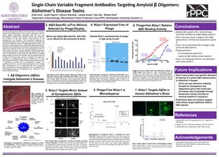
5. Phage-Free NUsc1 is
Monodisperse
1. Aβ Oligomers (AβOs)
Instigate Alzheimer’s Disease
Abstract
Amyloid beta oligomers (AβOs) accumulate early in Alzheimer’s
disease and experimentally cause memory dysfunction and the
major cellular pathologies associated with AD (e.g., tau
abnormalities, synapse loss, oxidative damage, etc.). However,
the structures of the AβO species most germane to AD
pathogenesis are ill-defined. This uncertainty regarding the
pathophysiologically relevant AβO structures has diminished the
perceived therapeutic value of targeting Aβ-derived toxins. Our
long-term research goals are to identify AβO species germane to
AD onset and to determine the structural characteristics of these
AβOs that contribute to their role in the pathogenesis of AD. To
help achieve this goal, we have identified multiple single-chain
variable fragment (scFv) antibodies with high specificity for AβOs
(and minimal affinity for Aβ monomers and fibrils) by panning
phage-displayed human scFv libraries. We have determined that
at least one of these scFvs, NUsc1, is specific for a small sub-
population of synapse-binding AβOs. Furthermore, we have
demonstrated that NUsc1 retains its AβO binding activity when it
is expressed in soluble form, not attached to phage. The
specificity of NUsc1, and the other AβO-specific scFvs, makes
them promising tools for (1) determining the role of individual
AβO conformations in AD pathogenesis; and (2) application as
brain imaging probes for AD diagnostics (e.g., Viola et al., 2015,
Nature Nanotechnology).
Acknowledgements
This research was funded in part by NIH grants R21
AG041953 and T32 AG20506, as well as anonymous
donations to the Klein lab.
Conclusions
Multiple AβO-specific scFvs, termed NUscs,
have been identified by phage-display (panel 2)
NUsc1 has been found to target a minor AβO
species that is >50 kDa and synaptotoxic (panel
3)
NUsc1 can be expressed free of phage in high
purity and yield (panel 4)
Phage-free NUsc1:
is monodisperse (panel 5) &
retains its AβO binding activity (panel 6)
NUsc1 can distinguish AD from non-demented
brain tissue (panel 7)
7. NUsc1 Targets AβOs in
Human Alzheimer‘s Brain
6. Phage-free NUsc1 Retains
AβO Binding Activity
NUsc1 may enable very specific detection
& tracking of a potent AβO species active
in Alzheimer’s disease
scFvs are also attractive antibody
formats for therapeutics and
diagnostics given their small size
(increases ease of passage through
blood-brain barrier) and lack of
immune-reactive Fc sequence
Preliminary data (not shown) suggest
other NUscs target additional distinct
AβO species
Future Implications
1.Lambert MP, et al. Proc Natl Acad Sci U S A. 1998;95(11):
6448-6453.
2.Velasco PT, et al. ACS Chem Neurosci. 2012;3(11):972-81.
3.Lambert MP, et al. J Neurochem. 2007;100(1):23-35.
References
AβO cascade for
Alzheimer’s disease
(AD) pathogenesis.
Soluble AβOs, and
not amyloid plaques,
instigate the neuron
damage leading to
dementia.1
Pathological characteristics of Alzheimer’s disease as a result
of Aβ oligomers. AβOs are potent neurotoxins that accumulate in
the central nervous system (CNS) of humans with AD and in
transgenic rodent AD models.
NUscs are highly AβO-specific, with little
-to-no affinity for Aβ monomers & fibrils
AβO-specific scFvs (NUscs) exhibit little-to-no affinity for the
less toxic forms of Aβ, monomers and fibrils. scFvs were
selected by phage-display from the Tomlinson Human Single Fold
scFv Libraries (MRC, Cambridge, UK) by panning with synthetic
AβOs. The specificity of these scFvs (termed NUscs) for AβOs over
Aβ monomer and fibrils was demonstrated by ELISA (shown above),
compared to the commercially-available pan-Aβ antibody 6E10.
< 50 kDa > 50 kDa
A
B
IP :
Residual ligand activity following IP
NU2
NUsc1
IgG
4G8
Buffer
only
IP
No IP
NUsc1 targets AβO sub-population > 50 kDa and capable of binding
neuronal synapses2
. A) AβOs remaining after immunoprecipitation (IP),
are incubated with cultured neurons. Antibodies for IP are: no IP (None),
NU2 (AβO mAb), NUsc1, IgG, 4G8 (Aβ mAb), and buffer. B) AβOs are
separated into <50 kDa (left) or >50 kDa (right) using molecular weight
cutoff ultrafiltration and incubated with cultured neurons .
α-cmyc α-His34 kDa
25 kDa
34 kDa
25 kDa
Phage-free NUsc1 expression confirmed by SDS-PAGE. NUsc1
was expressed free of phage in TG1 E.coli strain and purified by
Protein A. Size and purity was confirmed by Coomassie stain of
SDS-PAGE (left). The expected molecular weight of NUsc1 is 27
kDa. The presence of the expected affinity tags, cmyc and His, was
confirmed by Western immunoblotting (right).
Soluble NUsc1 expressed free of phage
in high purity & yield
-5
0
5
10
15
20
25
0 5 10 15 20 25
A280(mAu)
Elution Volume (ml)
Native FPLC-SEC
~23 kDa
Void Volume
(large aggregates)
~46 kDa
90% of
NUsc1 is
monomeric
Monodispersity of phage-free NUsc1 is confirmed by native
FPLC-SEC. Phage-free NUsc1 was eluted by FPLC-SEC using a non
-denaturing mobile phase. According to a calibration curve established
from molecular weight standards, NUsc1 primarily eluted at ~23 kDa
(expected molecular weight 27 kDa). Minor peaks were observed at
46 kDa (dimer) and the column void volume (large aggregates).
Integrating under the curve demonstrated that phage-free NUsc1 is
90% monomeric.
NU2 (mAb)
(High AβOs)
NUsc1
(Low AβOs)
NU2 (mAb)
(Low AβOs)
NUsc1
(High AβOs)
Phage-free NUsc1 exhibits AβO dose-dependent response in
ELISA assay. AβOs prepared at high concentrations (100 μM
peptide1
; red and blue) or lower, physiologically-relevant
concentrations (30 nM Aβ peptide2
; purple or green) were titrated in
an indirect ELISA assay. The activity of phage-free NUsc1 (blue and
green) was compared to the AβO-specific monoclonal antibody
NU2.3
NUsc1 seems to exhibit lower affinity for AβOs than full-length
NU2, as is often reported in the literature for scFvs.
NUsc1 detects AβOs in human brain in AD-dependent manner.
NUsc1 detects AβOs in human AD, but not non-demented control,
brain in both intact tissue (top; immunofluorescence) and aqueous
extracts (bottom; ELISA assay).
Western blotCoomassie