2. in vitro as the presence of more than one discrete EPSC response
in a Purkinje cell when the white matter is stimulated at various
intensities to activate the climbing fibers (Kano et al., 1995;
Hashimoto and Kano, 2003; Nishiyama and Linden, 2004). No
other abnormalities have been observed in the cerebellum of the
PKC␥Ϫ/Ϫ
mice, despite extensive analysis. Within the cerebellar
cortex and its targets, PKC␥ is expressed only in Purkinje cells
(Bareggi et al., 1996; Garcia and Harlan, 1997; Barmack et al.,
2000; Shutoh et al., 2003). The morphology, synaptic density,
EPSCs, Ca2ϩ
currents and short-term plasticity of Purkinje cells
are normal, as well as the regional projections of climbing fibers
to the vestibulocerebellum, targets of the vestibulocerebellum,
and eye movements evoked by vestibulocerebellar stimulation
(Chen et al., 1995; Kano et al., 1995; Shutoh et al., 2003). Notably,
long-term depression (LTD) of the parallel fiber–Purkinje cell
synapses, a form of synaptic plasticity implicated in cerebellum-
dependent learning, depends on the ␣ isoform of PKC and can be
induced to normal levels in vitro in PKC␥Ϫ/Ϫ
mice (Chen et al.,
1995; Leitges et al., 2004). Thus, by all measures, the effect of
PKC␥ deletion on the cerebellar circuit appears to be specific for
the development of the climbing fiber–Purkinje cell synapses.
At the behavioral level, broad-spectrum PKC inhibitors in the
cerebellum as a whole (Shutoh et al., 2003) or specifically in
Purkinje cells (De Zeeuw et al., 1998b; Koekkoek et al., 2003)
have been reported to abolish cerebellum-dependent learning. In
contrast, PKC␥Ϫ/Ϫ
mice have a more subtle behavioral pheno-
type, with sparing of some cerebellum-dependent behaviors. Al-
though PKC␥Ϫ/Ϫ
mice have impaired motor coordination on
rotorod tests, eyeblink conditioning is enhanced in these mice.
This led to the hypothesis that single climbing fiber innervation
of Purkinje cells is critical for complex, coordinated movements
but not for the modification of simple, elemental movements by
the cerebellum (Chen et al., 1995). However, a more recent study
reported that adaptation of the optokinetic reflex (OKR), another
simple, cerebellum-dependent motor learning task, is partially
impaired in PKC␥Ϫ/Ϫ
mice (Shutoh et al., 2003). Thus, the dif-
ferential effects of PKC␥Ϫ/Ϫ
deletion cannot be fully explained by
the complexity of the motor task.
One possibility is that PKC␥Ϫ/Ϫ
deletion and multiple climb-
ing fiber innervation have a variable effect on the function of
different cerebellar regions. A recent study reported that Purkinje
cells in cerebellar sulci exhibit some degree of multiple climbing
fiber innervation, even in adult, wild-type mice, suggesting that
some cerebellar regions may function well with multiple climbing
fiber innervation (Nishiyama and Linden, 2004). Eyeblink con-
ditioning, the OKR, and rotorod performance all depend on dif-
ferent regions of the cerebellum (Le Marec and Lalonde, 1997;
Katoh et al., 1998; Gerwig et al., 2005; Villarreal and Steinmetz,
2005). Thus, a region-dependent effect of multiple climbing fiber
innervation could differentially affect these tasks.
Here, we examine motor learning in the vestibulo-ocular re-
flex (VOR) in the PKC␥Ϫ/Ϫ
mice, because this task depends on
the flocculus, the same region of the cerebellum that mediates
OKR adaptation (Ito et al., 1982b; Nagao, 1983; Lisberger et al.,
1984; Luebke and Robinson, 1994; Katoh et al., 1998; McElligott
et al., 1998; Rambold et al., 2002). The VOR and OKR also share
circuitry downstream of the cerebellum, including the vestibular
and oculomotor nuclei (De Zeeuw et al., 1998a). The VOR and
OKR circuitry upstream of the cerebellum are different, but nei-
ther of the upstream pathways express PKC␥ (Garcia and Harlan,
1997; Barmack et al., 2000). We examined whether PKC␥ dele-
tion and the resultant multiple climbing fiber innervation had the
same effects on motor learning in the VOR as it does on motor
learning in the OKR.
Materials and Methods
Subjects. Experiments were performed on male adult (Ն8 weeks) mice
deficient in the ␥ isoform of PKC (n ϭ 42) and wild-type animals (n ϭ
40) with the genetic background from which the PKC␥Ϫ/Ϫ
mice were
derived (B6129PF2/J; The Jackson Laboratory, Bar Harbor, ME) (Abe-
liovich et al., 1993b). All mice used for the experiments had dark eyes.
Some of the wild-type mice used for the experiments described in this
paper were also used in another study (Kimpo et al., 2005). All animal
protocols were approved by the Stanford University Administrative
Panel for Laboratory Animal Care.
Surgical preparation. Mice were prepared for behavioral experiments
as described previously (Boyden and Raymond, 2003; Kimpo et al.,
2005). In summary, while the mouse was under ketamine–isoflurane
anesthesia, a head post was attached to the top of the skull using anchor
screws and dental acrylic, and a scleral search coil (IET, Marly, Switzer-
land) weighing ϳ50 mg was implanted on the temporal side of the right
eye, beneath the conjunctiva. The search coil leads were run subcutane-
ously to a two-pin connector. Mice were allowed to recover from surgery
for 5–7 d before behavioral testing, and those with eye scarring after
surgery were excluded from the study (five PKC␥Ϫ/Ϫ
and one wt).
Experiments. The head of the mouse was immobilized by attaching the
implanted head post to a restrainer. The restrainer was placed on a turn-
table (Carco IGTS, Pittsburgh, PA) that delivered a vestibular stimulus by
rotating the mouse about the vertical axis. The restrained mouse was
positioned in the center of a magnetic field generated by a set of 18-inch
magnetic coils (CNC Engineering, Seattle, WA) that was fixed to the
turntable. The magnetic coils generated signals in the scleral search coil
related to the eye position. An analog differentiator and filter with a 300
Hz corner frequency were used to obtain eye velocity from eye position.
The eye coil method (Robinson, 1963; Judge et al., 1980; Koekkoek et al.,
1997) was chosen because it provides a stable and precise measurement
of mouse eye movements and is therefore particularly reliable for mea-
suring learned changes in the VOR (Stahl et al., 2000; Boyden and Ray-
mond, 2003).
Visual image motion was delivered by an optokinetic drum, a translu-
cent half-dome with black and white vertical stripes, each of which sub-
tended 7.5° of visual angle. The optokinetic drum was backlit by two 60
W lamps. A silvered acrylic plate attached to the turntable helped to
ensure that the motion of the drum filled most of the mouse’s field of
view (approximately two-thirds of the sphere subtended by the optoki-
netic dome and its reflection).
Mice were first acclimatized to the head restrainer, during which the
eye coils were calibrated by moving the magnetic coils relative to the
stationary mouse using sinusoidal velocity profiles, each with a frequency
of 0.5 or 2 Hz with Ϯ10°/s peak velocity. This calibration was performed
with the mouse in total darkness.
Motor learning in the VOR was induced by pairing rotation of the
mouse (vestibular stimulus) with rotation of the visual stimulus. For all
training conditions, the vestibular stimulus had a sinusoidal velocity
profile with Ϯ10°/s peak velocity and a frequency of 0.5 or 2 Hz. The
visual stimulus moved at the same frequency as the head, with a peak
velocity relative to the head of 0, 2.5, 5, 7.5, 12.5, 15, or 20°/s, to create
training conditions with ideal VOR gains (to stabilize the visual image) of
0, 0.25, 0.5, 1.5, 1.75, or 2, respectively, which we refer to as ϫ0 (or
gain-down), ϫ0.25, ϫ0.5, ϫ1.25, ϫ1.5, ϫ1.75, and ϫ2 (or gain-up),
respectively. Before and after training, the VOR was measured in the dark
by collecting 5–10 18-s data files, with a bright light flashed briefly be-
tween collections to keep the animal alert, followed by an 8–10 s pause
before the next collection. The OKR was also measured at the training
frequency before training.
Individual mice were run on more than one training condition. To
allow the VOR gain to return to baseline, mice were placed in their home
cages in a normal visual-vestibular environment between experiments
for at least 48 h after an increase in VOR gain and at least 72 h after a
decrease in VOR gain. These time periods were sufficient to allow the
VOR gain to return to baseline (Kimpo et al., 2005). In general, training
Kimpo and Raymond • Impaired VOR Motor Learning in PKC␥Ϫ/Ϫ
Mice J. Neurosci., May 23, 2007 • 27(21):5672–5682 • 5673
3. conditions that increased or decreased the VOR gain were alternated for
each mouse.
Data analysis. All signals were digitized at a sampling frequency of 500
Hz. In wild-type mice, ϳ6% of the 2 Hz stimulus cycles contained sac-
cades, and ϳ21% of the 0.5 Hz stimulus cycles contained saccades. In
PKC␥Ϫ/Ϫ
mice, ϳ11 and 22% of the 2 and 0.5 Hz cycles, respectively,
contained saccades. When possible, saccades were interpolated by a
straight line. Stimulus cycles with saccades that subtended too much of
the cycle to be interpolated or with other movement artifacts were de-
leted. The remaining eye velocity cycles were aligned on head velocity or
optokinetic drum position and averaged. Using Fourier analysis, the am-
plitude and phase of the fundamental frequency of both eye and head
velocity were measured. The gain of the VOR was defined as the ratio of
the amplitudes of eye and head velocity. The phase of the VOR was
calculated as the difference between the eye velocity phase and head
velocity phase, minus 180°. A compensatory VOR would thus have a
phase of 0°. Positive VOR phase values indicate phase lag of the eye
velocity relative to the head velocity, and negative values indicate phase
lead.
Data from experiments with unreliable VOR measurements were ex-
cluded from analysis. Two criteria were used to exclude experiments.
One was if too few sinusoidal cycles of eye velocity remained for averag-
ing after the exclusion of cycles with motion artifacts. We required at least
10 total cycles of eye velocity for the Fourier analysis measurements of the
VOR before training and at least 10 cycles after training. The second
criterion for exclusion was if measurements of the VOR before training,
after training, or both were exceptionally variable. We required that the
SD of the VOR gain across the 5–10 data files was Ͻ50% of the average
VOR gain across these files. For PKC␥Ϫ/Ϫ
mice and wild-type experi-
ments, 1 of 275 and 2 of 132 experiments, respectively, were excluded
because of too few cycles, and 27 of 275 and 12 of 132 experiments,
respectively, were excluded because of highly variable VOR gain mea-
surements. The variability in VOR gain measurements did not reflect any
consistent trend for the VOR gain to increase or decrease with repeated
testing; in PKC␥Ϫ/Ϫ
mice, the baseline gain was higher in the last data file
than in the first data file in 46% of the rejected experiments, and, in the
other 54%, the gain was lower. For the analysis of VOR phase changes, we
also excluded experiments in which the SD was Ͼ100° before training,
after training, or both (17 of 275 in PKC␥Ϫ/Ϫ
and 4 of 132 in wt mice).
Retinal slip is defined as the velocity of the visual stimulus relative to
the eye. The peak speed and phase of retinal slip were calculated from the
amplitude and phase of the averaged eye and visual stimulus velocities
during the first minute of visual-vestibular training.
Statistical analysis. Each mouse was subjected to one to three experi-
ments using a given training condition. Data used for analysis were
weighed equally across mice by averaging all data from the same training
condition for each mouse before comparing between genotypes. One-
sample t tests were used to determine whether the change in VOR gain or
phase for each training condition was significantly different from zero.
Our conclusions on the learned changes in VOR for the full population of
PKC␥Ϫ/Ϫ
and wild-type mice were not affected by the application of a
Bonferroni-corrected threshold p value. Changes in VOR gain or phase
could result from associative learning, which depends on the relative
directions of head and visual stimulus movements during training or
from nonassociative processes, such as habituation, which would occur
equally during gain-up and gain-down training. To test for associative
learning, changes in VOR gain and phase after gain-up training were
compared with changes after gain-down training for a given training
frequency and genotype by paired t test. To determine differences in
learning between PKC␥Ϫ/Ϫ
and wild-type mice, unpaired t tests were
performed for each training condition. Our conclusions regarding com-
parisons between the two genotypes were unaffected by the Bonferroni
correction for the whole and matched populations (for description of
how matched subpopulations were identified, see next paragraph). To
test for any significant differences between PKC␥Ϫ/Ϫ
and wild-type mice
in baseline VOR and OKR gain and phase across different frequencies, we
performed two-factor ANOVA tests for genotype and frequency. When
genotype had a significant effect on the baseline performance, we per-
formed Bonferroni-corrected unpaired t tests between PKC␥Ϫ/Ϫ
and
wild-type at each frequency. We performed correlation analyses with
Bonferroni correction for multiple tests to test for any significant rela-
tionship between baseline performance parameters and changes in VOR
gain. Unless noted otherwise, the criterion significance level was p Ͻ
0.05.
To identify subpopulations of PKC␥Ϫ/Ϫ
and wild-type mice with sim-
ilar baseline VOR gains, we first ranked mice tested on a given training
condition according to VOR gain. Then, for each training condition,
wild-type mice with high and PKC␥Ϫ/Ϫ
mice with low baseline VOR
gains were excluded until the range was similar, and the average value was
not significantly different between wild-type and PKC␥Ϫ/Ϫ
mice, as de-
termined by unpaired t test ( p Ͼ 0.05). Similar procedures were used to
identify subpopulations of PKC␥Ϫ/Ϫ
and wild-type mice with similar
baseline OKR gains or matched retinal slip speed for each training con-
dition. Note that for these analyses, the baseline gains and retinal slip
were measured on the day of the experiment, whereas the VOR and OKR
data for each mouse shown in Figure 2 were averaged across several days,
for all training conditions.
A previous study (Shutoh et al., 2003) observed higher baseline VOR
and OKR gains in PKC␥Ϫ/Ϫ
mice than those measured in this study.
However, the vestibular stimuli used in that previous study had higher
peak velocities, which elicit higher VOR gains, and the optokinetic stim-
uli had lower velocities and frequencies, which elicit higher OKR gains
(Iwashita et al., 2001; Van Alphen et al., 2001).
Results
Impaired motor learning in the VOR in PKC␥Ϫ/Ϫ
mice
The VOR helps to stabilize visual images on the retina by moving
the eyes in the opposite direction of head movement. The gain of
the VOR is defined as the ratio of eye speed to head speed during
head movements in total darkness. The gain of the VOR can be
modified by a cerebellum-dependent form of motor learning.
Motor learning in the VOR was induced by presenting PKC␥Ϫ/Ϫ
and wild-type mice with visual and vestibular stimuli paired in a
way to increase (gain-up) or decrease (gain-down) the gain of the
VOR. Wild-type mice exhibited adaptive changes in VOR gain,
whereas the PKC␥Ϫ/Ϫ
mice did not.
In all experiments, the head movements used to measure the
VOR and to induce learning had sinusoidal velocity profiles with
peak velocity of Ϯ10°/s. During gain-up training, the visual stim-
ulus moved at the same speed as the head but in the opposite
direction, so that the ideal VOR gain was 2 (this training condi-
tion is often referred to as ϫ2). In wild-type mice, 30 min of
gain-up training using a visual-vestibular stimulus frequency of 2
Hz induced a significant, adaptive increase in the gain of the VOR
(Fig. 1a, white bars) ( p Ͻ 0.0001, one sample t test). During
gain-down training, the visual stimulus moved exactly with the
head, so that the ideal VOR gain was zero (this training condition
is often referred to as ϫ0). In wild-type mice, 30 min of gain-
down training at 2 Hz induced a significant, adaptive decrease in
the gain of the VOR (Fig. 1a, white bars) ( p Ͻ 0.0001). In con-
trast, PKC␥Ϫ/Ϫ
mice did not show any significant changes in
VOR gain after gain-up or gain-down training (Fig. 1a, gray bars)
( p Ͼ 0.15), and the changes were significantly different between
wild-type and PKC␥Ϫ/Ϫ
mice for both gain-up and gain-down
training ( p Ͻ 0.0001, t test).
Previous work has suggested that motor learning in the VOR
may be less dependent on climbing-fiber-triggered plasticity
mechanisms, such as LTD of the parallel fiber–Purkinje cell syn-
apses, when learning is induced using visual-vestibular stimuli at
lower sinusoidal frequencies such as 0.5 Hz (Raymond and Lis-
berger, 1998; Boyden et al., 2006). We therefore examined motor
learning in the PKC␥Ϫ/Ϫ
mice using 0.5 Hz visual-vestibular
stimuli but found that they were still profoundly impaired. After
30 min of 0.5 Hz gain-up training, wild-type mice showed a sig-
5674 • J. Neurosci., May 23, 2007 • 27(21):5672–5682 Kimpo and Raymond • Impaired VOR Motor Learning in PKC␥Ϫ/Ϫ
Mice
4. nificant increase in VOR gain (Fig. 1b, white bars) ( p Ͻ 0.01), but
PKC␥Ϫ/Ϫ
mice did not exhibit a significant change in VOR gain
(Fig. 1b, gray bars) ( p Ͼ 0.34). After 30 min of 0.5 Hz gain-down
training, wild-type mice showed a significant decrease in VOR
gain (Fig. 1b, white bars) ( p Ͻ 0.0001), whereas PKC␥Ϫ/Ϫ
mice
exhibited a significant increase in VOR gain, which is the mal-
adaptive direction (Fig. 1b, gray bars) ( p Ͻ 0.05). For both
gain-up and gain-down training at 0.5 Hz, the changes in the
PKC␥Ϫ/Ϫ
mice were significantly different from wild type ( p Ͻ
0.01, t test). Thus, PKC␥Ϫ/Ϫ
mice are impaired in the ability to
undergo both learned increases and decreases in VOR gain.
Wild-type mice exhibited a significant increase in VOR phase
lead after gain-down training at 0.5 Hz (Fig. 1b, right, white bars)
( p Ͻ 0.01), as reported in previous studies (Iwashita et al., 2001;
Van Alphen and De Zeeuw, 2002; Boyden and Raymond, 2003).
This change in phase was absent in PKC␥Ϫ/Ϫ
mice (Fig. 1b, gray
bars) ( p Ͼ 0.15), although the difference between wild-type and
PKC␥Ϫ/Ϫ
mice did not reach statistical significance ( p Ͼ 0.07, t
test). Neither wild-type nor PKC␥Ϫ/Ϫ
mice exhibited any signif-
icant changes in the phase of the VOR after gain-up training at 0.5
Hz (Fig. 1b) ( p Ͼ 0.08) or after gain-up or gain-down training at
2 Hz (Fig. 1a) ( p Ͼ 0.10).
Impairment of PKC␥Ϫ/Ϫ
mice on the learning task cannot be
explained by sensory or motor performance deficits
We evaluated whether the impaired VOR motor learning of the
PKC␥Ϫ/Ϫ
mice reflected an impairment of learning per se, or
whether it might be a secondary consequence of sensory or motor
performance deficits in the mutants. For
animals to exhibit motor learning in the
VOR, they must have functional vestibu-
lar, visual, and oculomotor systems. The
baseline (pretraining) performance of the
VOR provides a measure of vestibular and
oculomotor functions, and the baseline
performance of the OKR provides a mea-
sure of visual and oculomotor functions.
In the PKC␥Ϫ/Ϫ
mice, there were impair-
ments of both the baseline VOR and OKR,
but these deficits were not sufficient to ac-
count for the learning deficit.
PKC␥Ϫ/Ϫ
mice had significantly lower
average baseline VOR gains than wild-
type mice, measured using a range of head
movement frequencies (F(1,210) ϭ 40.0;
p Ͻ 0.0001 for genotype, two-factor
ANOVA) (Fig. 2a). This was true at each
of four frequencies tested, 0.5, 1, 2, and 5
Hz ( p Ͻ 0.0125, Bonferroni-corrected t
test). The baseline phase of the VOR also
was altered in PKC␥Ϫ/Ϫ
mice compared
with wild-type mice (F(1,209) ϭ 12.9; p Ͻ
0.01) (Fig. 2b), although this difference
was only significant when tested at 0.5 Hz
( p Ͻ 0.0001). Thus, deletion of PKC␥ af-
fects baseline VOR performance. How-
ever, the baseline VOR performance did
not appear to be responsible for the learn-
ing deficit. For each training condition,
there was no significant correlation be-
tween the baseline VOR gain of individual
mice and their learned changes in VOR
gain, in either PKC␥Ϫ/Ϫ
or wild-type mice
(Fig. 3, Table 1). The baseline VOR phase did not significantly
correlate with the learned changes in VOR gain either (Table 1).
In addition, there was considerable overlap in the baseline per-
formance of the two populations of animals (Fig. 2a,b). There-
fore, we compared learning in subpopulations of the PKC␥Ϫ/Ϫ
and wild-type mice that had similar distributions of baseline
VOR gain (Fig. 3, shaded areas) ( p Ͼ 0.31 for each training
condition, t test; see Materials and Methods). The results for these
subpopulations of mice were similar to those for the full popula-
tions: the wild-type mice exhibited adaptive changes in VOR gain
for each of the four training conditions tested (gain-up and gain-
down training at 0.5 and 2 Hz) (Fig. 1, white diagonal hatched
bars) ( p Ͻ 0.05), whereas PKC␥Ϫ/Ϫ
mice did not exhibit any
significant changes in VOR gain (Fig. 1, gray diagonal hatched
bars) ( p Ͼ 0.15). Therefore, both the correlation and matched
population analyses indicate that impaired motor learning in the
PKC␥Ϫ/Ϫ
mice cannot be accounted for by their lower average
baseline VOR gains.
The OKR performance of the PKC␥Ϫ/Ϫ
mice was also differ-
ent from wild-type mice. The OKR gain, measured at 0.5, 1, and
2 Hz, was significantly lower in the PKC␥Ϫ/Ϫ
mice (F(1,137) ϭ
81.1; p Ͻ 0.0001 for genotype) (Fig. 2c), and the phase lagged that
of wild-type mice (F(1,137) ϭ 10.0; p Ͻ 0.01) (Fig. 2d). Neverthe-
less, there was no significant correlation between the baseline
OKR gain or phase of individual mice and their learned changes
in VOR gain (Table 1). Moreover, in subpopulations of wild-type
and PKC␥Ϫ/Ϫ
mice with similar baseline OKR gains ( p Ͼ 0.13, t
test), the wild-type mice still exhibited significant changes in
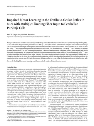
Figure 1. Impaired VOR motor learning in PKC␥Ϫ/Ϫ
mice. Left, Average percentage change in VOR gain in wild-type (wt;
white bars) and PKC␥Ϫ/Ϫ
mice (gray bars) after gain-up (ϫ2) and gain-down (ϫ0) training at 2 Hz (a) and 0.5 Hz (b). Right,
ChangesinVORphaseinducedbyeachtrainingcondition.Openbarsindicatedatafromthefullpopulation.Hatchedbarsaredata
fromsubpopulationsofwild-typeandPKC␥Ϫ/Ϫ
micematchedforbaselineVORgain(diagonalhatchedbars),baselineOKRgain
(cross-hatched bars), or peak speed of retinal slip during training (horizontal hatched bars). Error bars represent SE. Numbers
aboveorbelowthebarsindicatethenumberofmiceineachpopulationorsubpopulation.*pϽ0.05,**pϽ0.01,onesamplet
test. #
p Ͻ 0.05 significant change in VOR gain but in the maladaptive direction.
Kimpo and Raymond • Impaired VOR Motor Learning in PKC␥Ϫ/Ϫ
Mice J. Neurosci., May 23, 2007 • 27(21):5672–5682 • 5675
5. VOR gain (Fig. 1, white cross-hatched
bars) ( p Ͻ 0.05), whereas PKC␥Ϫ/Ϫ
mice
did not (Fig. 1, gray cross-hatched bars)
( p Ͼ 0.11). Thus, the impaired motor
learning in the VOR cannot be attributed
to the lower average OKR gain or larger
OKR phase lag in the PKC␥Ϫ/Ϫ
mice.
The VOR gain changes in the PKC␥Ϫ/Ϫ
mice were significantly different from
wild-type mice with matched baseline
VOR gain or OKR gain for all training
conditions ( p Ͻ 0.01, t test) except
gain-up at 0.5 Hz ( p Ͼ 0.14). The latter
reflected a tendency for the VOR gain at
0.5 Hz to increase in the PKC␥Ϫ/Ϫ
mice
during both gain-up and gain-down train-
ing ( p Ͼ 0.16, paired t test of gain-up vs
gain-down in n ϭ 16 and 7 PKC␥Ϫ/Ϫ
mice
with baseline VOR or OKR gain, respec-
tively, matched to wild-type mice), which
therefore appeared to be nonassociative.
VOR motor learning is impaired for a
wide range of retinal slip speeds
The motion of a visual stimulus on the
retina, referred to as retinal slip, is thought
to play a key role in the induction of motor
learning in the VOR and OKR adaptation.
Furthermore, both cerebellar tasks de-
pend on the flocculus and share the same
motor output circuitry. A previous study
of OKR adaptation in PKC␥Ϫ/Ϫ
mice re-
vealed an altered dependency of this form
of motor learning on the speed of retinal
slip, such that OKR adaptation was im-
paired in PKC␥Ϫ/Ϫ
mice when the speed
of retinal slip during training was high, but
OKR adaptation was completely normal
when the speed of retinal slip was low,
namely Յ2.6°/s (Shutoh et al., 2003).
Therefore, we examined the retinal slip
during each of the training conditions
used to induce motor learning in the VOR.
Unlike OKR adaptation, motor learning
in the VOR was impaired in the PKC␥Ϫ/Ϫ
mice even when the speed of retinal slip
was low.
During the four training conditions
shown in Figure 1, PKC␥Ϫ/Ϫ
mice experi-
enced retinal slip that was, on the whole,
similar to that experienced by wild-type
mice. During gain-down training, retinal
slip was in the same direction as head
movements (close to 0°) (Fig. 4b), and its
average peak speed was Ͻ2.6°/s at both
training frequencies. During gain-up
training, both wild-type and PKC␥Ϫ/Ϫ
mice experienced peak retinal slip speeds
Ͼ10°/s in the opposite direction (close to
180°) from head movement (Fig. 4a).
Although the retinal slip experienced
by the PKC␥Ϫ/Ϫ
and wild-type mice was
generally similar, there were some differ-
Figure2. ThebaselineVORandOKRofPKC␥Ϫ/Ϫ
micearedifferentfromthoseofwild-typemice.Barsrepresenttheaverage
baseline VOR gain (a), VOR phase (b), OKR gain (c), and OKR phase (d) in wt (ϩ, open bars) and PKC␥Ϫ/Ϫ
(E, gray bars) mice
measured at different frequencies of head rotation or visual stimulus rotation. Each symbol represents averaged data from one
mouse, and the number below each bar indicates the number of mice for each measurement.
Figure 3. The learned change in VOR gain as a function of the baseline VOR gain of wild-type (ϩ) and PKC␥Ϫ/Ϫ
(E)
mice for each training condition. Each symbol represents data from one mouse. The regression lines for each population of
miceareshown:PKC␥Ϫ/Ϫ
,solidlines;wild-typemice,dashedlines.ShadedareasindicatetherangesofbaselineVORgain
inthesubpopulationofmiceusedforcomparinglearninginwild-typeandPKC␥Ϫ/Ϫ
micewithsimilarbaselineVORgains.
For number of mice, see Table 1.
5676 • J. Neurosci., May 23, 2007 • 27(21):5672–5682 Kimpo and Raymond • Impaired VOR Motor Learning in PKC␥Ϫ/Ϫ
Mice
6. ences. For the training conditions shown in Figure 1, PKC␥Ϫ/Ϫ
mice experienced larger retinal slip speed than wild-type mice,
except during 2 Hz gain-down training, when the retinal slip
speed was smaller in PKC␥Ϫ/Ϫ
mice (Fig. 4) ( p Ͻ 0.05, t test). To
determine whether these differences in retinal slip speed could
account for the impaired VOR motor learning in PKC␥Ϫ/Ϫ
mice,
we compared subpopulations of wild-type and PKC␥Ϫ/Ϫ
mice
with similar peak retinal slip speed for each training condition
(see Materials and Methods; p Ͼ 0.12, t test). We found that,
within these subpopulations, wild-type mice still exhibited signif-
icant learned changes in VOR gain for each training condition
(Fig. 1, white horizontal hatched bars) ( p Ͻ 0.05), whereas
PKC␥Ϫ/Ϫ
mice did not (Fig. 1, gray horizontal hatched bars)
( p Ͼ 0.14). The PKC␥Ϫ/Ϫ
mice were significantly different from
wild-type mice matched for retinal slip for all training conditions
( p Ͻ 0.01, t test) except for gain-up training at 0.5 Hz ( p Ͼ 0.07),
again reflecting the tendency for PKC␥Ϫ/Ϫ
mice to increase their
VOR gain regardless of training condition at 0.5 Hz ( p Ͼ 0.11,
paired t test; n ϭ 8).
In Figure 4, the retinal slip shown is the average across the
population of mice. Across individual wild-type and PKC␥Ϫ/Ϫ
mice, the retinal slip speed and the percentage change in VOR
gain were not significantly correlated for any training condition
(Table 1). There were also no significant correlations between the
phase of the retinal slip and motor learning (Table 1).
To test VOR motor learning of PKC␥Ϫ/Ϫ
mice across a
broader range of retinal slip speeds, we used several additional
training conditions (ϫ0.25, ϫ0.5, ϫ0.75, ϫ1.25, ϫ1.5, and
ϫ1.75; see Materials and Methods). Figure 5, a and c, plots the
average peak retinal slip velocity for each training condition and
the corresponding average learned change in VOR gain for the
population. Figure 5, b and d, plots the peak retinal slip velocity
and the corresponding learned change in VOR gain from indi-
vidual mice. The different training conditions produced a wide
range of retinal slip velocities, but none induced a significant
change in VOR gain in the PKC␥Ϫ/Ϫ
mice ( p Ͼ 0.32, one sample
t test). These results confirm that the impairment of VOR motor
learning in these mice is independent of retinal slip velocity.
Three different analyses (Table 1, Figs. 1, 5) all lead to the same
conclusion: the impaired VOR motor learning of PKC␥Ϫ/Ϫ
mice
cannot be explained by the retinal slip experienced during train-
ing. Furthermore, the same range of low retinal slip speeds that
supported intact OKR adaptation failed to support VOR motor
learning in these mice.
Discussion
Motor learning in the VOR is profoundly impaired in PKC␥Ϫ/Ϫ
mice. This impairment could not be accounted for by the retinal
slip present during training, the baseline VOR, or the baseline
visual or oculomotor function of these mice. Hence, impairment
on this task appears to result from perturbation of the neural
processes involved in motor learning per se rather than a second-
ary consequence of a sensory or motor performance deficit.
Previously, it was shown that nonspecific inhibition of all PKC
isoforms in Purkinje cells, by expression of a transgene for a
peptide inhibitor of PKC (L7–PKCI), profoundly impairs VOR
motor learning (De Zeeuw et al., 1998b). In those L7–PKCI mice,
it was reported that no multiple climbing fiber innervation was
present in adults, perhaps because of incomplete inhibition of
PKC␥ by the transgene (Goossens et al., 2001; Ghoumari et al.,
2002). The learning impairment in the L7–PKCI mice was attrib-
uted to the documented impairment of LTD at the parallel fiber
to Purkinje cell synapses (pf–Pk LTD) in those mice. In contrast,
specific deletion of the ␥ isoform of PKC results in multiple
climbing fiber innervation in adult mice and no disruption of
pf–Pk LTD (Chen et al., 1995; Kano et al., 1995). Despite exten-
sive analysis, no other disruption of the cerebellar circuit has been
identified in the PKC␥Ϫ/Ϫ
mice, apart from the multiple climbing
fiber innervation of Purkinje cells. The dendritic arborization of
cultured Purkinje cells from PKC␥Ϫ/Ϫ
mice is slightly en-
Table 1. Correlation between baseline performance and percentage change in VOR gain
Gain-up at 2 Hz Gain-down at 2 Hz Gain-up at 0.5 Hz Gain-down at 0.5 Hz
Parameter Wt PKC␥Ϫ/Ϫ
wt PKC␥Ϫ/Ϫ
wt PKC␥Ϫ/Ϫ
wt PKC␥Ϫ/Ϫ
RS max Velocity 0.28 (20) Ϫ0.36 (25) Ϫ0.04 (21) ؊0.50 (21) 0.24 (18) Ϫ0.28 (22) 0.06 (22) Ϫ0.18 (20)
RS phase Ϫ0.38 (20) 0.12 (25) 0.01 (21) 0.12 (21) Ϫ0.03 (18) Ϫ0.20 (22) Ϫ0.13 (22) 0.51 (20)
VOR gain Ϫ0.32 (20) 0.01 (25) Ϫ0.02 (21) Ϫ0.24 (21) 0.01 (21) 0.12 (22) 0.11 (21) Ϫ0.02 (20)
VOR phase Ϫ0.18 (20) 0.21 (24) Ϫ0.23 (21) Ϫ0.13 (20) 0.18 (20) 0.18 (21) 0.58 (20) Ϫ0.20 (19)
OKR gain ؊0.54 (14) Ϫ0.18 (24) Ϫ0.29 (15) 0.61 (19) Ϫ0.01 (21) 0.23 (18) 0.38 (20) 0.46 (16)
OKR phase 0.15 (14) 0.13 (24) Ϫ0.14 (15) Ϫ0.01 (19) 0.30 (21) Ϫ0.04 (18) 0.20 (20) 0.04 (16)
NumbersarecorrelationcoefficientsbetweeneachparameterandthepercentagechangeinVORgainforeachtrainingconditionforwtandPKC␥Ϫ/Ϫ
mice.Numbersinparenthesesindicatenumberofmiceforeachanalysis.Noneofthe
coefficients are significant when a Bonferroni’s correction for multiple comparisons is applied. Without the correction, the coefficients in bold are significant (p Ͻ 0.05); however, these coefficients have low values, indicating weak
correlations, and are scattered throughout the table.
Figure 4. Retinal slip experienced by wild-type and PKC␥Ϫ/Ϫ
mice during training. Histo-
grams show the average peak speed of retinal slip (left) and average phase of the retinal slip
relative to head velocity (right) for wt (ϩ, white bars) and PKC␥Ϫ/Ϫ
(E, gray bars) mice
during gain-up (a) and gain-down (b) training at 2 and 0.5 Hz. Each symbol represents data
from one mouse. For number of mice, see Table 1. *p Ͻ 0.05, **p Ͻ 0.01, t test.
Kimpo and Raymond • Impaired VOR Motor Learning in PKC␥Ϫ/Ϫ
Mice J. Neurosci., May 23, 2007 • 27(21):5672–5682 • 5677
7. larged, although this has not been con-
firmed in vivo (Kano et al., 1995;
Schrenk et al., 2002). Within the cere-
bellar cortex and its targets, PKC␥ is ex-
pressed only in Purkinje cells (Garcia
and Harlan, 1997; Barmack et al., 2000;
Shutoh et al., 2003). Therefore, although
other mechanisms cannot be ruled out,
it is likely that the altered cerebellum-
dependent learning in the PKC␥Ϫ/Ϫ
mice reflects the multiple climbing fiber
innervation of Purkinje cells.
The multiple climbing fiber innerva-
tion of Purkinje cells could perturb several
forms of climbing fiber-related plasticity
in vivo. Climbing fibers regulate the in-
duction of both LTD and LTP at the par-
allel fiber-to-Purkinje cell synapses (Coes-
mans et al., 2004). Also, the synapses from
climbing fibers to Purkinje cells undergo
homosynaptic LTD, and this LTD is PKC
dependent, although it is not known
which PKC isoform is involved (Hansel
and Linden, 2000). In addition, climbing
fiber activity has been shown to regulate
the frequency and pattern of Naϩ
spike
(“simple spike”) firing in Purkinje cells,
which could influence the induction of
plasticity at the synapses of the Purkinje
cells or at downstream sites (Loewenstein
et al., 2005; McKay et al., 2007). Finally,
the calcium transients associated with sur-
plus climbing fiber synapses are more re-
stricted to the proximal dendrites of the
Purkinje cell than the calcium transients
associated with normal climbing fiber in-
puts, and this may alter their function
(Hashimoto and Kano, 2003).
Although eyeblink conditioning, the
rotorod task, and motor learning in the OKR and VOR all depend
on the cerebellum, the deletion of PKC␥ had different effects on
each form of motor learning. Therefore, the usage of climbing
fiber-dependent plasticity mechanisms and hence the effect of
multiple climbing fiber innervation may vary significantly across
these tasks.
Extracerebellar effects of PKC␥ deletion
Delay eyeblink conditioning is enhanced in the PKC␥Ϫ/Ϫ
mice,
whereas the other three cerebellum-dependent tasks that have
been tested are impaired (Chen et al., 1995; Shutoh et al., 2003)
(Fig. 1). Extracerebellar effects of PKC␥ deletion could contrib-
ute to this difference. In the VOR and OKR circuit, PKC␥ is not
expressed anywhere except in Purkinje cells. In contrast, PKC␥ is
expressed in the hippocampus, and, in rodents, lesions of the
hippocampus facilitate the acquisition of delay eyeblink condi-
tioning (Lee and Kim, 2004). PKC␥Ϫ/Ϫ
mice have deficits in
hippocampal function that may, like a surgical lesion, facili-
tate eyeblink conditioning (Abeliovich et al., 1993a,b). The
hippocampus is not thought to contribute to OKR and VOR
adaptation. Hence, the difference between eyeblink and ocu-
lomotor learning in PKC␥Ϫ/Ϫ
mice could reflect an extracer-
ebellar effect on eyeblink conditioning.
Inhibitory and excitatory signaling in cerebellar circuits
A second factor that could contribute to the variable effects of
PKC␥ deletion across the different cerebellum-dependent learn-
ing tasks is the organization of the relevant neural circuits down-
stream of the cerebellar cortex and, in particular, how activity in
the cerebellar cortex influences performance of the learned
behavior.
During eyeblink conditioning, Purkinje cells develop an in-
hibitory response to the conditioned stimulus (Kotani et al.,
2006; Jirenhed et al., 2007). This conditioned decrease in Pur-
kinje cell firing below the spontaneous rate should disinhibit the
target neurons of the Purkinje cells in the interpositus nucleus,
which drive the production of the conditioned blink responses.
The conditioned inhibitory response in the Purkinje cells has
been attributed to pf–Pk LTD, and Chen et al. (1995) suggested
that the facilitated eyeblink conditioning in PKC␥Ϫ/Ϫ
mice re-
flects enhanced pf–Pk LTD. If multiple climbing fiber innerva-
tion enhances the induction of pf–Pk LTD in vivo, that could lead
to an enhanced inhibitory response of the Purkinje cells to the
conditioned stimulus, enhanced disinhibition of interpositus
neurons, and hence facilitation of blink responses (i.e., enhanced
eyeblink conditioning). Likewise, because climbing fiber activity
can suppress simple spike activity in the Purkinje cells (Colin et
al., 1980; Demer et al., 1985; Luebke and Robinson, 1994), any
Figure5. PKC␥Ϫ/Ϫ
miceareimpairedinVORmotorlearningregardlessoftheretinalslipvelocity.Averagepercentagechange
intheVORgainplottedasafunctionoftheaveragepeakretinalslipvelocityforeachtrainingconditionfor2Hz(a)and0.5Hz(c)
trainingfrequencies,respectively,inwt(opensymbols)andPKC␥Ϫ/Ϫ
(blacksymbols)mice.b,d,PercentagechangeinVORgain
andpeakretinalslipspeedforindividualmicerunonthedifferenttrainingconditions(seesymbollegend).Eachsymbolrepresents
datafromonemouse.Positivevelocityvaluesindicatethattheretinalslipvelocityisinthesamedirectionastheeyevelocity,and
negative values indicate that the retinal slip velocity is in the opposite direction. Gray areas indicate peak retinal slip velocities
between Ϫ3 and ϩ3°/s. Error bars indicate SE. *p Ͻ 0.05 and **p Ͻ 0.0001, one-sample t test.
5678 • J. Neurosci., May 23, 2007 • 27(21):5672–5682 Kimpo and Raymond • Impaired VOR Motor Learning in PKC␥Ϫ/Ϫ
Mice
8. enhancement in the rate of climbing fiber input to the Purkinje
cells attributable to multiple climbing fiber innervation could
suppress spontaneous simple spiking in the Purkinje cells, which
would also disinhibit the interpositus nucleus and facilitate the
production of blinks.
Studies of several lines of mutant mice that lack pf–Pk LTD
have provided evidence for a role of pf–Pk LTD, not just in eye-
blink conditioning, but also OKR adaptation, motor learning in
the VOR, and the rotorod task (Chen et al., 1996; Kishimoto et al.,
2001; Shutoh et al., 2002; Koekkoek et al., 2003; Katoh et al.,
2005). However, enhanced pf–Pk LTD would not necessarily en-
hance performance on these tasks. In the eyeblink circuit, one
might expect that anything that decreases Purkinje cell firing
could facilitate production of the blink. In contrast, VOR and
OKR performance and learning use both decreased and increased
firing rate signals in the Purkinje cells and their target neurons in
the vestibular nuclei. During the VOR and OKR, activity in the
left vestibular nucleus increases for rightward eye movements
and decreases for leftward eye movements, whereas activity in the
right vestibular nucleus increases for leftward eye movements
and decreases for rightward eye movements (Lisberger et al.,
1994). Because of this push–pull arrangement of the oculomotor
circuitry, a general, bilateral suppression of activity in Purkinje
cells and disinhibition of the vestibular nuclei would not neces-
sarily facilitate the VOR or OKR responses or the learning-related
changes in those responses. Furthermore, disinhibition of the
target neurons of the Purkinje cells could put those downstream
neurons out of the dynamic range that is optimal for learning. For
such tasks, an optimal amount of pf–Pk LTD may be required for
learning (Koekkoek et al., 2005), and enhanced pf–Pk LTD may
impair learning. Thus, enhanced LTD may not always facilitate
learning but sometimes facilitate and sometimes impair
cerebellum-dependent learning, depending on the circuit level
organization. More broadly, we expect that VOR motor learning,
OKR adaptation, and rotorod tasks depend on the appropriate
balance of pf–Pk LTD and LTP. Recently, it has been shown that
climbing fiber activity can suppress pf–Pk LTP (Van Beugen et
al., 2006); therefore, the appropriate balance of pf–Pk LTP and
LTD may be disrupted by multiple climbing fiber innervation of
Purkinje cells.
The timing demands are different between cerebellar tasks
Several factors discussed above, including region-specific effects
of PKC␥ deletion within the cerebellum, extracerebellar effects of
PKC␥ deletion, and organization of the relevant circuits down-
stream of the cerebellar cortex, could contribute to the different
effects of PKC␥ deletion on eyeblink conditioning compared
with the other tasks that have been tested. However, none of these
factors can account for the difference between the two oculomo-
tor learning tasks. OKR adaptation and motor learning in the
VOR both depend on the same cerebellar region and share much
of the same extracerebellar circuitry (De Zeeuw et al., 1998a).
Nevertheless, motor learning in the VOR is considerably more
impaired. OKR adaptation was intact for slower retinal slip
speeds, whereas motor learning in the VOR was impaired across
the whole range of retinal slip speeds tested, including speeds at
which OKR adaptation was normal (Shutoh et al., 2003). This
discrepancy must arise from other differences in the features of
the tasks.
One difference among the four learning tasks that correlates
with the extent to which they are impaired in the PKC␥Ϫ/Ϫ
mice
is their temporal requirements. The VOR is a very fast reflex, with
a latency of ϳ10 ms, good function at head rotation frequencies
up to 25 Hz or higher (Lisberger, 1984; Huterer and Cullen, 2002;
Ramachandran and Lisberger, 2005), and adaptive changes in the
VOR response at frequencies up to at least 25 Hz (Raymond and
Lisberger, 1996; Ramachandran and Lisberger, 2005). It is diffi-
cult to quantify the temporal requirements of the complex move-
ments measured by the rotorod task, but it is reasonable to as-
sume they also are fast. In contrast, the OKR is much slower, with
a latency of ϳ70 or higher (Haddad et al., 1980; Marsh and Baker,
1997; Van Alphen et al., 2001) and with the gain of the response
falling off at frequencies above ϳ1 Hz (Godaux et al., 1983;
Schweigart et al., 1997; Iwashita et al., 2001; Beck et al., 2004;
Katoh et al., 2007). The temporal requirements of eyeblink con-
ditioning are even less stringent. Conditioning does not occur at
interstimulus intervals (ISI) much less than 150 ms. Although the
blink response is timed to occur around the time of the uncon-
ditioned stimulus, the onset latency is ϳ125 ms after onset of the
conditioned stimulus, even for the shortest effective ISIs (Schnei-
derman and Gormezano, 1964; Smith et al., 1969; Mauk and
Ruiz, 1992). In addition, the peak of the conditioned response
can occur anywhere between 80 ms before to 50 ms after the time
point at which the onset of the unconditioned stimulus occurred
during training (Schneiderman and Gormezano, 1964; Smith et
al., 1969; Mauk and Ruiz, 1992; Perrett et al., 1993; Ivkovich and
Thompson, 1997; Gruart et al., 2000; Ivarsson and Svensson,
2000; Bao et al., 2002; Lee and Kim, 2004). Thus, in the PKC␥Ϫ/Ϫ
mice, the learning tasks with the highest temporal demands (ro-
torod and motor learning in the VOR) were most impaired, and
those with lower temporal demands (OKR adaptation and eye-
blink conditioning) were least impaired. Hence, multiple climb-
ing fiber innervation may disrupt the temporal precision of sig-
naling necessary for learning.
General conclusion
Although eyeblink conditioning, the rotorod task, and motor
learning in the OKR and VOR all depend on the cerebellum, the
deletion of PKC␥ had different effects on each form of motor
learning. Therefore, the usage of climbing fiber-dependent plas-
ticity mechanisms and hence the effect of multiple climbing fiber
innervation may vary significantly across these tasks. The finding
of different effects of PKC␥ deletion on different cerebellum-
dependent learning tasks is consistent with recent studies that
suggested that there is more than one way to store memories in
the cerebellum and that different components of motor learning
depend on different signaling pathways in the cerebellum and
related circuitry (Boyden et al., 2006; Hansel et al., 2006). Like-
wise, mutants with impaired hippocampal LTP have impaired
learning for some, but intact learning for other, hippocampus-
dependent learning tasks (Reisel et al., 2002; Bannerman et al.,
2003). A better understanding of such differences in the perfor-
mance of mutants on tasks that depend on the same brain struc-
ture or circuits are likely to yield fundamental insights into circuit
level properties of learning. The current results provide a step
in this direction by identifying the organization of relevant ex-
tracerebellar circuitry and the precision of timing required
for each task as circuit-level organizational features that could
influence the usage of particular plasticity mechanisms in the
cerebellum.
References
Abeliovich A, Chen C, Goda Y, Silva AJ, Stevens CF, Tonegawa S (1993a)
Modified hippocampal long-term potentiation in PKC␥-mutant mice.
Cell 75:1253–1262.
Abeliovich A, Paylor R, Chen C, Kim JJ, Wehner JM, Tonegawa S (1993b)
Kimpo and Raymond • Impaired VOR Motor Learning in PKC␥Ϫ/Ϫ
Mice J. Neurosci., May 23, 2007 • 27(21):5672–5682 • 5679
9. PKC␥ mutant mice exhibit mild deficits in spatial and contextual learn-
ing. Cell 75:1263–1271.
Albus JS (1971) A theory of cerebellar function. Math Biosci 10:25–61.
Bannerman DM, Deacon RM, Seeburg PH, Rawlins JN (2003) GluR-A-
Deficient mice display normal acquisition of a hippocampus-dependent
spatial reference memory task but are impaired during spatial reversal.
Behav Neurosci 117:866–870.
Bao S, Chen L, Kim JJ, Thompson RF (2002) Cerebellar cortical inhibition
and classical eyeblink conditioning. Proc Natl Acad Sci USA
99:1592–1597.
Bareggi R, Narducci P, Grill V, Lach S, Martelli AM (1996) Selective distri-
bution of multiple protein kinase C isoforms in mouse cerebellar cortex.
Biol Cell 87:55–63.
Barmack NH, Qian Z, Yoshimura J (2000) Regional and cellular distribu-
tion of protein kinase C in rat cerebellar Purkinje cells. J Comp Neurol
427:235–254.
Beck JC, Gilland E, Tank DW, Baker R (2004) Quantifying the ontogeny of
optokinetic and vestibuloocular behaviors in zebrafish, medaka, and
goldfish. J Neurophysiol 92:3546–3561.
Ben-Shlomo H, Sigmund O, Stabel S, Reiss N, Naor Z (1991) Preferential
release of catecholamine from permeabilized PC12 cells by alpha- and
beta-type protein kinase C subspecies. Biochem J 280:65–69.
Boyden ES, Raymond JL (2003) Active reversal of motor memories reveals
rules governing memory encoding. Neuron 39:1031–1042.
Boyden ES, Katoh A, Pyle JL, Chatila TA, Tsien RW, Raymond JL (2006)
Selective engagement of plasticity mechanisms for motor memory sorage.
Neuron 51:823–834.
Brandon NJ, Uren JM, Kittler JT, Wang H, Olsen R, Parker PJ, Moss SJ
(1999) Subunit-specific association of protein kinase C and the receptor
for activated C kinase with GABA type A receptors. J Neurosci
19:9228–9234.
Cambray-Deakin M, Adu J, Burgoyne R (1990) Neuritogenesis in cerebellar
granule cells in vitro: a role for protein kinase C. Brain Res Dev Brain Res
53:40–46.
Chen C, Kano M, Abeliovich A, Chen L, Bao S, Kim J, Hashimoto K, Thomp-
son RF, Tonegawa S (1995) Impaired motor coordination correlates
with persistent multiple climbing fiber innervation in PKC␥ mutant mice.
Cell 83:1233–1242.
Chen L, Bao S, Lockard J, Kim J, Thompson R (1996) Impaired classical
eyeblink conditioning in cerebellar-lesioned and Purkinje cell degenera-
tion (pcd) mutant mice. J Neurosci 16:2829–2838.
Coesmans M, Weber JT, De Zeeuw CI, Hansel C (2004) Bidirectional par-
allel fiber plasticity in the cerebellum under climbing fiber control. Neu-
ron 44:691–700.
Coffey E, Sihra T, Nicholls D (1993) Protein kinase C and the regulation of
glutamate exocytosis from cerebrocortical synaptosomes. J Biol Chem
268:21060–21065.
Colin F, Manil J, Desclin JC (1980) The olivocerebellar system. I. Delayed
and slow inhibitory effects: an overlooked salient feature of cerebellar
climbing fibers. Brain Res 187:3–27.
De Zeeuw CI, van Alphen AM, Koekkoek SK, Buharin E, Coesmans M, Mor-
purgo MM, van den Burg J (1998a) Recording eye movements in mice:
a new approach to investigate the molecular basis of cerebellar control of
motor learning and motor timing. Otolaryngol Head Neck Surg
119:193–203.
De Zeeuw CI, Hansel C, Bian F, Koekkoek SK, van Alphen AM, Linden DJ,
Oberdick J (1998b) Expression of a protein kinase C inhibitor in Pur-
kinje cells blocks cerebellar LTD and adaptation of the vestibulo-ocular
reflex. Neuron 20:495–508.
Demer JL, Echelman DA, Robinson DA (1985) Effects of electrical stimula-
tion and reversible lesions of the olivocerebellar pathway on Purkinje cell
activity in the flocculus of the cat. Brain Res 346:22–31.
Eccles JC, Ito M, Szentagothai J (1967) The cerebellum as a neuronal learn-
ing machine. Berlin: Springer.
Eilers J, Takechi H, Finch EA, Augustine GJ, Konnerth A (1997) Local den-
dritic Ca2ϩ
signaling induces cerebellar long-term depression. Learn
Mem 4:159–168.
Fan H-Y, Tong C, Li M-Y, Lian L, Chen D-Y, Schatten H, Sun Q-Y (2002)
Translocation of the classic protein kinase C isoforms in porcine oocytes:
implications of protein kinase C involvement in the regulation of nuclear
activity and cortical granule exocytosis. Exp Cell Res 277:183–191.
Garcia MM, Harlan RE (1997) Protein kinase C in central vestibular, cere-
bellar, and precerebellar pathways of the rat. J Comp Neurol 385:26–42.
Gerwig M, Hajjar K, Dimitrova A, Maschke M, Kolb FP, Frings M, Thilmann
AF, Forsting M, Diener HC, Timmann D (2005) Timing of conditioned
eyeblink responses is impaired in cerebellar patients. J Neurosci
25:3919–3931.
Ghoumari AM, Wehrle R, De Zeeuw CI, Sotelo C, Dusart I (2002) Inhibi-
tion of protein kinase C prevents Purkinje cell death but does not affect
axonal regeneration. J Neurosci 22:3531–3542.
Gilbert PF, Thach WT (1977) Purkinje cell activity during motor learning.
Brain Res 128:309–328.
Godaux E, Gobert C, Halleux J (1983) Vestibuloocular reflex, optokinetic
response, and their interactions in the alert cat. Exp Neurol 80:42–54.
Goossens J, Daniel H, Rancillac A, van der Steen J, Oberdick J, Crepel F, De
Zeeuw CI, Frens MA (2001) Expression of protein kinase C inhibitor
blocks cerebellar long-term depression without affecting Purkinje cell
excitability in alert mice. J Neurosci 21:5813–5823.
Gruart A, Schreurs BG, Dominguez del Toro E, Delgado-Garcia JM (2000)
Kinetic and frequency-domain properties of reflex and conditioned eyelid
responses in the rabbit. J Neurophysiol 83:836–852.
Gundlfinger A, Kapfhammer J, Kruse F, Leitges M, Metzger M (2003) Dif-
ferent regulation of Purkinje cell dendritic development in cerebellar slice
cultures by protein kinase C alpha and beta. J Neurobiol 57:95–109.
Haddad GM, Demer JL, Robinson DA (1980) The effect of lesions of the
dorsal cap of the inferior olive on the vestibulo-ocular and optokinetic
systems of the cat. Brain Res 185:265–275.
Hansel C, Linden DJ (2000) Long-term depression of the cerebellar climb-
ing fiber–Purkinje neuron synapse. Neuron 26:473–482.
Hansel C, de Jeu M, Belmeguenai A, Houtman SH, Buitendijk GH, Andreev
D, De Zeeuw CI, Elgersma Y (2006) ␣CaMKII is essential for cerebellar
LTD and motor learning. Neuron 51:835–843.
Hartell NA (1996) Strong activation of parallel fibers produces localized
calcium transients and a form of LTD that spreads to distant synapses.
Neuron 16:601–610.
Hashimoto K, Kano M (2003) Functional differentiation of multiple climb-
ing fiber inputs during synapse elimination in the developing cerebellum.
Neuron 38:785–796.
Hashimoto T, Ase K, Sawamura S, Kikkawa U, Saito N, Tanaka C, Nishizuka
Y (1988) Postnatal development of a brain-specific subspecies of pro-
tein kinase C in rat. J Neurosci 8:1678–1683.
Hesslow G, Ivarsson M (1996) Inhibition of the inferior olive during condi-
tioned responses in the decerebrate ferret. Exp Brain Res 110:36–46.
Huang F, Young W, Yoshida Y, Huang K (1990) Developmental expression
of protein kinase C isozymes in rat cerebellum. Brain Res Dev Brain Res
52:121–130.
Huterer M, Cullen KE (2002) Vestibuloocular reflex dynamics during high-
frequency and high-acceleration rotations of the head on body in Rhesus
monkey. J Neurophysiol 88:13–28.
Ito M (1972) Neural design of the cerebellar motor control system. Brain
Res 40:81–84.
Ito M, Sakurai M, Tongroach P (1982a) Climbing fibre induced depression
of both mossy fibre responsiveness and glutamate sensitivity of cerebellar
Purkinje cells. J Physiol (Lond) 324:113–134.
Ito M, Jastreboff PJ, Miyashita Y (1982b) Specific effects of unilateral lesions
in the flocculus upon eye movements in albino rabbits. Exp Brain Res
42:233–242.
Ivarsson M, Svensson P (2000) Conditioned eyeblink response consists of
two distinct components. J Neurophysiol 83:796–807.
Ivkovich D, Thompson RF (1997) Motor cortex lesions do not affect learn-
ing or performance of the eyeblink response in rabbits. Behav Neurosci
111:727–738.
Iwashita M, Kanai R, Funabiki K, Matsuda K, Hirano T (2001) Dynamic
properties, interactions and adaptive modifications of vestibulo-ocular
reflex and optokinetic response in mice. Neurosci Res 39:299–311.
Jirenhed D-A, Bengtsson F, Hesslow G (2007) Acquisition, extinction, and
reacquisition of a cerebellar cortical memory trace. J Neurosci
27:2493–2502.
Jorntell H, Ekerot CF (2002) Reciprocal bidirectional plasticity of parallel
fiber receptive fields in cerebellar Purkinje cells and their afferent inter-
neurons. Neuron 34:797–806.
Judge SJ, Richmond BJ, Chu FC (1980) Implantation of magnetic search
5680 • J. Neurosci., May 23, 2007 • 27(21):5672–5682 Kimpo and Raymond • Impaired VOR Motor Learning in PKC␥Ϫ/Ϫ
Mice
10. coils for measurement of eye position: an improved method. Vision Res
20:535–538.
Kano M, Hashimoto K, Chen C, Abeliovich A, Aiba A, Kurihara H, Watanabe
M, Inoue Y, Tonegawa S (1995) Impaired synapse elimination during
cerebellar development in PKC gamma mutant mice. Cell 83:1223–1231.
Katoh A, Kitazawa H, Itohara S, Nagao S (1998) Dynamic characteristics
and adaptability of mouse vestibulo-ocular and optokinetic response eye
movements and the role of the flocculo-olivary system revealed by chem-
ical lesions. Proc Natl Acad Sci USA 95:7705–7710.
Katoh A, Yoshida T, Himeshima Y, Mishina M, Hirano T (2005) Defective
control and adaptation of reflex eye movements in mutant mice deficient
in either the glutamate receptor 2 subunit or Purkinje cells. Eur J Neurosci
21:1315–1326.
Katoh A, Jindal JA, Raymond JL (2007) Motor deficits in homozygous and
heterozygous P/Q-type calcium channel mutants. J Neurophysiol
97:1280–1287.
Kim JJ, Krupa DJ, Thompson RF (1998) Inhibitory cerebello-olivary pro-
jections and blocking effect in classical conditioning. Science
279:570–573.
Kimpo RR, Boyden ES, Katoh A, Ke MC, Raymond JL (2005) Distinct pat-
terns of stimulus generalization of increases and decreases in VOR gain.
J Neurophysiol 94:3092–3100.
Kishimoto Y, Kawahara S, Fujimichi R, Mori H, Mishina M, Kirino Y (2001)
Impairment of eyeblink conditioning in GluRdelta2-mutant mice de-
pends on the temporal overlap between conditioned and unconditioned
stimuli. Eur J Neurosci 14:1515–1521.
Koekkoek SK, van Alphen AM, van den Burg J, Grosveld F, Galjart N, De
Zeeuw CI (1997) Gain adaptation and phase dynamics of compensatory
eye movements in mice. Genes Funct 1:175–190.
Koekkoek SK, Hulscher HC, Dortland BR, Hensbroek RA, Elgersma Y,
Ruigrok TJ, De Zeeuw CI (2003) Cerebellar LTD and learning-
dependent timing of conditioned eyelid responses. Science 301:
1736–1739.
Koekkoek SKE, Yamaguchi K, Milojkovic BA, Dortland BR, Ruigrok TJH,
Maex R, De Graaf W, Smit AE, VanderWerf F, Bakker CE (2005) Dele-
tion of FMR1 in Purkinje cells enhances parallel fiber LTD, enlarges
spines, and attenuates cerebellar eyelid conditioning in Fragile X syn-
drome. Neuron 47:339–352.
Kotani S, Kawahara S, Kirino Y (2006) Purkinje cell activity during classical
eyeblink conditioning in decerebrate guinea pigs. Brain Res 1068:70–81.
Le Marec N, Lalonde R (1997) Sensorimotor learning and retention during
equilibrium tests in Purkinje cell degeneration mutant mice. Brain Res
768:310–316.
Lee T, Kim JJ (2004) Differential effects of cerebellar, amygdalar, and hip-
pocampal lesions on classical eyeblink conditioning in rats. J Neurosci
24:3242–3250.
Leitges M, Kovac J, Plomann M, Linden DJ (2004) A unique PDZ ligand in
PKC␣ confers induction of cerebellar long-term synaptic depression.
Neuron 44:585–594.
Lisberger SG (1984) The latency of pathways containing the site of motor
learning in the monkey vestibulo-ocular reflex. Science 225:74–76.
Lisberger SG, Miles FA, Zee DS (1984) Signals used to compute errors in
monkey vestibuloocular reflex: possible role of flocculus. J Neurophysiol
52:1140–1153.
Lisberger SG, Pavelko TA, Broussard DM (1994) Responses during eye
movements of brain stem neurons that receive monosynaptic inhibition
from the flocculus and ventral paraflocculus in monkeys. J Neurophysiol
72:909–927.
Loewenstein Y, Mahon S, Chadderton P, Kitamura K, Sompolinsky H, Yarom
Y, Hausser M (2005) Bistability of cerebellar Purkinje cells modulated
by sensory stimulation 8:202–211.
Luebke AE, Robinson DA (1994) Gain changes of the cat’s vestibulo-ocular
reflex after flocculus deactivation. Exp Brain Res 98:379–390.
Marr D (1969) A theory of cerebellar cortex. J Physiol (Lond) 202:437–470.
Marsh E, Baker R (1997) Normal and adapted visuooculomotor reflexes in
goldfish. J Neurophysiol 77:1099–1118.
Martin WJ, Malmberg AB, Basbaum AI (2001) PKC␥ contributes to a sub-
set of the NMDA-dependent spinal circuits that underlie injury-induced
persistent pain. J Neurosci 21:5321–5327.
Mauk MD, Ruiz BP (1992) Learning-dependent timing of Pavlovian eyelid
responses: differential conditioning using multiple interstimulus inter-
vals. Behav Neurosci 106:666–681.
Mauk MD, Steinmetz JE, Thompson RF (1986) Classical conditioning using
stimulation of the inferior olive as the unconditioned stimulus. Proc Natl
Acad Sci USA 83:5349–5353.
McElligott JG, Beeton P, Polk J (1998) Effect of cerebellar inactivation by
lidocaine microdialysis on the vestibuloocular reflex in goldfish. J Neuro-
physiol 79:1286–1294.
McKay BE, Engbers JD, Mehaffey WH, Gordon GRJ, Molineux ML, Bains JS,
Turner RW (2007) Climbing fiber discharge regulates cerebellar func-
tions by controlling the intrinsic characteristics of Purkinje cell output.
J Neurophysiol 97:2590–2604.
Moriya M, Tanaka S (1994) Prominent expression of protein kinase C␥
mRNA in the dendrite-rich neuropil of mice cerebellum at the critical
period for synaptogenesis. NeuroReport 5:929–932.
Nagao S (1983) Effects of vestibulocerebellar lesions upon dynamic charac-
teristics and adaptation of the vestibulo-ocular and optokinetic responses
in pigmented rabbits. Exp Brain Res 53:36–46.
Nishiyama H, Linden DJ (2004) Differential maturation of climbing fiber
innervation in cerebellar vermis. J Neurosci 24:3926–3932.
Perrett SP, Ruiz BP, Mauk MD (1993) Cerebellar cortex lesions disrupt
learning-dependent timing of conditioned eyelid responses. J Neurosci
13:1708–1718.
Ramachandran R, Lisberger SG (2005) Normal performance and expres-
sion of learning in the vestibulo-ocular reflex (VOR) at high frequencies.
J Neurophysiol 93:2028–2038.
Rambold H, Churchland A, Selig Y, Jasmin L, Lisberger SG (2002) Partial
ablations of the flocculus and ventral paraflocculus in monkeys cause
linked deficits in smooth pursuit eye movements and adaptive modifica-
tion of the VOR. J Neurophysiol 87:912–924.
Raymond JL, Lisberger SG (1996) Behavioral analysis of signals that guide
learned changes in the amplitude and dynamics of the vestibulo-ocular
reflex. J Neurosci 16:7791–7802.
Raymond JL, Lisberger SG (1998) Neural learning rules for the vestibulo-
ocular reflex. J Neurosci 18:9112–9129.
Reisel D, Bannerman DM, Schmitt WB, Deacon RM, Flint J, Borchardt T,
Seeburg PH, Rawlins JN (2002) Spatial memory dissociations in mice
lacking GluR1. Nat Neurosci 5:868–873.
Robinson DA (1963) Method of measuring eye movement using a scleral
search coil in a magnetic field. IEEE Trans Biomed Electronics
10:137–145.
Saito N, Shirai Y (2002) Protein kinase C␥ (PKC␥): function of neuron
specific isotype. J Biochem (Tokyo) 132:683–687.
Saito N, Kikkawa U, Nishizuka Y, Tanaka C (1988) Distribution of protein
kinase C-like immunoreactive neurons in rat brain. J Neurosci
8:369–382.
Schneiderman N, Gormezano I (1964) Conditioning of the nictitating
membrane of the rabbit as a function of CS-US interval. J Comp Physiol
Psychol 57:188–195.
Schrenk K, Kapfhammer JP, Metzger F (2002) Altered dendritic develop-
ment of cerebellar Purkinje cells in slice cultures from protein kinase
C␥-deficient mice. Neuroscience 110:675–689.
Schweigart G, Mergner T, Evdokimidis I, Morand S, Becker W (1997) Gaze
stabilization by optokinetic reflex (OKR) and vestibulo-ocular reflex
(VOR) during active head rotation in man. Vision Res 37:1643–1652.
Sears LL, Steinmetz JE (1991) Dorsal accessory inferior olive activity dimin-
ishes during acquisition of the rabbit classically conditioned eyelid re-
sponse. Brain Res 545:114–122.
Shutoh F, Katoh A, Kitazawa H, Aiba A, Itohara S, Nagao S (2002) Loss of
adaptability of horizontal optokinetic response eye movements in
mGluR1 knockout mice. Neurosci Res 42:141–145.
Shutoh F, Katoh A, Ohki M, Itohara S, Tonegawa S, Nagao S (2003) Role of
protein kinase C family in the cerebellum-dependent adaptive learning of
horizontal optokinetic response eye movements in mice. Eur J Neurosci
18:134–142.
Silinsky EM, Searl TJ (2003) Phorbol esters and neurotransmitter release:
more than just protein kinase C? Br J Pharmacol 138:1191–1201.
Smith MC, Coleman SR, Gormezano I (1969) Classical conditioning of the
rabbit’s nictitating membrane response at backward, simultaneous, and
forward CS-US intervals. J Comp Physiol Psychol 69:226–231.
Stahl JS, van Alphen AM, De Zeeuw CI (2000) A comparison of video and
magnetic search coil recordings of mouse eye movements. J Neurosci
Methods 99:101–110.
Kimpo and Raymond • Impaired VOR Motor Learning in PKC␥Ϫ/Ϫ
Mice J. Neurosci., May 23, 2007 • 27(21):5672–5682 • 5681
11. Tanaka C, Saito N (1992) Localization of subspecies of protein kinase C
in the mammalian central nervous system. Neurochem Int 21:
499–512.
Tatsukawa T, Chimura T, Miyakawa H, Yamaguchi K (2006) Involvement
of basal protein kinase C and extracellular signal-regulated kinase 1/2
activities in constitutive internalization of AMPA receptors in cerebellar
Purkinje cells. J Neurosci 26:4820–4825.
Van Alphen AM, De Zeeuw CI (2002) Cerebellar LTD facilitates but is not
essential for long-term adaptation of the vestibulo-ocular reflex. Eur
J Neurosci 16:486–490.
Van Alphen AM, Stahl JS, De Zeeuw CI (2001) The dynamic characteristics
of the mouse horizontal vestibulo-ocular and optokinetic response. Brain
Res 890:296–305.
Van Beugen BJ, Nagaraja RY, Hansel C (2006) Climbing fiber-evoked en-
docannabinoid signaling heterosynaptically suppresses presynaptic cere-
bellar long-term potentiation. J Neurosci 26:8289–8294.
Van Der Zee EA, Luiten PGM, Disterhoft JF (1997) Learning-induced alter-
ations in hippocampal PKC-immunoreactivity: a review and hypothesis
of its functional significance. Prog Neurorsychopharmacol Biol Psychia-
try 21:531–572.
Villarreal RP, Steinmetz JE (2005) Neuroscience and learning: lessons from
studying the involvement of a region of cerebellar cortex in eyeblink
classical conditioning. J Exp Anal Behav 84:631–652.
Watanabe E (1984) Neuronal events correlated with long-term adaptation
of the horizontal vestibulo-ocular reflex in the primate flocculus. Brain
Res 297:169–174.
5682 • J. Neurosci., May 23, 2007 • 27(21):5672–5682 Kimpo and Raymond • Impaired VOR Motor Learning in PKC␥Ϫ/Ϫ
Mice