VIP Call Girls Pune Vani 9907093804 Short 1500 Night 6000 Best call girls Ser...
Shoulder dystocia
1. SHOULDER DYSTOCIA
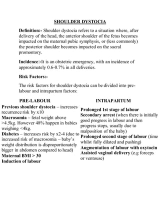
Definition:- Shoulder dystocia refers to a situation where, after
delivery of the head, the anterior shoulder of the fetus becomes
impacted on the maternal pubic symphysis, or (less commonly)
the posterior shoulder becomes impacted on the sacral
promontory.
Incidence:-It is an obstetric emergency, with an incidence of
approximately 0.6-0.7% in all deliveries.
Risk Factors:-
The risk factors for shoulder dystocia can be divided into pre-
labour and intrapartum factors:
PRE-LABOUR INTRAPARTUM
Previous shoulder dystocia – increases
recurrence risk by x10
Macrosomia – fetal weight above
>4.5kg. However 48% happen in babies
weighing <4kg.
Diabetes – increases risk by x2-4 (due to
increased risk of macrosomia – baby’s
weight distribution is disproportionately
bigger in abdomen compared to head)
Maternal BMI > 30
Induction of labour
Prolonged 1st stage of labour
Secondary arrest (when there is initially
good progress in labour and then
progress stops, usually due to
malposition of the baby)
Prolonged second stage of labour (time
whilst fully dilated and pushing)
Augmentation of labour with oxytocin
Assisted vaginal delivery (e.g forceps
or ventouse)
2. Clinical Features:-
Shoulder dystocia is defined by a delay in delivery of the
shoulders following the head during a vaginal delivery with the
next contraction after using normal traction.
On examination, signs that may occur to aid the diagnosis are:
Difficulty in delivery of the fetal head or chin.
Failure of restitution – the fetal remains in the occipital-
anterior position after delivery by extension and therefore does
not ‘turn to look to the side’.
‘Turtle Neck‘ sign – the fetal head retracts slightly back into the
pelvis, so that the neck is no longer visible, akin to a turtle
retreated into its shell.
Management:-
The immediate steps in the management of shoulder dystocia
include:
Call for help – shoulder dystocia is an obstetric emergency (will
need senior obstetrician, senior midwife and paediatrician in
attendance).
Advise the mother to stop pushing – this can worsen the
impaction.
Avoid downwards traction on the fetal head (increases risk of
brachial plexus injury) – only use “routine” axial traction (i.e.
keep the head in line with the baby’s spine), and do not apply
fundal pressure (increases the risk of uterine rupture).
Consider episiotomy – this will not relieve obstruction but can
make access for maneuvers easier.
3. First Line Manoeuvres
McRoberts manoeuvre – hyperflex maternal hips (knees
to chest position) and tell the patient to stop pushing. This
widens the pelvic outlet by flattening the sacral promontory
and increasing the lumbosacral angle. This single
manoeuvre has a success rate of about 90% and is even
higher when combined with ‘suprapubic pressure’.
Suprapubic pressure is applied in either a sustained or
rocking fashion to apply pressure behind the anterior
shoulder to disimpact it from underneath the maternal
symphysis.
Second Line (‘Internal’) Manoeuvres
Posterior arm – insert hand posteriorly into sacral hollow
and grasp posterior arm to deliver.
Internal rotation (“corkscrew manoeuvre”) – apply
pressure simultaneously in front of one shoulder and behind
the other to move baby 180 degrees or into an oblique
position.
If the above manoeuvres fail then roll patient onto all
fours and repeat (this may widen the pelvic outlet as the
legs are abducted and flexed).
Further Manoeuvers
These are only to be considered when the above measures have
been unsuccessful, and are very rarely used in the UK:
Cleidotomy – fracturing the fetal clavicle.
Symphysiotomy – cutting the pubic symphysis.
4. Zavenelli – returning the fetal head to the pelvis for
delivery of the baby via caesarean section.
Post-Delivery
After delivery of the fetus, active management of the 3rd
stage of labour is recommended (due to increased risk of
PPH). A PR examination should be performed to exclude a
3rd degree tear.
Shoulder dystocia can be a traumatic
experience, particularly if the women does not have
regional anaesthesia.
Debrief the mother and birth partner(s), and advise them of
the risk of recurrence with any subsequent delivery.
Consider a physiotherapist review before discharge, as
women are at increased risk of pelvic floor
weakness/3rd
degree tear, musculoskeletal pain and
temporary nerve damage.
a paediatric review is recommended before discharge to
assess for brachial plexus injury, humeral fracture or
hypoxic brain injury.
Complications
Maternal
Postpartum hemorrhage
Rectovaginal fistula
Symphyseal separation or diathesis, with or without
5. transient femoral neuropathy
Third- or fourth-degree episiotomy or tear
Uterine rupture
Fetal
Brachial plexus palsy
Clavicle fracture
Fetal death
Fetal hypoxia, with or without permanent neurologic
damage
Fracture of the humerus
McRoberts maneuver and suprapubic pressure