1. Journées Plénières du GDR 3070 Physique de la cellule au tissu - 2015
Title:
A microfluidic platform to study vascular endothelial cell mechanotransduction
Authors:
Julie Lafaurie-Janvore, Elizabeth E. Antoine, Sidney J. Perkins, Avin Babataheri, Abdul I. Barakat
Abstract:
Atherosclerosis develops preferentially at arterial branches and bifurcations where blood flow is multi-
directional and highly disturbed. Elucidating the role that arterial blood flow plays in the development of the
disease requires understanding endothelial cell mechanotransduction and, in particular, how flow-derived
forces regulate endothelial cell function. An interesting in vivo observation is that endothelial cells are
elongated in athero-protected arterial regions and cuboidal in athero-prone regions. Probing relations among
cell shape, flow-derived mechanical forces, and predilection for atherosclerosis requires experimental
platforms that simultaneously enable control of cell shape, exposure of the cells to controlled flow
environments, and real-time imaging of cellular responses. To this end, we have built a microfluidic-based
system that allows independent control of various parameters governing cell mechanotransduction, including
the amplitude and direction of the flow, cell density, cell shape, and cell polarization relative to the flow. In
this platform, cell shape control is accomplished at either the single-cell or monolayer levels by using
adhesive micro-patterns of different dimensions, thus allowing the study of single cells as well as collective
cell behavior. The microfabricated substrate is coupled to a microfluidic chamber that allows precise control
of the flow field and thus of the shear stress exerted by the flow on endothelial cells. A key feature of the
platform is the ability to perform high resolution live-cell imaging during flow experiments. Intracellular
calcium mobilization upon flow application is an important endothelial cell response to flow-derived forces.
As a proof of concept, we have used the microfluidic platform to compare the responses of cuboidal primary
Bovine Aortic Endothelial Cells cultured on unpatterned surfaces with those of elongated cells cultured on
15 µm-wide adhesive line patterns that are oriented either parallel or orthogonal to the flow direction.
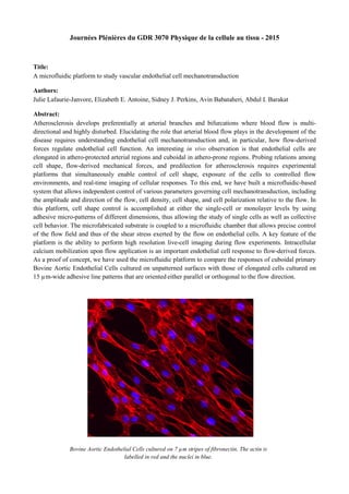
Bovine Aortic Endothelial Cells cultured on 7 µm stripes of fibronectin. The actin is
labelled in red and the nuclei in blue.