2. cellular players in glomerular disease, using selected
illustrative examples. Figure 2 summarizes some of
the ways in which glomerular injury can occur, with
individual diseases that will be discussed below and
in later articles in this series.
The Cellular Composition of the Glomerulus: Intrinsic
Glomerular Cells
Mesangial Cells: Matrix Homeostasis and a Glomerular
Scaffold
Mesangial cells provide support for the glomerular cap-
illary network and help maintain the homeostasis of the
mesangial matrix by secreting soluble factors. When injured,
mesangial cells can develop an activated phenotype or die
(via apoptosis or other mechanisms) (5). Circulating soluble
factors or metabolites can induce these responses directly, or
cause mesangial cells to secrete factors that elicit these
responses in an autocrine manner (6). In a process analogous
to wound healing, mesangial cell injury without ongoing
injurious stimuli may result in healthy remodelling of the
glomerulus, with mesangial cell migration, proliferation of
mesangial cell precursors in the juxta-glomerular apparatus,
and production of appropriate mesangial matrix (7).
Mesangial cell activation commonly results in hyper-
trophy and proliferation, excessive matrix production, and
the production of reactive oxygen species (5). Activated
mesangial cells produce chemokines and cytokines, which
act on mesangial cells themselves and on other resident
glomerular cells or leukocytes. These nearby cells in turn
secrete mediators that act on mesangial cells, forming a
paracrine loop (3). PDGFB is a potent mesangial cell mito-
gen. Its production by glomerular endothelial cells is es-
sential for mesangial cell development (5) and its
expression is upregulated in IgA nephropathy and other
proliferative forms of GN (8). Mesangial matrix expansion
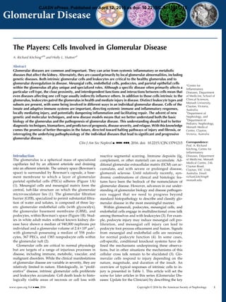
Figure 1. | Basic structure of the glomerulus and the glomerular filtration barrier. (A) Each glomerulus is composed of an afferent arteriole,
which supplies the glomerular capillaries, and an efferent arteriole, into which they drain. Mesangial cells and mesangial matrix provide
structural support for the glomerular capillaries, lined by specialized fenestrated endothelium, and then the glomerular basement membrane.
On the urinary side of the glomerular basement membrane are podocytes, with foot processes that wrap around the glomerular capillaries. The
urinary space is lined by a cup-like layer of parietal epithelial cells which adhere to the basement membrane of Bowman’s capsule. (B) The
glomerular filtration barrier is a specialized molecular sieve, with properties that aid filtration of small solutes from the blood to the urine, while
limiting the passage of macromolecules such as albumin.
2 Clinical Journal of the American Society of Nephrology
3. and the release of vasoactive mediators results in de-
creased glomerular surface area and altered glomerular
hemodynamics, with decreased GFR (3,5). If mesangial
cell activation is ongoing, ECM accumulation in the inter-
stitial space leads to interstitial fibrosis, followed by glo-
merulosclerosis (9).
Mesangial cells are targets both in immunologic injury
and in metabolic disease. Mesangial IgA deposition is the
hallmark of IgA nephropathy. In this disease, current models
imply a multihit pathogenesis with immune complexes
of anti-glycan autoantibodies and galactose-deficient
IgA1 being deposited in the mesangium, resulting in
mesangial cell injury and proliferation (10). Mesangial cell
hypertrophy and matrix expansion are histologic fea-
tures of diabetic nephropathy, mediated by metabolic
and hemodynamic changes in the setting of diabetes.
These include hyperglycemia, advanced glycation end
products, oxidized free fatty acids, and angiotensin II
(5). Acquired or genetic abnormalities of the GBM may
also influence the phenotype and activation status of
mesangial cells, with mesangial matrix expansion and
subsequent glomerular sclerosis, as in experimental Alport
disease (11).
Glomerular Endothelial Cells: Adapted for Selective
Permeability
Glomerular endothelial cells are uniquely adapted for
selective permeability and filtration. Although the glomerular
endothelium is continuous, it contains fenestrations, which
cover up to 50% of the glomerular surface area (12). On
conventional electron microscopy these fenestrations
appear as ovoid transcellular “holes”, 60–70 nm in
diameter (12). However, the fenestrations (and the glo-
merular endothelium itself) are covered by glycocalyx, a
carbohydrate-rich, gel-like mesh with important roles in
capillary permeability, regulation of the interactions be-
tween leukocytes and endothelial cells, and transduction
of shear stress (12).
In some disease states, endothelial injury leads to altered
microvascular permeability and albuminuria. Inflamma-
tory stimuli increase permeability by widening endothelial
cell-cell junctions, and in some instances, inducing trans-
cellular holes (13). Endothelial disease is a feature of rap-
idly progressive forms of GN, including ANCA-associated
GN, anti-GBM GN, and class 3 and 4 lupus nephritis.
However, reduced GFR also occurs in association with a
loss of endothelial fenestral area in other diseases, such as
preeclampsia (14) and diabetic nephropathy (15). Glomerular
endothelial cells are particularly vulnerable to injury medi-
ated by complement dysregulation, for example in atypical
hemolytic uremic syndrome (16). Podocyte-derived
vascular endothelial growth factor (VEGF) is critical
to the maintenance of endothelial cell structure and
function. Anti-VEGF therapies are associated with pro-
teinuria in humans, and mice with a podocyte-
specific VEGF deletion develop proteinuria (17). The
glomerular endotheliosis and proteinuria seen in preeclamp-
sia is associated with increased placental production of
soluble fms-like tyrosine kinase-1, an endogenous VEGF
antagonist (18).
The endothelial glycocalyx is predominantly composed
of proteoglycans, with their negatively charged glycos-
aminoglycan (GAG) chains covalently bound to the cell
surface, and sialoproteins, with adsorbed components
Table 1. Key functions and responses of intrinsic glomerular cells
Cell Type
Normal Function and
Features
Responses to Injury
Relevant Glomerular
Diseases (Examples)
Mesangial
cells
Maintain structural architecture
of glomerulus
Lysis with healthy remodeling IgA nephropathy
Mesangial matrix homeostasis
Apoptosis Diabetic nephropathy
Regulate filtration surface area
Hypertrophy
Phagocytose apoptotic cells
Proliferation and matrix expansion
leading to glomerulosclerosis
Glomerular
endothelial
cells
Fenestrations and glycocalyx
facilitate selective permeability
and filtration
Apoptosis ANCA-associated GN
Loss of fenestrations Lupus nephritis
(class 3 and 4)Widening of cell-cell junctions,
transcellular holes Hemolytic uremic
syndrome
Glycocalyx damage, loss of GAG
synthesis
Diabetic nephropathy
Podocytes Foot processes wrap around
capillaries
Apoptosis Minimal change disease
Adherence to GBM
Foot process effacement FSGS
Slit diaphragm regulates filtration
Detachment from GBM, podocyte
loss
Diabetic nephropathy
Loss of slit diaphragm
Parietal
epithelial
cells
Line Bowman’s capsule Apoptosis Crescentic GN
Several subsets of cells likely
with different functions
Migration to glomerular tuft,
production of ECM proteins
leading to glomerulosclerosis
FSGS
Subset of cells may be able to
differentiate into podocytes
and play a reparative function
Proliferation leading to crescent and
pseudocrescent formation
GAG, glycosaminoglycan; GBM, glomerular basement membrane; ECM, extracellular matrix.
Clin J Am Soc Nephrol ▪: ccc–ccc, ▪▪▪, 2016 Cells Involved in Glomerular Disease 3
4. Figure 2. | Simplified diagrammatic representation of a selection of mechanisms of glomerular injury. (A) Antibody-mediated glomerular
injury. From left to right, (i) neutrophils (shown) and macrophages induce injury after anti-a3(IV)NC1 autoantibodies bind to the GBM in anti-
GBM GN; (ii) in membranous glomerulopathy autoantibodies against PLA2R1 (and other antigens) on podocytes are deposited subepithelially,
with the involvement of complement; (iii) antibodies can bind to antigens lodged in the glomerulus (grey dots) with recruitment of macrophages
(shown) and neutrophils, and the activation of complement; (iv) circulating immune complexes can be deposited in glomeruli, activate
complement, and recruit leukocytes; (v) ANCA, (with complement) activates neutrophils and enables their recruitment to the glomerulus. Not
shown, but important, is IgA deposition in mesangial areas. (B) Cell-mediated immune mechanisms. (i) Effector CD41 cells (often Th1 or Th17
type) recognize antigens that can be intrinsic to or planted in the glomeruli. This occurs via their T cell receptor recognizing MHC class II
peptide complexes (several cell types could possibly be involved in this process). Activated T cells produce cytokines (IL-17A and IFN-g as
examples) that have direct effects on intrinsic kidney cells and activate, together with costimulatory molecules (e.g., CD154/CD40), innate
leukocytes such as macrophages. Not shown are interactions between intrinsic renal cells and T cells that include costimulation and cytokines.
(ii) CD81 cells can recognize antigenic peptides with MHC class I on intrinsic cells and secrete cytokines or induce cell death. (C) Metabolic,
vascular, and other mechanisms of injury. Podocyte and foot process injury and dysfunction occurs due to (i) genetic abnormalities of slit
diaphragm proteins and (ii) in minimal change disease and FSGS due to circulating permeability factors. Metabolic factors such as (iii) systemic
and intraglomerular hypertension and (iv) hyperglycemia and its consequences are common, and affect both the cells and the structural
components of the glomerulus. Both glomerular endothelial cell and podocyte injury are important consequences of preeclampsia, involved a
number of mediators including soluble fms-like tyrosine kinase-1. C3 glomerulopathy, as well as some types of atypical hemolytic uremic
syndrome (vi), can be induced by autoantibodies to, or genetic abnormalities in, complement regulatory proteins, resulting in complement
activation. a3(IV)NC1, the non-collagenous domain of the a3 chain of type IV collagen; FLT1, fms-like tyrosine kinase-1; GBM, glomerular
basement membrane; Mac, macrophage; M-type PLA2R1, phospholipase A2 receptor 1; Th, T helper; VEGF, vascular endothelial growth
factor.
4 Clinical Journal of the American Society of Nephrology
5. such as albumin (12). The net negative charge of the glyco-
calyx is thought to play a role in the charge selectivity of
the GFB, helping restrict the passage of the negatively
charged macromolecule, albumin (12). While some data
does not support this ‘charge selectivity’ theory (19), it is
likely that the glycocalyx, functioning as a hydrogel,
forms a physical barrier that is important for permselectiv-
ity (12,20). The development of albuminuria in experimen-
tal models of diabetes is associated with changes in the
glycocalyx, including loss of GAGs (12), which may occur
due to hyperglycemia-induced disruption of GAG synthe-
sis (21). Damage to any of the three layers of the GFB (en-
dothelium, GBM, or podocyte) can result in the presence of
high molecular weight proteins in the urine (22). However,
because changes in component of the GFB often affect the
other elements, the contribution of each individual com-
ponent to the filtration barrier is not easy to discern (22).
Additionally, even in health, a significant amount of al-
bumin may pass through the GFB and be retrieved by the
proximal tubule, mediated by the neonatal Fc receptor,
FcRn (19,23). The relative importance of these two
pathways in glomerular diseases remains the subject
of debate and may differ in individual diseases and
patients (23).
Glomerular endothelial cell injury may promote tubu-
lointerstitial fibrosis, leading to ESRD in CKD (24). Shear
stress is necessary for glomerular endothelial function and
with poor glomerular perfusion, decreased survival sig-
nals from endothelial cells induce regression and rarefac-
tion of the peritubular capillary network (24). This reduced
microvascular blood flow results in chronic tubulointersti-
tial hypoxia and fibrosis, with inflammation from damaged
tubular epithelial cells propagating further endothelial cell
injury (24).
Podocytes: Critical to Maintaining the Filtration Barrier
Podocytes possess multiple foot processes that wrap
around the glomerular capillaries, with filtration slits
between adjacent processes (25). Although sometimes de-
scribed as a specialized epithelial cell, the podocyte is a
uniquely differentiated cell with some mesenchymal fea-
tures (4,26). Diseased podocytes may exhibit actin cytoskel-
etal rearrangement, loss of the slit diaphragm, a more
cuboidal morphology (27), and may dedifferentiate in dis-
ease along epithelial or mesenchymal pathways, termed
‘podocyte disease transformation’ (26). Podocytes have lim-
ited capacity for repair or regeneration, with podocyte loss
being a feature of many conditions that lead to glomerulo-
sclerosis (28). As podocytes are lost from glomeruli, the
GFB is compromised; experimentally, when .20% of po-
docytes are lost, progressive glomerulosclerosis ensues
(29). Evidence that the extent of podocyte loss determines
outcome is consistent with the “podocyte depletion” hy-
pothesis (30). This hypothesis unifies a variety of glomeru-
lar diseases by postulating that the degree of podocyte
depletion induced by injurious processes is a critical deter-
minant of progression to glomerulosclerosis.
The term ‘podocytopathy’ describes disease that feature
podocyte dysfunction as the primary manifestation of the
disease process and that result in significant proteinuria.
Podocytopathies commonly occur due to circulating fac-
tors or inherited deficiencies of podocyte genes (4,31), and
include minimal change disease and FSGS. However, po-
docyte injury and loss can also occur due to other envi-
ronmental cues, including indirectly, due to abnormalities
in the paracrine, ECM, and GBM-mediated signaling nec-
essary for normal podocyte function (4,25). Both intact
junctions between podocyte foot processes at the slit di-
aphragm and adhesion of podocytes to the GBM are es-
sential for the function of the GFB (25). Podocyte
detachment occurs due to increased shear stress either in
glomerular hypertension or hyperfiltration, or to impaired
podocyte adhesion, more commonly occurring in inflam-
matory glomerular diseases (28). Podocyte adhesion to the
GBM involves a range of regulatory and scaffold proteins.
As well as serving as a mechanical anchor, adhesion is
an interface for the bidirectional transmission of signals
concerning cell growth, differentiation, and survival (32).
Podocyte transmembrane receptors bind to the GBM, with
the extracellular domain binding to a specific GBM protein
(e.g., collagen or laminin), and the intracellular domain re-
cruiting effector proteins linked to components of the podo-
cyte cytoskeleton, most commonly actin. Integrins are a
major family of such transmembrane receptors, with muta-
tions in integrins and changes in activation status implicated
in a number of diseases, including FSGS (33,34).
Podocyte foot processes are connected by the podocyte
intercellular junction, commonly called the slit diaphragm
due to its appearance on electron microscopy. This junction
is a necessary component of the GFB that determines
glomerular permeability characteristics. Within the kid-
ney, nephrin and podocin are unique to the podocyte and
essential for its function (25). Inactivating mutations of the
genes encoding nephrin and podocin causes nephrotic
syndrome with diffuse collapse of podocyte foot processes
(35,36). Both proteins are also involved in signal transduc-
tion (37), and signaling via adaptor proteins has a major
influence on the function of the podocyte actin cytoskele-
ton (25). Lastly, podocytes may act as “immune cells” (38)
that under some circumstances, present antigenic pep-
tides to CD41 T cells (39).
Glomerular PECs: Not Merely Lining Cells
PECs adhere in a monolayer to Bowman’s capsule, and
in humans are morphologically similar to squamous epi-
thelial cells (1). Several subpopulations of PECs exist in
humans, expressing combinations of podocyte, progenitor,
or tubular markers, although no consensus has been
reached on the nomenclature and function of these cell
types (1). Although podocytes and PECs are derived
from a common mesenchymal progenitor, PECs proliferate
under normal conditions, whereas terminally differenti-
ated podocytes have limited ability to regenerate (1).
Some studies suggest that some PECs can differentiate
into podocytes (1,40), supported by in vivo imaging studies
in mice showing not only migration of PECs to the visceral
layer of the glomerular tuft, but also nanotube “bridges”
between podocytes and PECs, and podocyte migration to
the Bowman’s capsule epithelial layer (41). While PECs
may be able to play a reparative role, they can also con-
tribute to injury after conversion. Activated PECs can pro-
liferate and contribute to crescent formation in rapidly
progressive GN (1), or participate in the formation of pseu-
docrescents and sclerotic lesions, as in certain forms of
Clin J Am Soc Nephrol ▪: ccc–ccc, ▪▪▪, 2016 Cells Involved in Glomerular Disease 5
6. FSGS, with PECs migrating to the glomerular tuft and pro-
ducing ECM matrix proteins (1).
Leukocytes and the Glomerulus in Health and Disease
Leukocytes participate in many forms of glomerular
disease. They can influence disease by inducing, damp-
ening, or resolving systemic immune responses that lead to
glomerular disease. Alternately, leukocytes may act locally
within the glomerulus to mediate inflammation, or poten-
tially to resolve inflammation and mediate repair, or
contribute to homeostasis within normal glomeruli. Fig-
ure 3 shows leukocytes within glomeruli in GN. The sys-
temic actions of a range of innate and adaptive leukocytes
are critical to glomerular diseases discussed in articles
later in this series. The pathogenesis of these diseases
are intimately related to leukocyte behavior and activity
systemically, for example, the loss of tolerance and the
generation of nephritogenic autoimmunity in secondary
lymphoid organs (42–46).
Leukocytes in the Normal Glomerulus
Cross-sections of glomeruli from humans without renal
disease show few leukocytes, suggesting that the glomerulus
may not, under steady state conditions, support substantial
leukocyte recruitment. However, viewing normal mouse
glomeruli in vivo in three dimensions over time shows that
leukocytes patrol the normal glomerulus (Figure 4A) (47).
Patrolling monocytes in glomeruli are phenotypically sim-
ilar to monocytes that survey other blood vessels, and
although they may have homeostatic functions, they
could also promote renal and glomerular inflammation
by sensing danger (48).
Glomerular Leukocyte Recruitment: Specialized and
Potentially Damaging
A variety of leukocytes are recruited to glomeruli in
inflammatory glomerular disease. Generally speaking,
leukocyte recruitment is mediated by adhesion molecules
and chemokines (Figure 4B). Adhesion of leukocytes in glo-
merular capillaries does not follow the traditional selectin-
mediated rolling paradigm that exists in postcapillary
venules in other tissues. On recruitment, leukocytes halt
suddenly in glomerular capillaries (49), with some re-
maining static and others migrating bidirectionally (47).
The specific adhesion molecules that mediate this arrest
and migration are context-dependent, both in terms of
the leukocyte type involved and the process inducing the
recruitment. Also critical to leukocyte recruitment are
chemokines, with some specialization in that specific
chemokines are associated with specific chemokine
receptor-expressing leukocytes (e.g., CXCL8/IL-8 and
CXCR2-expressing neutrophils). Activated products of
complement, C3a and C5a, are chemoattractants, while
other mediators are more specialized, e.g., leukocytes that
express Fcg receptors, monocyte/macrophages, and neutro-
phils, are recruited via IgG deposited in glomeruli (50).
Neutrophils and the Glomerulus: First Responders with a
Damaging Phenotype
Neutrophils, the most abundant immune cells, are first-
line responders in inflammation and in a variety of
Figure 3. | Leukocytes in glomeruli of patients with rapidly progressive GN. High powered photomicrographs as illustrative examples of
leukocyteswithinglomeruliinANCA-associatedGN. Inallpanelscell nucleiarestainedwith49,6-diamidino-2-phenylindole(blue).(A)CD451cells
(green, CD45isacommonleukocytemarker)in theglomerulus,(B) CD31Tcells(green), (C)CD681macrophages(green),and (D)myeloperoxidase
(red) expressing leukocytes (neutrophils or macrophages) in a segmental glomerular lesion with local loss of CD34 (green), an endothelial cell lesion.
Nephrin (marking podocytes) is in white. Magnification: (A), (C ) and (D) x400; (B) x600. Photomicrographs from Ms. Kim O’Sullivan.
6 Clinical Journal of the American Society of Nephrology
7. glomerular diseases. On activation, neutrophils produce
and release reactive oxygen species, proteases, cytokines,
and chemokines. Neutrophils are activated systemically
(51,52), or locally by proinflammatory mediators gener-
ated or deposited in the kidney, such as immune com-
plexes (47). Proinflammatory products of neutrophils
directly damage the GFB. In addition, in some forms of
GN (such as ANCA-associated GN and lupus nephritis)
neutrophils generate neutrophil extracellular traps, web-like
structures of histones decorated with proteases, peptides,
and enzymes that are also likely to be injurious (53). In
vivo microscopy reveals that a neutrophil can influence in-
jury over a considerable proportion of the glomerulus by
crawling within glomerular capillaries, in one disease
model covering an average of 150 mm/hour (47).
Monocytes, Macrophages, and the Glomerulus: Diverse
and Important
Innate cells of the monocyte/macrophage lineage have
multiple roles in tissues. Macrophages can adopt proin-
flammatory (M1) or anti-inflammatory (M2) phenotypes,
although these phenotypes exist on a continuum (46).
These phenotypes are plastic in vivo, with macrophages
responding to cues in the tissue microenvironment. In
the initial phases of experimental AKI, due to ischemia
reperfusion injury, macrophages in the interstitium are
proinflammatory and damaging (54). After the injurious
stimulus subsides, the same cells take on a healing and
reparative role (54). There is no intrinsic reason why intra-
glomerular leukocytes, in some situations, are not also
anti-inflammatory. While these reparative roles are
healthy if injury is self-limited, when pathologic processes
are ongoing, macrophages function as “frustrated” healing
cells and contribute to progressive fibrotic injury. In auto-
immune kidney disease, both humoral and cell-mediated
effector responses use macrophages as effector cells. Glo-
merular IgG activates macrophages via Fc-FcgR interac-
tions, while effector T helper 1 (Th1) and T helper 17
(Th17) cells activate these cells both by cytokines and via
cell-cell contact.
Figure 4. | Leukocyte recruitment and behavior in the glomerulus. (A) In health, neutrophils and monocytes (and potentially other leukocytes)
patrol the glomerulus. While some are static, the majority migrate bidirectionally within glomerular capillaries. (B) In acute disease, leukocytes
can be recruited and retained in glomeruli via a number of molecular processes. Examples are given with reference to the neutrophil (from left
to right). Neutrophils can be recruited via direct FcgR-Fc interactions. Adhesion molecules participate in recruitment, migration, and retention,
with, for example, Mac-1 (CD11b/CD18) on neutrophils slowing migration and inducing retention. P-selectins is not constitutively expressed
by glomerular endothelial cells, but in some situations P-selectin can participate in forming bridges with recruited platelets (pink oval) to
recruit neutrophils. Lastly, chemokines, for example CXCL8 (IL-8) secreted by endothelial cells, podocytes, mesangial cells (not shown), or
other leukocytes (not shown), attract leukocytes expressing appropriate chemokine receptors down a concentration gradient. Other
mechanisms, for example complement (not shown), can also attract leukocytes to the glomerulus. FcgR, Fcg receptor; ICAM-1, intercellular
adhesion molecule 1.
Clin J Am Soc Nephrol ▪: ccc–ccc, ▪▪▪, 2016 Cells Involved in Glomerular Disease 7
8. Dendritic Cells: The Interstitium, and the Glomerulus As
Well?
Systemically, dendritic cells (DCs), specialized leuko-
cytes that present antigen to T cells, play a key role in
adaptive immune responses that cause GN. In the kidney,
DCs form an extensive interstitial network (55), where they
help maintain tolerance to filtered self-peptides (56) and
act innately as pro- or anti-inflammatory renal mononu-
clear phagocytes. These interstitial DCs are largely acutely
protective. However, in inflammatory states, recruited in-
flammatory monocytes differentiate into proinflammatory
DCs, while activated resident DCs carry antigen to drain-
ing lymph nodes and activate naïve T cells. In contrast to
the interstitial DC network, DCs are rarely present in nor-
mal glomeruli (57) and only uncommonly in acute disease.
While human biopsy studies are divided as to the presence
of DCs in glomeruli in more established injury, in estab-
lished rapidly progressive GN, DCs within injured glo-
meruli may participate in T cell recruitment and
activation (43).
Mast Cells: More than Just Mediators of Allergy
While mast cells traditionally are considered as media-
tors of anaphylaxis and allergy, they are tunable cells that
contribute to, and regulate inflammation and autoimmu-
nity. In addition to histamine and other mediators of
allergy, mast cells secrete both stored and newly synthe-
sized pro- and anti-inflammatory cytokines. Mast cells
are present within the kidney in GN in both the tubuloin-
terstitium and the glomerulus. Intrarenal mast cells are
proinflammatory, with the capacity to degranulate (58),
while in secondary lymphoid organs they are anti-
inflammatory, acting with regulatory T cells to help maintain
tolerance (59,60).
T Lymphocytes: Subsets Directing Immunity and
Glomerular Disease
Conventional CD41 and CD81 T lymphocytes have a
wide range of ab T cell receptors that can recognize a huge
variety of antigenic peptides. T cells are instrumental in
protection from infectious threats but in disease, T cells
mediate a number of forms of inflammatory and autoim-
mune glomerular disease. CD41 cells orchestrate adaptive
immunity, differentiating into subsets that direct the sub-
sequent components of the immune response. These sub-
sets, while somewhat plastic, are definable by their
signature cytokines, transcription factors, and to some
degree, chemokine receptors (44). In glomerular disease,
T follicular helper cells promote T cell–dependent anti-
body production. In contrast, Th1 and Th17 cells recognize
antigens intrinsic to, or planted in glomeruli, and act as
local effectors of glomerular injury in experimental rapidly
progressive forms of GN (61). Th1 cells are particularly
associated with macrophage activation, and Th17 cells
are associated with neutrophil recruitment and activation.
Temporally, at least in responding to foreign antigens,
Th17 cells act earlier in immune responses and over time
convert to a mixed Th1/Th17 or Th1 phenotype (44). Th2
cells support eosinophils, important in allergic interstitial
nephritis and in glomerular disease in sub-Saharan Africa
(62). CD81 cells are cytotoxic, killing cells that express
their cognate peptide with MHC class 1, but also secrete
cytokines and may act in a proinflammatory manner (63).
B Lymphocytes: Antibodies As Key Mediators of Glomerular
Disease
B cells and their terminally differentiated progeny, antibody-
secreting plasma cells, are critical cells in humoral im-
munity. Within lymphoid organs, they produce antibodies
that impact the glomerulus in different ways in many forms
of GN. In autoimmunity and after some infections, Igs can
form circulating immune complexes that are deposited in
glomeruli, including IgA1 complexes in IgA nephropathy.
Alternately, in situ immune complexes form when anti-
bodies bind to antigens that are intrinsic to the glomerulus
or antigens that have been “planted” in the glomerulus. In
malignant conditions, such as multiple myeloma, single
clones of antibodies or light chains can be deposited in
glomeruli, while cryoglobulins (both IgM and IgG) lodge
in glomeruli in infectious, autoimmune, and malignant
diseases. Antibody production by the B cell/plasma cell
lineage is not the only possible role for B cells. In GN, B cell
aggregates in the interstitium could secrete cytokines and,
especially in established autoimmune responses, autoreactive
B cells may assist in antigen recognition by concentrating
autoantigenic peptides derived from autoantigens internalized
by their own cognate B cell receptor (64).
Immunoregulatory Immune Cells: T Cells, Macrophages,
and More
A variety of immune cells, including some CD41 and
CD81 T cells, macrophages, and DCs, dampen both sys-
temic immunity and local inflammation. The best de-
scribed subset is the CD41CD251foxp31 regulatory T
cell (Treg) that maintains tolerance and dampens inflam-
mation via soluble mediators and cell-cell contact. Both
endogenous and exogenous Tregs regulate experimental
glomerular disease (65,66) by inhibiting systemic immu-
nity, and probably also via local effects. Studies using reg-
ulatory CD81 T cells in experimental membranous
nephropathy illustrate their anti-inflammatory role (67),
with transfer of regulatory macrophages and DCs also lim-
iting experimental glomerular disease (68,69), including
progressive and chronic fibrotic injury. Given the potential
plasticity of many of these cells, future cell therapy trials
would need to consider the question of the stability of
transferred immune cells.
Innate Lymphoid Cells and Other T Lymphocytes:
Unconventional and Interesting
Other potentially important leukocytes include natural
killer (NK) cells, other innate lymphoid cells, and uncon-
ventional T cells: gd T cells, NK T cells, MR1-restricted
mucosal associated invariant T cells, and MHC class
1b–reactive T cells (70). Some unconventional T cells inter-
act with MHC class I/II-peptide complexes, but in a manner
distinct to that of classical T cells, while others recognize
glycolipids, vitamin B metabolites, and modified peptides.
Unlike conventional ab cells, whose T cell receptors un-
dergo somatic recombination, unconventional T cells have
relatively restricted patterns of antigen recognition (70).
Little is known about these cells in glomerular disease.
8 Clinical Journal of the American Society of Nephrology
9. However, for example, NK cells and their ligands are pre-
sent in the kidney in human lupus nephritis (71). Experi-
mentally, deficiency of invariant natural killer T cells
exacerbated GN (72,73), while proinflammatory gd T cells
are present in kidneys after passive antibody transfer (74).
Some of these subsets are particularly relevant to mucosal
immunity, with clear potential relevance not only for IgA
nephropathy but also for other forms of glomerular disease.
Intrinsic Glomerular Cells and Leukocytes Interact in
Glomerular Disease
Interactions between leukocytes and resident glomerular
cells in immune glomerular disease are dynamic and sub-
stantial, going beyond the simplistic paradigm of glomerular
cells merely being the target of leukocyte-mediated injury.
Cytokines, chemokines, and costimulatory molecules from
intrinsic glomerular cells activate (and regulate) leukocytes
locally, and it is possible that effector T cells recognize
antigenic peptides displayed by intrinsic glomerular cells.
There are many examples of these interactions: chemokines
derived from toll-like receptor 4–expressing glomerular
endothelial cells recruit neutrophils in experimental
ANCA-associated GN (75), while cytokines and costimula-
tory molecules expressed by intrinsic renal cells participate
in local activation of effector CD41 T cells in experimental
GN (76,77).
Summary and Conclusions
The glomerulus is vulnerable to injury from inflamma-
tory, metabolic, and other disease processes, which not
uncommonly involve the “dark side” of innate and adap-
tive immunity. In recent years we have made great prog-
ress in understanding glomerular disease. We now better
understand systemic disease processes that direct glomer-
ular injury; the local cellular, humoral, metabolic, genetic,
and molecular responses that induce injury; the detail of
the pathologic appearances of human glomerular disease;
and some of the prognostic indicators. This knowledge
allows us to take a more integrated view of glomerular
disease. Key challenges in the future include not only
identifying common local pathways for intervention and
regulating systemic processes that mediate glomerular
disease, but also developing more specific and less toxic
therapies specific to individual glomerular diseases, and
subsets of patients (or even individual patients) with a
specific disease.
Acknowledgments
Ms. Kim O’Sullivan is thanked for the photomicrographs in
Figure 3. We apologize to the many excellent researchers whose
work we have not discussed and cited.
Research performed by A.R.K in glomerular disease is funded by
Project Grants from the National Health and Medical Research
Council of Australia (NHMRC) (nos. 1048575, 1045065, 1046585,
1064112, and 1084869). H.L.H. is funded by an NHMRC Post-
graduate Scholarship (no. 1075304).
Disclosures
A.R.K. has been a member of an advisory board for Roche
Products, Australia. No Roche studies (that the authors are aware
of) are reported or discussed in the manuscript. H.L.H. declares no
conflicts of interest.
References
1. Shankland SJ, Smeets B, Pippin JW, Moeller MJ: The emergence
of the glomerular parietal epithelial cell. Nat Rev Nephrol 10:
158–173, 2014
2. Puelles VG, Douglas-Denton RN, Cullen-McEwen LA, Li J,
Hughson MD, Hoy WE, Kerr PG, Bertram JF: Podocyte Number in
Children and Adults: Associations with Glomerular Size and
Numbers of Other Glomerular Resident Cells. J Am Soc Nephrol
26: 2277–2288, 2015
3. Schlo¨ndorff D, Banas B: The mesangial cell revisited: no cell is an
island. J Am Soc Nephrol 20: 1179–1187, 2009
4. Saleem MA: One hundred ways to kill a podocyte. Nephrol Dial
Transplant 30: 1266–1271, 2015
5. Abboud HE: Mesangial cell biology. Exp Cell Res 318: 979–985,
2012
6. Simonson MS, Ismail-Beigi F: Endothelin-1 increases collagen
accumulation in renal mesangial cells by stimulating a chemo-
kine and cytokine autocrine signaling loop. J Biol Chem 286:
11003–11008, 2011
7. Watson S, Cailhier JF, Hughes J, Savill J: Apoptosis and glomer-
ulonephritis. Curr Dir Autoimmun 9: 188–204, 2006
8. Floege J, Eitner F, Alpers CE: A new look at platelet-derived
growth factor in renal disease. J Am Soc Nephrol 19: 12–23, 2008
9. Qian Y, Feldman E, Pennathur S, Kretzler M, Brosius FC 3rd: From
fibrosis to sclerosis: mechanisms of glomerulosclerosis in di-
abetic nephropathy. Diabetes 57: 1439–1445, 2008
10. Magistroni R, D’Agati VD, Appel GB, Kiryluk K: New develop-
ments in the genetics, pathogenesis, and therapy of IgA ne-
phropathy. Kidney Int 88: 974–989, 2015
11. Tanaka M, Asada M, Higashi AY, Nakamura J, Oguchi A, Tomita
M, Yamada S, Asada N, Takase M, Okuda T, Kawachi H,
Economides AN, Robertson E, Takahashi S, Sakurai T,
Goldschmeding R, Muso E, Fukatsu A, Kita T, Yanagita M: Loss of
the BMP antagonist USAG-1 ameliorates disease in a mouse
model of the progressive hereditary kidney disease Alport syn-
drome. J Clin Invest 120: 768–777, 2010
12. Salmon AH, Satchell SC: Endothelial glycocalyx dysfunction in
disease: albuminuria and increased microvascular permeability.
J Pathol 226: 562–574, 2012
13. Kumar P, Shen Q, Pivetti CD, Lee ES, Wu MH, Yuan SY: Molecular
mechanisms of endothelial hyperpermeability: implications in
inflammation. Expert Rev Mol Med 11: e19, 2009
14. Lafayette RA, Druzin M, Sibley R, Derby G, Malik T, Huie P,
Polhemus C, Deen WM, Myers BD: Nature of glomerular
dysfunction in pre-eclampsia. Kidney Int 54: 1240–1249,
1998
15. Toyoda M, Najafian B, Kim Y, Caramori ML, Mauer M: Podocyte
detachment and reduced glomerular capillary endothelial
fenestration in human type 1 diabetic nephropathy. Diabetes
56: 2155–2160, 2007
16. Nester CM, Barbour T, de Cordoba SR, Dragon-Durey MA,
Fremeaux-Bacchi V, Goodship TH, Kavanagh D, Noris M,
Pickering M, Sanchez-Corral P, Skerka C, Zipfel P, Smith RJ:
Atypical aHUS: State of the art. Mol Immunol 67: 31–42, 2015
17. Eremina V, Jefferson JA, Kowalewska J, Hochster H, Haas M,
Weisstuch J, Richardson C, Kopp JB, Kabir MG, Backx PH,
Gerber HP, Ferrara N, Barisoni L, Alpers CE, Quaggin SE: VEGF
inhibition and renal thrombotic microangiopathy. N Engl J Med
358: 1129–1136, 2008
18. Fan X, Rai A, Kambham N, Sung JF, Singh N, Petitt M, Dhal S,
Agrawal R, Sutton RE, Druzin ML, Gambhir SS, Ambati BK,
Cross JC, Nayak NR: Endometrial VEGF induces placental sFLT1
and leads to pregnancy complications. J Clin Invest 124: 4941–
4952, 2014
19. Comper WD: The limited role of the glomerular endothelial cell
glycocalyx as a barrier to transglomerular albumin transport.
Connect Tissue Res 55: 2–7, 2014
20. Dane MJ, van den Berg BM, Lee DH, Boels MG, Tiemeier GL,
Avramut MC, van Zonneveld AJ, van der Vlag J, Vink H, Rabelink
TJ: A microscopic view on the renal endothelial glycocalyx. Am J
Physiol Renal Physiol 308: F956–F966, 2015
Clin J Am Soc Nephrol ▪: ccc–ccc, ▪▪▪, 2016 Cells Involved in Glomerular Disease 9
10. 21. Singh A, Fride´n V, Dasgupta I, Foster RR, Welsh GI, Tooke JE,
Haraldsson B, Mathieson PW, Satchell SC: High glucose
causes dysfunction of the human glomerular endothelial
glycocalyx. Am J Physiol Renal Physiol 300: F40–F48,
2011
22. Garg P, Rabelink T: Glomerular proteinuria: a complex interplay
between unique players. Adv Chronic Kidney Dis 18: 233–242,
2011
23. Dickson LE, Wagner MC, Sandoval RM, Molitoris BA: The
proximal tubule and albuminuria: really! J Am Soc Nephrol 25:
443–453, 2014
24. Ballermann BJ, Obeidat M: Tipping the balance from angiogen-
esis to fibrosis in CKD. Kidney Int Suppl (2011) 4: 45–52, 2014
25. Lennon R, Randles MJ, Humphries MJ: The importance of
podocyte adhesion for a healthy glomerulus. Front Endocrinol
(Lausanne) 5: 160, 2014
26. May CJ, Saleem M, Welsh GI: Podocyte dedifferentiation: a
specialized process for a specialized cell. Front Endocrinol
(Lausanne) 5: 148, 2014
27. Sedor JR, Madhavan SM, Kim JH, Konieczkowski M: Out on a
LIM: chronic kidney disease, podocyte phenotype and the
Wilm’s tumor interacting protein (WTIP). Trans Am Clin Climatol
Assoc 122: 184–197, 2011
28. Kriz W, Lemley KV: A potential role for mechanical forces in the
detachment of podocytes and the progression of CKD. J Am Soc
Nephrol 26: 258–269, 2015
29. Wharram BL, Goyal M, Wiggins JE, Sanden SK, Hussain S,
Filipiak WE, Saunders TL, Dysko RC, Kohno K, Holzman LB,
Wiggins RC: Podocyte depletion causes glomerulosclerosis:
diphtheria toxin-induced podocyte depletion in rats expressing
human diphtheria toxin receptor transgene. J Am Soc Nephrol
16: 2941–2952, 2005
30. Wiggins RC: The spectrum of podocytopathies: a unifying view of
glomerular diseases. Kidney Int 71: 1205–1214, 2007
31. Lal MA, Young KW, Andag U: Targeting the podocyte to treat
glomerular kidney disease. Drug Discov Today 20: 1228–1234,
2015
32. Hynes RO: The extracellular matrix: not just pretty fibrils.
Science 326: 1216–1219, 2009
33. Has C, Sparta` G, Kiritsi D, Weibel L, Moeller A, Vega-Warner V,
Waters A, He Y, Anikster Y, Esser P, Straub BK, Hausser I,
Bockenhauer D, Dekel B, Hildebrandt F, Bruckner-Tuderman L,
Laube GF: Integrin a3 mutations with kidney, lung, and skin
disease. N Engl J Med 366: 1508–1514, 2012
34. Karamatic Crew V, Burton N, Kagan A, Green CA, Levene C,
Flinter F, Brady RL, Daniels G, Anstee DJ: CD151, the first
member of the tetraspanin (TM4) superfamily detected on
erythrocytes, is essential for the correct assembly of human
basement membranes in kidney and skin. Blood 104: 2217–
2223, 2004
35. Kestila¨ M, Lenkkeri U, Ma¨nnikko¨ M, Lamerdin J, McCready P,
Putaala H, Ruotsalainen V, Morita T, Nissinen M, Herva R,
Kashtan CE, Peltonen L, Holmberg C, Olsen A, Tryggvason K:
Positionally cloned gene for a novel glomerular protein–
nephrin–is mutated in congenital nephrotic syndrome. Mol Cell
1: 575–582, 1998
36. Boute N, Gribouval O, Roselli S, Benessy F, Lee H, Fuchshuber A,
Dahan K, Gubler MC, Niaudet P, Antignac C: NPHS2, encoding
the glomerular protein podocin, is mutated in autosomal re-
cessive steroid-resistant nephrotic syndrome. Nat Genet 24:
349–354, 2000
37. New LA, Martin CE, Jones N: Advances in slit diaphragm sig-
naling. Curr Opin Nephrol Hypertens 23: 420–430, 2014
38. Reiser J, von Gersdorff G, Loos M, Oh J, Asanuma K, Giardino L,
Rastaldi MP, Calvaresi N, Watanabe H, Schwarz K, Faul C,
Kretzler M, Davidson A, Sugimoto H, Kalluri R, Sharpe AH,
Kreidberg JA, Mundel P: Induction of B7-1 in podocytes is as-
sociated with nephrotic syndrome. J Clin Invest 113: 1390–1397,
2004
39. Goldwich A, Burkard M, Olke M, Daniel C, Amann K, Hugo C,
Kurts C, Steinkasserer A, Gessner A: Podocytes are non-
hematopoietic professional antigen-presenting cells. J Am Soc
Nephrol 24: 906–916, 2013
40. Appel D, Kershaw DB, Smeets B, Yuan G, Fuss A, Frye B, Elger M,
Kriz W, Floege J, Moeller MJ: Recruitment of podocytes from
glomerular parietal epithelial cells. J Am Soc Nephrol 20:
333–343, 2009
41. Hackl MJ, Burford JL, Villanueva K, Lam L, Suszta´k K, Schermer B,
Benzing T, Peti-Peterdi J: Tracking the fate of glomerular epithelial
cells in vivo using serial multiphoton imaging in new mouse
models with fluorescent lineage tags. Nat Med 19: 1661–1666,
2013
42. Holdsworth SR, Kitching AR, Tipping PG: Th1 and Th2 T helper
cell subsets affect patterns of injury and outcomes in glomeru-
lonephritis. Kidney Int 55: 1198–1216, 1999
43. Kitching AR: Dendritic cells in progressive renal disease: some
answers, many questions. Nephrol Dial Transplant 29: 2185–
2193, 2014
44. Kitching AR, Holdsworth SR: The emergence of TH17 cells as
effectors of renal injury. J Am Soc Nephrol 22: 235–238, 2011
45. Kitching AR, Holdsworth SR, Hickey MJ: Targeting leukocytes in
immune glomerular diseases. Curr Med Chem 15: 448–458,
2008
46. Rogers NM, Ferenbach DA, Isenberg JS, Thomson AW, Hughes J:
Dendritic cells and macrophages in the kidney: a spectrum of
good and evil. Nat Rev Nephrol 10: 625–643, 2014
47. Devi S, Li A, Westhorpe CL, Lo CY, Abeynaike LD, Snelgrove SL,
Hall P, Ooi JD, Sobey CG, Kitching AR, Hickey MJ: Multiphoton
imaging reveals a new leukocyte recruitment paradigm in the
glomerulus. Nat Med 19: 107–112, 2013
48. Carlin LM, Stamatiades EG, Auffray C, Hanna RN, Glover L,
Vizcay-Barrena G, Hedrick CC, Cook HT, Diebold S, Geissmann
F: Nr4a1-dependent Ly6C(low) monocytes monitor endothelial
cells and orchestrate their disposal. Cell 153: 362–375, 2013
49. Kuligowski MP, Kitching AR, Hickey MJ: Leukocyte recruitment
to the inflamed glomerulus: a critical role for platelet-derived
P-selectin in the absence of rolling. J Immunol 176: 6991–6999,
2006
50. Coxon A, Cullere X, Knight S, Sethi S, Wakelin MW, Stavrakis G,
Luscinskas FW, Mayadas TN: Fc gamma RIII mediates neutrophil
recruitment to immune complexes. a mechanism for neutrophil
accumulation in immune-mediated inflammation. Immunity 14:
693–704, 2001
51. Falk RJ, Terrell RS, Charles LA, Jennette JC: Anti-neutrophil cy-
toplasmic autoantibodies induce neutrophils to degranulate and
produce oxygen radicals in vitro. Proc Natl Acad Sci U S A 87:
4115–4119, 1990
52. Xiao H, Heeringa P, Hu P, Liu Z, Zhao M, Aratani Y, Maeda N,
Falk RJ, Jennette JC: Antineutrophil cytoplasmic autoantibodies
specific for myeloperoxidase cause glomerulonephritis and
vasculitis in mice. J Clin Invest 110: 955–963, 2002
53. Kessenbrock K, Krumbholz M, Scho¨nermarck U, Back W, Gross
WL, Werb Z, Gro¨ne HJ, Brinkmann V, Jenne DE: Netting neu-
trophils in autoimmune small-vessel vasculitis. Nat Med 15:
623–625, 2009
54. Lee S, Huen S, Nishio H, Nishio S, Lee HK, Choi BS, Ruhrberg C,
Cantley LG: Distinct macrophage phenotypes contribute to
kidney injury and repair. J Am Soc Nephrol 22: 317–326, 2011
55. Soos TJ, Sims TN, Barisoni L, Lin K, Littman DR, Dustin ML,
Nelson PJ: CX3CR11 interstitial dendritic cells form a contigu-
ous network throughout the entire kidney. Kidney Int 70: 591–
596, 2006
56. Lukacs-Kornek V, Burgdorf S, Diehl L, Specht S, Kornek M, Kurts C:
The kidney-renal lymph node-system contributes to cross-
tolerance against innocuous circulating antigen. J Immunol
180: 706–715, 2008
57. Schwarz M, Taubitz A, Eltrich N, Mulay SR, Allam R, Vielhauer V:
Analysis of TNF-mediated recruitment and activation of glo-
merular dendritic cells in mouse kidneys by compartment-
specific flow cytometry. Kidney Int 84: 116–129, 2013
58. Scandiuzzi L, Beghdadi W, Daugas E, Abrink M, Tiwari N,
Brochetta C, Claver J, Arouche N, Zang X, Pretolani M, Monteiro
RC, Pejler G, Blank U: Mouse mast cell protease-4 deteriorates
renal function by contributing to inflammation and fibrosis in
immune complex-mediated glomerulonephritis. J Immunol 185:
624–633, 2010
59. Eller K, Wolf D, Huber JM, Metz M, Mayer G, McKenzie AN,
Maurer M, Rosenkranz AR, Wolf AM: IL-9 production by regu-
latory T cells recruits mast cells that are essential for regulatory T
cell-induced immune suppression. J Immunol 186: 83–91, 2011
10 Clinical Journal of the American Society of Nephrology
11. 60. Gan PY, Summers SA, Ooi JD, O’Sullivan KM, Tan DS, Muljadi
RC, Odobasic D, Kitching AR, Holdsworth SR: Mast cells
contribute to peripheral tolerance and attenuate autoimmune
vasculitis. J Am Soc Nephrol 23: 1955–1966, 2012
61. Summers SA, Steinmetz OM, Li M, Kausman JY, Semple T,
Edgtton KL, Borza DB, Braley H, Holdsworth SR, Kitching AR:
Th1 and Th17 cells induce proliferative glomerulonephritis. J Am
Soc Nephrol 20: 2518–2524, 2009
62. Walker A, Ellis J, Irama M, Senkungu J, Nansera D, Axton J,
Coward RJ, Peat DS, Bode HH, Mathieson PW: Eosinophilic
glomerulonephritis in children in Southwestern Uganda. Kidney
Int 71: 569–573, 2007
63. Reynolds J, Norgan VA, Bhambra U, Smith J, Cook HT, Pusey CD:
Anti-CD8 monoclonal antibody therapy is effective in the pre-
vention and treatment of experimental autoimmune glomerulo-
nephritis. J Am Soc Nephrol 13: 359–369, 2002
64. Steinmetz OM, Velden J, Kneissler U, Marx M, Klein A,
Helmchen U, Stahl RA, Panzer U: Analysis and classification of
B-cell infiltrates in lupus and ANCA-associated nephritis. Kidney
Int 74: 448–457, 2008
65. Wolf D, Hochegger K, Wolf AM, Rumpold HF, Gastl G, Tilg H,
Mayer G, Gunsilius E, Rosenkranz AR: CD41CD251 regulatory
T cells inhibit experimental anti-glomerular basement mem-
brane glomerulonephritis in mice. J Am Soc Nephrol 16: 1360–
1370, 2005
66. Ooi JD, Snelgrove SL, Engel DR, Hochheiser K, Ludwig-Portugall I,
Nozaki Y, O’Sullivan KM, Hickey MJ, Holdsworth SR, Kurts C,
Kitching AR: Endogenous foxp3(1) T-regulatory cells suppress
anti-glomerular basement membrane nephritis. Kidney Int 79:
977–986, 2011
67. Wang YM, Zhang GY, Hu M, Polhill T, Sawyer A, Zhou JJ, Saito M,
Watson D, Wu H, Wang Y, Wang XM, Wang Y, Harris DC,
Alexander SI: CD81 regulatory T cells induced by T cell vacci-
nation protect against autoimmune nephritis. J Am Soc Nephrol
23: 1058–1067, 2012
68. Zheng D, Cao Q, Lee VW, Wang Y, Zheng G, Wang Y, Tan TK,
Wang C, Alexander SI, Harris DC, Wang Y: Lipopolysaccharide-
pretreated plasmacytoid dendritic cells ameliorate experimental
chronic kidney disease. Kidney Int 81: 892–902, 2012
69. Wang Y, Wang YP, Zheng G, Lee VW, Ouyang L, Chang DH,
Mahajan D, Coombs J, Wang YM, Alexander SI, Harris DC: Ex
vivo programmed macrophages ameliorate experimental
chronic inflammatory renal disease. Kidney Int 72: 290–299,
2007
70. Godfrey DI, Uldrich AP, McCluskey J, Rossjohn J, Moody DB: The
burgeoning family of unconventional T cells. Nat Immunol 16:
1114–1123, 2015
71. Spada R, Rojas JM, Pe´rez-Yagu¨e S, Mulens V, Cannata-Ortiz P,
Bragado R, Barber DF: NKG2D ligand overexpression in lupus
nephritis correlates with increased NK cell activity and differ-
entiation in kidneys but not in the periphery. J Leukoc Biol 97:
583–598, 2015
72. Yang JQ, Wen X, Liu H, Folayan G, Dong X, Zhou M, Van Kaer L,
Singh RR: Examining the role of CD1d and natural killer T cells in
the development of nephritis in a genetically susceptible lupus
model. Arthritis Rheum 56: 1219–1233, 2007
73. Mesnard L, Keller AC, Michel ML, Vandermeersch S, Rafat C,
Letavernier E, Tillet Y, Rondeau E, Leite-de-Moraes MC: Invariant
natural killer T cells and TGF-beta attenuate anti-GBM glomer-
ulonephritis. J Am Soc Nephrol 20: 1282–1292, 2009
74. Turner JE, Krebs C, Tittel AP, Paust HJ, Meyer-Schwesinger C,
Bennstein SB, Steinmetz OM, Prinz I, Magnus T, Korn T, Stahl RA,
Kurts C, Panzer U: IL-17A production by renal gd T cells promotes
kidney injury in crescentic GN. J Am Soc Nephrol 23: 1486–
1495, 2012
75. Summers SA, van der Veen BS, O’Sullivan KM, Gan PY, Ooi JD,
Heeringa P, Satchell SC, Mathieson PW, Saleem MA,
Visvanathan K, Holdsworth SR, Kitching AR: Intrinsic renal cell
and leukocyte-derived TLR4 aggravate experimental anti-MPO
glomerulonephritis. Kidney Int 78: 1263–1274, 2010
76. Ruth AJ, Kitching AR, Semple TJ, Tipping PG, Holdsworth SR:
Intrinsic renal cell expression of CD40 directs Th1 effectors in-
ducing experimental crescentic glomerulonephritis. J Am Soc
Nephrol 14: 2813–2822, 2003
77. Timoshanko JR, Kitching AR, Holdsworth SR, Tipping PG: In-
terleukin-12 from intrinsic cells is an effector of renal injury in
crescentic glomerulonephritis. J Am Soc Nephrol 12: 464–471,
2001
Published online ahead of print. Publication date available at www.
cjasn.org.
Clin J Am Soc Nephrol ▪: ccc–ccc, ▪▪▪, 2016 Cells Involved in Glomerular Disease 11