Recommended

Recommended
More Related Content
What's hot
What's hot (18)
Similar to Tumor activation alters endothelial cell mechano-responsiveness and drug sensitivity
Similar to Tumor activation alters endothelial cell mechano-responsiveness and drug sensitivity (20)
Tumor activation alters endothelial cell mechano-responsiveness and drug sensitivity
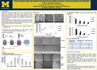
- 1. Tumor activation alters the mechano-responsiveness, capillary formation, and drug sensitivity of endothelial cells in synthetic matrices Yang Wu, Bingxin Guo, and Gargi Ghosh Bioengineering Program, Department of Mechanical Engineering University of Michigan - Dearborn Introduction Solid cancers induce the formation of new blood vessels to promote growth and metastasis. Past efforts have been focused on characterizing the altered growth factor signaling pathway in tumor capillary endothelial cells; however, the mechanical microenvironment of tumor also plays a significant role in regulating the formation of vascular patterns. Here, we used synthetic hydrogel based cell culture platforms to probe how activation of human umbilical endothelial cells (HUVECs) by tumor secreted factors alters the responses to matrix modulus and in turn the capillary network formation and drug sensitivity. 2).Poro size 3). Diffusion 4) . Morphology images in HR and HCR media 5) . Sprout images on with and W/O cancer cells scaffolds Goals Understand how tumor activation affect mechanosensitivity of endothelial cells Understand how matrix stiffness and tumor activation affect response of endothelial cells to vandetanib inhibition Materials and Methods Cancer cells Pre-polymer solution Cancer cell laden scaffolds UV curing Addition of HUVECs on the top of cell laden scaffolds Capillary sprouts + Results 11 kPa 78 kPa36 kPa 0 200 400 600 800 1000 1200 0 10 20 30 Cummulativerelease ofFITC-dextran(ng) Time (hour) 11 kPa 36 kPa 78 kPa Conclusion/ Future Studies Acknowledgement Authors would like to thank University of Michigan, Dearborn and Office of Vice President of Research, University of Michigan, Ann Arbor for their financial support Our study revealed that while in absence of activation, HUVECs prefer a substrate of appropriate stiffness for optimal capillary network formation; tumor activation disrupts the mechano- responsive behavior of HUVECs. on reducing the capillary network was also investigated. The response of HUVECs to the anti-angiogenic agent was substrate modulus dependent displaying increased sensitivity on the compliant gels. 6). Vandetanib inhibition on with and W/O cancer cells scaffolds 7). IC50 of Vandetanib (µM) 1. Scaffold characterization 1). Compression modulus and swelling ratio 0 5 10 15 20 25 11 36 78 SwellingRatio Compression Modulus (kPa) 0 10 20 30 40 50 60 70 80 90 5% 10% 15% Compression modulus(KPa) Concentration of PEGDA Compression Modulus (kPa) Without cancer cells With cancer cells 11 0.14 0.24 36 0.16 0.36 78 0.21 0.57 11 KPa 36 KPa 78 KPa 36 KPa11 KPa 78 KPa * * * * * * * * * * * * * * * ** * * * * * * 11 KPa 36 KPa 78 KPa 36 KPa11 KPa 78 KPa HR media HCR media With cancer cells Without cancer cells 0 1 2 3 4 5 0 0.5 1 2 Sproutspernodes (withcancercells) Drug concentration ( µM) 11 kPa 36 kPa 78 kPa 0 0.5 1 1.5 2 2.5 3 3.5 4 0 0.5 1 2 Sproutspernodes (withoutcancercells) Drug concentrtion (µM) 11 kPa 36 kPa 78 kPa