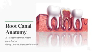
2. Introduction
The entire internal space or central cavity
within a tooth is entirely enclosed by dentin
except at the apical foramen
It is divided into-
• Coronal Portion- PulpChamber
• Radicular Portion – RootCanal
Pulp
Chamber
Root
Canals
2
4. Coronal portion i.e pulp chamber
reflects the external form of crown
Pulp Horns : Pulp horns are landmarks
present occlusal to pulp chamber
The roof of pulp chamber consists of
dentin covering the pulp chamber
occlusally or incisally
The floor of pulp chamber merges into
the root canal at the orifices.Thus, canal
orifices are the openings in the floor of
pulp chamber leading into the root
canals
PulpChamber
4
5. Canal Orifice: Canal orifices are openings in
the floor of pulp chamber leading into root
canals
PulpChamber
5
6. The root canal extends from canal orifice to
the apical foramen
RootCanal
6
7. It is based on anatomic and histological
landmarks in the apical part of the root
canal
• ApicalConstriction ( Minor Diameter)
• Apical Foramen (Major Diameter)
• Cementodentinal junction
• Apical Delta
• Accessory Foramen
• LateralCanals
• Bifurcation/TrifurcationCanals
Apical Root Anatomy
7
8. BEST FOR You
O R G A N I C S C O M P A N Y
Apical Constriction
» It is the apical portion of the root
canal having the narrowest
diameter which is located 0.5-
1mm short of the apical foramen
8
9. BEST FOR You
O R G A N I C S C O M P A N Y
Apical Foramen
» It is the main apical opening on the
root surface through which blood
vessels enter into the root canal
»The shape of the space between
the major and minor diameter has
described as-
• Funnel shaped
• Hyperbolic
• Morning glory
9
10. BEST FOR You
O R G A N I C S C O M P A N Y
Cementodentinal
junction
» It is the point in the canal where
cementum and dentin are united.
» It is approximately 0.1mm away
from the apical foramen
1
0
11. BEST FOR You
O R G A N I C S C O M P A N Y
Apical Delta
» Opening of accessory and lateral
canals in the root surface
AccessoryForamen
» It is a triangular area of the root
surrounded by main canal, accessory
canal and periradicular tissue
11
12. BEST FOR You
O R G A N I C S C O M P A N Y
AccessoryCanal
» Canal that branches from the main root canal.
» Most commonly seen in the apical third
» May also occur in bifurcation and trifurcation
area of multirooted tooth which are known as
furcation canal
Lateral canal
» Canals that are located approximately at right
angle to the main root canal
1
2
13. Clinical Significance of ApicalThird
Most of the curvature occurs in the
apical third and so must be prepared
very carefully
Should be prepared adequately so that the
irrigant can chemically debride the accessory
canal as instruments cannot reach there
13
14. Clinical Significance of ApicalThird
During obturation, the filling should end at the
apical constriction otherwise periapical
healing will be impaired
During periapical surgery apical 3mm of root
should be resected to eliminate the accessory
canals which lodge microorganism
14
17. Weine’s Classification
A single canal extends
from the pulp chamber to
the apex
Two separate canals
leaving the pulp chamber
but exiting as one canal
Two separate canals leaving
the chamber and exiting as
two separate foramina
One canal leaving the
chamber but dividing into
two separate canals and
exiting in two separate
foramina 17
19. Vertucci’s Classification
A single canal extends
from the pulp chamber to
the apex
Two separate canals leave
the pulp chamber and join
short of the apex to form
one canal
One canal leave the pulp
chamber and divides into two
in the root, the two then
merge to exit as one canal
Two separate, distinct
canals extends from the
pulp chamber to the apex
19
20. Vertucci’s Classification
One canal leaves the pulp
chamber divides and then
rejoins in the body of the root
and finally redivides into two
distinct canals short of the apex
Three separate, distinct
canals extend from the
pulp chamber to the
apex
Two separate canals leave
the pulp chamber, merge in
the body of the root and
redivide short of the apex to
exit as two distinct canals
One canal leaves the pulp
chamber and divides short
of the apex two separate,
distinct canals with
separate apical foramina 20
21. BEST FOR You
O R G A N I C S C O M P A N Y
1. Clinical methods
• Anatomystudies
• Radiographs
• Exploration
Methodsof determining pulpanatomy
21
22. BEST FOR You
O R G A N I C S C O M P A N Y
Methodsof determining pulpanatomy
2. InVitro methods
• Sectioning of teeth byCBCT
• Use of dyes
Pulpal tissue remnants fluorescing under blue curing
light, marking the presence of the canal orifices
22
Sectioning of tooth byCBCT
23. BEST FOR You
O R G A N I C S C O M P A N Y
23
Variations of pulp space
1. Variations in development
Fusion Concrescence Taurodontism
Dilacerations
Dentogenesis imperfectas
2. Variations in shape of pulp cavity
C-shaped canal
Curved canal Bayonet-shaped canal
24. BEST FOR You
O R G A N I C S C O M P A N Y
Variations of pulp space
1. Variations in pulp cavity due to pathology 1. Variations in apical third
Pulpstones Calcifications
Internal resorption External resorption
Accessoryand lateral canals
24
25. BEST FOR You
O R G A N I C S C O M P A N Y
Maxillary Central Incisor
Length of tooth
(mm)
Canal Lateral canals Root Curvature (%)
Average length 22.5 One canal 99.4% 24% Straight 75
Maximum length
25.6
Two canals 0.6% Distal curved 8
Minimum length
21.0
Mesial curved 4
Range 4.6 Labial curved 9
Lingual curved 4
25
26. Maxillary Central
Incisor
Pulp Chamber
Located in the center of the crown with equal
distance from the dentinal walls
Mesiodistally,The pulp chamber is ovoid in
shape
Buccopalatally, it is narrow
In young patient,Central incisor has three pulp
horns
PulpCanal
Pulp horn
22.5mm
26
27. BEST FOR You
O R G A N I C S C O M P A N Y
Root Canal
» It has one root with one root canal
» Root canal is broad labio-palatally,
conical in shape and centrally
located
» 17% cases show labial or palatal
curvature of the root
» Lateral canals present in about 24% ,
usually in the apical third area
27
28. BEST FOR You
O R G A N I C S C O M P A N Y
In cross-section,
• Cervical level:Canal is ovoid mesiodistally
• Middle root level:Canal is ovoid to round
• Apical third level:Canal is generally round
in shape
28
29. BEST FOR You
O R G A N I C S C O M P A N Y
Maxillary Lateral Incisors
Length of tooth (mm) Canal Lateral canals Root Curvature (%)
Average length 21 One canal 93.4% 10% Straight 30
Maximum length 25.1 Two canals 6.6% Distal curved 53
Minimum length 20.5 Mesial curved 3
Range 4.6 Labial curved 4
Bayonet and gradual
curve 6
29
30. Maxillary Lateral
Incisor
Pulp Chamber
The shape of the pulp chamber is similar to the
maxillary central incisor
It has two pulp horns, corresponding to the
development mammelons
21mm
30
31. BEST FOR You
O R G A N I C S C O M P A N Y
Root Canal
» Root canal has finer diameter than that
of central incisor through shape is
similar to that
» The canal is wider labiopalatally
» Apical region of the canal is usually
curved in a palatal direction
31
32. BEST FOR You
O R G A N I C S C O M P A N Y
In cross-section,
• Cervical level:Canal is ovoid labiopalatally
• Middle third level:Canal is ovoid
• Apical third level:Canal is generally round
in shape
32
33. BEST FOR You
O R G A N I C S C O M P A N Y
Maxillary Canines
Length of tooth
(mm)
Canal Lateral canals Root Curvature (%)
Average length 26.5 One canal 96.5% 24% Straight 39
Maximum length 28.9 Two canals 3.5% Distal curved 32
Minimum length 23.1 Mesial curved 0
Range 5.8 Labial curved 13
Lingual curved 7
Bayonet and gradual
curve 7
33
34. Maxillary Canines
Pulp Chamber
Labiopalatally, the pulp chamber is
almost triangular shape
Mesiodistally, it is narrow
Usually one pulp horn is present
26.5mm
34
35. BEST FOR You
O R G A N I C S C O M P A N Y
Root Canal
» There is single root canal which is
wider labiopalatally than in
mesiodistal aspect
» Canal is usually straight but may
show a distal apical curvature
35
36. BEST FOR You
O R G A N I C S C O M P A N Y
In cross-section,
• Cervical and middle third level:Canal is
ovoid in shape
• Apical third level:At apex it becomes
circular
36
37. BEST FOR You
O R G A N I C S C O M P A N Y
Maxillary First Premolars
23.8 foramen 13
18.8 foramen 72
Curvature of roots
Length of Canal (%) Direction Double roots
tooth (mm) Single root Buccal Palatal
Average length One canal one Straight
21 foramen 9
38 28 45
Maximum length Two canalsOne Distal curved 37 14 14
Minimum length Two canalsTwo Mesial curved 0 0 0
Three canals
Range 5 Three foramen Labial curved
6
15 14 28
Lingual curved 3 36 9
Bayonet curve 0 8 0
37
38. Maxillary First
Premolars
Pulp Chamber
Pulp chamber is wider buccopalatally two pulp
horns; corresponding to buccal and palatal cusps
The roof of the pulp chamber is coronal to the
cervical line
Floor is convex generally with two canal orifices
21 mm
38
39. BEST FOR You
O R G A N I C S C O M P A N Y
Root Canal
» Two roots
»When fused roots, a groove running in
occlusoapical direction divides the root
buccal and palatal portions each
containing a single root canal
» The root canals are usually straight and
divergent
39
40. BEST FOR You
O R G A N I C S C O M P A N Y
In cross-section,
• Cervical level:Canal is ovoid in shape
• Middle and apical third level:Canals show
circular shape
40
41. BEST FOR You
O R G A N I C S C O M P A N Y
Maxillary Second Premolars
Length of tooth (mm) Canal (%) Root Curvature (%)
Average length 21.5 One canalOne foramen 75 Straight 9.5
Maximum length 23 Two canals Two foramen 24 Distal curved 27
Minimum length 19 Three canals 1 Mesial curved 1.6
Range 4 Buccal curved 12.7
Lingual curved 4.0
Bayonet curve 20.6
41
42. Maxillary Second
Premolars
Pulp Chamber
Pulp chamber is wider buccopalatally
Narrower mesiodistally
Pulp horn under each cusp, buccal pulp
horn more prominent
21.5 mm
42
43. BEST FOR You
O R G A N I C S C O M P A N Y
Root Canal
» In more than 60% cases, single root with
single canal is found
» If there are two canals, they may be
separated or distinct along the entire
length of the root
» Canal is wider buccopalatally forming
ribbon like shape
43
44. BEST FOR You
O R G A N I C S C O M P A N Y
In cross-section,
• Cervical level:Canal is ovoid and narrow in
shape
• Middle third level:Canal is ovoid
• Apical third level:At apex it becomes circular
44
45. R You
BEST FO
O R G A N I C S C O M P A N Y
Maxillary First Molars
Length
of tooth
(mm)`
Mesiob
uccal
(mm)
Distobu
ccal
(mm)
Palatal
(mm) Canal
(%)
Directio
n
Average
length
19.9 19.4 20.6
Three
41.1
Straight
Maximum
length
21.6 21.2 22.5 Four 56.5
Distal
curved
Minimum
length
Mesial
curved
Range
18.2 17.6 17.6 Five 2.4
3.4 3.6 3.8
Buccal
curved
Lingual
curved
Bayonet
curve
Curvature of roots
Mesial (%) Distal (%) Palatal (%)
Canals in
m
e
s
i
o
b
u
c
c
a
l
r
o
o
t
46. Maxillary First Molars
Pulp Chamber
Largest pulp chamber
Four pulp horns ; mesiobuccal, mesiopalatal,
distobuccal and distopalatal
Roof ; Rhomboidal in shape
Roof converges, palatal wall disappears and
forms a triangular form
21 mm
46
47. Maxillary First Molars
Pulp Chamber
Anatomic dark lines in the floor connect the
orifices
Orifices are located in the 3 angles of the floor
Mesiobuccal orifice under mesiobuccal cusp
May have depression in the palatal end of the
mesiobuccal orifice where a 4th canal may be
present
MB2 canal is located mesial to or directly on a
line between the MB1 and palatal orifice
47
48. BEST FOR You
O R G A N I C S C O M P A N Y
Root Canal
» Generally three roots with three or four
canals
» Two canals in mesiobuccal root are
closely interconnected and sometimes
merge into one canal
48
49. BEST FOR You
O R G A N I C S C O M P A N Y
Root Canal
» Mesiobuccal canal:
• Narrowest of the three canals
• Flattened in mesiodistal direction at cervix
but becomes round as it reaches apically
» Distobuccal canal:
• Narrow, tapering canal
• Flattened in mesiodistal direction but
generally it is round in cross- section
49
50. BEST FOR You
O R G A N I C S C O M P A N Y
Root Canal
» Palatal canal:
• Largest diameter
• In cross-section, rounded triangular
coronally and round apically
» Palatal canal can curve buccally in the
apical one-third
» Lateral canals are found in 40 percent of
the molars at apical third and at
trifurcation area
50
51. BEST FOR You
O R G A N I C S C O M P A N Y
Length
of tooth
(mm)`
Mesiob
uccal
(mm)
Distobu
ccal
(mm)
Palatal
(mm) Canal
(%)
Directio
n
Curvature of roots
Mesial (%) Distal (%) Palatal (%) Canals in
mesiobuccal
root
Average
length
20.2 19.4 20.8 Three 54 Straight 22 54 63
One canal one
foramen 63
Maximum
length
22.2 21.3 22.6 Fused 46
Distal
curved
54 0
Two canals
One foramen
13
Minimum
length
18.2 17.5 19.0
Mesial
curved
0 17 0
Two canals
Two foramen
24
Range 4.0 3.8 3.6
Buccal
curved
37
Lingual
curved
0
51
Maxillary Second Molars
52. Maxillary Second
molars
Pulp Chamber
Similar to maxillary 1st molar, except
narrower mesiodistally
Roof- Rhomboidal in shape
Floor-Obtuse triangle
Mesiobuccal and distobuccal canals closer
together
21 mm
52
53. BEST FOR You
O R G A N I C S C O M P A N Y
Root Canal
» Mesiobuccal root:
• Broad buccolingually
• Prominent depression in mesial and distal
surfaces
• 1 or 2 canals
» Distobuccal root:
• Rounded/Ovoid, single canal
• Orifice appears on same line joining
mesiobuccal and palatal canals
» Palatal root:
• Broad mesiodistally
• Ovoid ,single canal
53
54. BEST FOR You
O R G A N I C S C O M P A N Y
Conclusion
»Through knowledge of root canal anatomy and access cavity preparation will
enable the clinician to produce endodontic treatments of high quality and
considerable longevity
» A successful treatment outcome depends on the complete debridement and
disinfections of all canals
54
55. BEST FOR You
O R G A N I C S C O M P A N Y
You
Thank
For your attention. . .