SlideShare a Scribd company logo
1 of 94
ANATOMY OF PULP CAVITY OF
MAXILLARY TEETH
PRESENTED BY:
DR.HUMAIRA JABEEN TAMANNA
INTRODUCTION
WHAT IS DENTAL
PULP?
PULP
Pulp is a soft tissue of mesenchymal origin
residing within the pulp cavity.
PULP CAVITY
ā€¢ The entire internal space or central cavity
enclosed by dentine except at the apical
foramen.
ā€¢ It consists of the following entities.
ļƒ˜Pulp chamber
ļƒ˜Root canals
PULP CHAMBER
PARTS:
ļƒ˜ Roof
ļƒ˜ Pulp horns
A pulp horn is an accentuation of the pulp chamber
directly under a cusp or a developmental lobe.
ļƒ˜ Floor
ļƒ˜ Canal orifice
ROOT CANAL
Portion of the pulp cavity from the canal orifice
to the apical foramen
Consists of three sections:
ā€¢ Coronal
ā€¢ Middle
ā€¢ Apical
LATERAL AND ACCESSORY
CANALS
ā€¢ A lateral canal is a canal that is located at approximately
right angles to main root canal.
ā€¢ According to Green , the incidence of accessory
foramina ranged from 10% in maxillary central
incisor and mandibular cuspids to 47% .
ROOT CANAL ANATOMY
ā€¢ The number of canals in a tooth correlates to the
number of roots in a tooth.
ā€¢ The space inside the root canals is filled with a
highly vascularized and highly innervated loose
connective tissue , called the dental pulp.
ā€¢ The pulp tissue is in communication with the
periodontium and the rest of the body through the
apical foramen.
ā€¢ Usually a root canal has curvature or constriction
before terminating at apex.
APICAL ROOT ANATOMY
It is based on following anatomic and
histological landmarks in the apical part of
the root canal.
APICAL CONSTRICTION (MINOR
DIAMETER)
ā€¢ It has an apical part of root canal having
the narrowest diameter short of the apical
foramina.
ā€¢ It may or may not coincide with CDJ.
APICAL FORAMEN (MAJOR
DIAMETER)
ā€¢ It is main apical opening on the surface of root
canal through which blood vessels enter the
canal.
ā€¢ Its diameter is almost double the apical
constriction giving it a funnel shape appearance.
CEMENTODENTINAL JUNCTION
ā€¢ Cementodentinal junction is the point in the canal
where cementum meets dentin.
ā€¢ The position of CDJ varies but usually it lies 0.5-
3.0mm short of the anatomic apex.
APICAL DELTA
It is a triangular area of root surrounded by
main canal ,accessory canals and
periradicular tissue.
SIGNIFICANCE OF APICAL THIRD
ā€¢ The root canal treatment of apical part of root is
difficult sometimes because of presence of
accessory and lateral canals, pulp stones , varying
amounts of irregular secondary dentin and area of
resorption.
ā€¢ Most of the curvature occur in apical third.
ā€¢ Obturation should end at apical constriction.
ā€¢ Apical 3mm of root is generally resected during
endodontic surgery in order to eliminate canal
aberrations.
ISTHMUS
It is defined as narrow passage or anatomic
part connecting two larger structures. It is
commonly found in teeth with multiple
canals.
ā€¢ Isthumus is a narrow , ribbon shaped
communication between two root canals
which can be complete or incomplete.
ā€¢ It contains pulp or pulpally derived tissue
and acts as store house of bacteria so the
isthmus should be well prepared and filled if
seen on resected surface.
CLASSIFICATION:
Type I: two or three canals with no visible
communication(incomplete isthmus)
Type II: two canals showing definite connection with
two main canals.
Type III: three canals showing definite connection
with main canals.
Type IV: it is similar to type II or type III with canals
extending to isthmus area.
Type V: it is true connection throughout the section
of root.
The pulp canal system is complex, and canals
may branch, divide, and rejoin
(apical foramen).
Weine categorized the root canal
systems into four basic types:
ā€¢ Type I- Single canal from pulp chamber to
apex.
ā€¢ Type II- Two separate canals leaving the pulp
chamber but merging short of the apex to
form only one canal.
ā€¢ Type III- Two separate canals leaving the
chamber and exiting the root in separate
apical foramina.
ā€¢ Type IV- One canal leaving the pulp chamber
but dividing short of the apex into two
separate and distinct canals with separate
apical foramina.
Vertucci et al, identified eight pulp
space configurations, which briefly
can be described as follows :
ā€¢ Type I : A single canal extends from the
pulp chamber to the apex
ā€¢ Type II: Two separate canals leave the
pulp chamber and join short of the apex to
form one canal
ā€¢ Type III: One canal leaves the pulp chamber and
divides into two in the root; the two then merge to
exit as one canal
ā€¢ Type IV: Two separate, distinct canals extend
from the pulp chamber to the apex
ā€¢ Type V: One canal leaves the pulp chamber and
divides short of the apex into two separate,
distinct canals with separate apical foramina
ā€¢ Type VI: Two separate canals leave the pulp
chamber, merge in the body of the root, and
redivide short of the apex to exit as two distinct
canals .
ā€¢ Type VII: One canal leaves the pulp chamber,
divides and then rejoins in the body of the root,
and finally redivides into two distinct canals short
of the apex.
ā€¢ Type VIII: Three separate, distinct canals extend
from the pulp chamber to the apex.
METHODS OF DETERMINING PULP
ANATOMY
1. CLINICAL METHOD
ā€¢ Anatomy studies
ā€¢ Radiographs
ā€¢ Exploration
ā€¢ High resolution computed tomography
ā€¢ Visualization endogram
ā€¢ Fiber optic endoscope
ā€¢ Magnetic resonance imaging
2. IN VITRO METHODS
ā€¢ Sectioning of teeth
ā€¢ Use of dyes
ā€¢ Clearing of teeth
ā€¢ Contrasting media
ā€¢ Scanning electron microscopic analysis
CLINICAL METHODS
ANATOMIC STUDIES
The knowledge of anatomy gained from
various studies and books is commonly
used method.
RADIOGRAPHS
They are also useful in assessing the root
canal anatomy . Since , it is a two
dimensional picture of three dimensional
object , one has to analyze the radiograph
carefully.
HIGH RESOLUTION COMPUTED
TOMOGRAPHY
It shows three-dimensionl picture of root canal
system using computer image processing.
FIBER OPTIC ENDOSCOPE
It is used to visualize canal anatomy.
VISUALIZATION ENDOGRAM
In this technique , an irrigant is used which helps
in visualization of the canals on radiograph.
This solution is called Ruddleā€™s solution. After
injecting this solution into canal system ,
radiograph is taken to visualize the canal
anatomy.
MAGNETIC RESAONANCE IMAGING
It produces data on computer which helps in
knowing canal morphology.
EXPLORATION
On reaching pulpal floor one finds the
grooves and anatomic dark lines which
connect the canal orifices , this is called
dentinal map.
IN VITRO METHODS
SECTIONING
In this, teeth are sectioned longitudinally for
visualization of root canal system.
USE OF DYES
Methylene blue or fluorescein sodium
dyes(commonly used ) help in location
pulp tissue present in pulp chamber
because dyes stain any vital tissue present
in pulp chamber or root canals.
CLEARING OF ROOTS
In this roots are initially decalcified using
either 5 percent nitric acid or 10 percent
hydrochloric acid and then dehydrated
using different concentrations of alcohols
and immersed in different clearing agents
like methyl salicylate or xylene. By this
treatment , tooth becomes transparent ,
then a dyes is injected and anatomy is
visualized .
HYPAQUE/CONTRASTING MEDIA
It is iodine containing media which is
injected into root canal space and
visualized on radiograph.
SCANNING ELECTRON
MICROSCOPIC (SEM) ANALYSIS
It also helps in evaluating root canal anatomy
FACTORS AFFECTING INTERNAL
ANATOMY
Internal anatomy of teeth ,reflects the tooth
form ,yet various enviromental factors
whether physiological or pathological affect
its shape and size because of pulpal and
dentinal reaction to them.
ā€¢ AGE
ā€¢ IRRITANTS
ā€¢ CALCIFICATIONS
ā€¢ RESORPTION
AGE
With advancing age , there is continued dentin
formation causing regression in shape and
size of pulp cavity. Clinically it may pose
problems in locating the pulp chamber and
canals.
IRRITANTS
Various irritants like caries, periodontal
disease , attrition, abrasion, erosion, cavity
preparation and other operative procedures
may stimulate dentin formation at the base
of tubules resulting in change in shape of
pulp cavity.
CALCIFICATIONS
pulp stones or diffuse calcifications are usually
present in chamber and the radicular pulp.
These alter the internal anatomy of teeth and may
make the process of canal location difficult.
RESORPTION
Chronic inflammation or for unknown cause internal
resorption may result in change of shape of pulp
cavity making the treatment of such teeth
challenging.
VARIATION IN DEVELOPMENT
ā€¢ GEMINATION-It arises from an attempt at
division of a single tooth germ by an invagination
resulting in incomplete formation of two teeth.
ā€¢ FUSION
it results in union two normally seperated tooth
germ. Fused teeth may show separated tooth germ.
Fused teeth may show separate or fused pulp space.
ā€¢ CONCRESCENCE-In this fusion occurs after the
root formation has completed. Teeth are joined by
cementum only.
ā€¢ TAURODONTISM
In this ,body of tooth is enlarged at the expense of
roots .Pulp chamber of this tooth is extremely large
with a greater apico-occlusal height .
Pulp lacks the normal costriction at cervical level of
tooth.
ā€¢ TALONā€™S CUSP
It resembles eagleā€™s talon.
ā€¢ In this , anamolous structure projects lingually from
the cingulum area of maxillary or mandibular
incisor .
ā€¢ DILACERATION-It is an extraordinary curving of
the roots of the teeth.Etiology of dilaceration is
usually related to trauma during the root
development in which movement of the crown and a
part of root may result in sharp angulation after
tooth completes development.
ā€¢ DENTINOGENESIS IMPERFECTA
It results in defective formation of dentin.
It shows partial or total precocious obliteration of
pulp chamber and root canals because of
continued formation of dentin .
ā€¢ DENTIN DYSPLASIA
It is characterized by formation of normal enamel
,atypical dentin and abnormal pulpal morphology.
In this root canals are obliterated so need special
care while instrumentation.
ā€¢ DENS IN DENTE OR DENS INVAGINATUS
This condition represents an exaggeration of the lingual
pit.Tooth with dens invaginatus has tendency for plaque
accumulation which predisposes it to early decay and thus
pulpitis.
ā€¢ DENS EVAGINATUS
In this condition an anomalous tubercle or cusp is located on
the occlusal surface .Because of occlusal abrasion, this
tubercle wears off fast causing early exposure of accessory
pulp horn that extends into the tubercle .
VARIATION IN SHAPE OF PULP
CAVITY
ā€¢ GRADUAL CURVE-It is most common condition in
which root canal gradually curves from orifice to the apical
foramen.
ā€¢ APICAL CURVE-In this root canal is generally straight
but at apex it shows curve.
ā€¢ C-SHAPED CANAL
This type of canal is usually found in mandibular molars.It is
named so because of its morphology.
Pulp chamber in C-shaped molar is single ribbon shaped with
180degree arc or more.
ā€¢ BAYONET-SHAPED CANAL
It is commonly seen in premolars.
ā€¢ SICKLE-SHAPED CANALS
In this, canal is sickle shaped.
Cross section of this canal shows ribbon
shape.
VARIATION IN PULP CAVITY DUE TO
PATHOLOGY
ā€¢ PULP STONES AND CALCIFICATION
Pulp stones are nodular calcified masses
present in either coronal and radicular pulp
or both these.
They are present in at least 50 percent of
teeth.
Presence of pulp stones may alter the internal
anatomy of the pulp cavity,making the
access cavity of the tooth difficult.
ā€¢ INTERNAL RESORPTION
It is unusual form of tooth resorption that
begins centrally within the tooth, initiated
in most cases by a peculiar inflammation
of the pulp .
It is characterized by oval shaped
enlargement of root canal space.
ā€¢ EXTERNAL RESORPTION
It is initiated in the periodontium and it
effects the external or lateral surface of the
root.
VARIATION IN APICAL THIRD
ā€¢ DIFFERENT LOCATIONS OF APICAL FORAMEN
Apical foramen may exist on mesial ,distal , buccal or
lingual surface of the root.
ā€¢ ACCESSORY AND LATERAL CANALS
They are the lateral branches of the main canal that form a
communication between the pulp and periodontium .
They can be seen anywhere from furcation to apex but tend
to be more common apical third and posterior teeth.
ā€¢ OPEN APEX
It also referred as blunderbuss canal .
ā€¢ In vital teeth with open apex, treatment should be apex-o-
genesis and in nonvital teeth, it is apexification.
VARIATION IN SIZE OF ROOT
ā€¢ MACRODONTIA
In this condition , pulp space and teeth are
enlarged throughout the dentition .
Commonly seen in giggantism.
ā€¢ MICRODONTIA
In this condition , pulpspace and teeth appear
smaller in size .
It is commonly seen in cases of dwarfism.
ANATOMY OF PULP
CAVITY OF MAXILLARY
TEETH
MAXILLARY CENTRAL INCISOR
ā€¢ Average length of the tooth: 23.3mm
ā€¢ Root curvature (most common to least
common): straight(75%), labial, distal
ā€¢ Single canal-99.4%
ā€¢ Two canals-0.6%
PULP CHAMBER:
ā€¢ The pulp chamber of the maxillary central
incisor is located in the center of the crown
equidistant from the dentinal walls
ā€¢ The pulp chamber usually follows the
contours of the crown and has three pulp
horns.
ā€¢ It is broad mesiodistally than
buccolingually, with its broadest part
incisally
ROOT CANAL
ā€¢ Central incisor has one root with one canal.
ā€¢ Coronally the root canal is wider
buccopalatally.
ā€¢ Lateral canals may be present (24% )
usually in the apical third.
ā€¢ Most of the time canal is found to be
straight
ANOMALIES
ā€¢ TALONā€™S CUSP- due to overdevelopment of the
dental tubercle on the lingual side.
A small cusp like projection with a pulp horn is
seen on the lingual side of the teeth.
ā€¢ Dens invaginatus
ā€¢ Fusion
ā€¢ Gemination
CLINICAL CONSIDERATION:
ā€¢ Placing the access cavity too far palatally
makes straight line access difficult.
ā€¢ Lateral canals are always found in apical
third.
ā€¢ Labial perforation is most commonly seen
during access cavity preparation.
ā€¢ Most of the canal is straight ,but 15 to 20
percent of roots show palatal curve .
Maxillary lateral incisor
ā€¢ Average length of the tooth: 21.8mm
ā€¢ Average age of calcification: 11years
ā€¢ Root curvature: distal(53%),
straight(30%)
ā€¢ Single canal 93.4%
ā€¢ Two canals-6.6%
PULP CHAMBER
ā€¢ The shape of the pulp chamber of the
maxillary lateral incisor is similar to that of
the maxillary central incisor but smaller.
ā€¢ It has two pulp horns, corresponding to
the developmental mammelons.
ā€¢ It is broad mesiodistally, with its broadest
part incisally.
ā€¢ The incisal outline of the pulp chamber
tends to be more rounded.
ROOT CANAL
ā€¢ Root canal has finer diameter than that of
central incisor though the shape is similar
to that.
ā€¢ Labiopalatally the canal is wider.
ā€¢ Canal is ovoid labiopalatally in cervical
third, ovoid in the middle third and round
in apical third.
ā€¢ Apical region of the canal is usually curved
in a palatal direction.
CLINICAL CONSIDERATION:
ā€¢ cervical constriction need to be removed
during coronal preparation to produce
smooth progression from pulp chamber to
root canal.
ā€¢ Lateral canals are most common than
maxillary central incisors.
ā€¢ Labial perforation is most common error
during access cavity preparation.
ANOMALIES
ā€¢ Peg lateral (gardnerā€™s syndrome)
ā€¢ Fusion (with central incisor)
ā€¢ Gemination
ā€¢ Dens Invaginatus:
MAXILLARY CANINE
ā€¢ Average length of the tooth: 26.5mm
ā€¢ Average age of calcification: 13-15 years
ā€¢ Root curvature: straight(39%),distal
(32%)
ā€¢ Single canal-96.5%
ā€¢ Two canals-3.5%
PULP CHAMBER
ā€¢ The pulp chambers of the maxillary cuspids are the
largest of any single-rooted teeth.
ā€¢ Labiopalatally, the chamber is triangular in shape,
with the apex pointed incisally.
ā€¢ Mesiodistally it is narrow.
ā€¢ In cross section, the chamber is ovoid in shape,
with the greater diameter labiopalatally.
ā€¢ One pulp horn is present, corresponding to one
cusp.
ā€¢ The external access outline form is oval or slot
shaped because no mesial or distal pulps horns are
present
ROOT CANAL
ā€¢ There is single root canal which is wider
labiopalatally than in mesiodistal aspect.
ā€¢ Cross section at cervical and middle third
show its oval shape, at apex it becomes
circular.
ā€¢ Canal is usually straight but may show a
distal apical curvature.
ANOMALIES
ā€¢ Dilaceration
ā€¢ Dens invaginatus
ā€¢ 2 canals 2 root
ā€¢ Facial talon cusp has been reported
CLINICAL CONSIDERATION
ā€¢ Cervical constriction needs to be shaped
during corornal flaring to produce
uniformly tapered preparations.
ā€¢ Surgical access sometimes become difficult
because of long length of the tooth.
ā€¢ lateral canals are present in apical third.
ā€¢ While preparing care must be taken to
avoid blockage.
MAXILLARY FIRST PREMOLAR
ā€¢ Average length: 21.8mm
ā€¢ No. of roots: 2roots(54.6%),
1 root(43%), 3roots(2.4%)
ā€¢ Root curvature:
Buccal- lingual(36%), straight(28%),bayonet
curve(8%)
Palatal- straight(45%), buccal(28%)
ā€¢ Single root- straight(38%), distal(37%),
buccal(15%)
ā€¢ Average time of calcification: 12-13years
PULP CHAMBER
ā€¢ The pulp chamber is wider buccopalatally with two
pulp horns,one under each cusp.
ā€¢ The buccal pulp horn is more prominent than the
palatal.
ā€¢ The floor of the pulp chamber is convex, usually
with two canal orifices, Palatal orifice is larger and
is kidney shaped.
ā€¢ In cross section, the pulp chamber is wide and
ovoid in a buccopalatal dimension.
ā€¢ Roof of the pulp chamber is coronal to the cervical
line.
ROOT CANAL
ā€¢ Most commonly two roots and two root
canals (buccal & palatal), three canals can
also occur rarely(palatal, mesiobuccal &
distobuccal)
ā€¢ The palatal canal is generally the larger of
the 2 canals.
ā€¢ Buccal canal is directly under the buccal
cusp and palatal canal under the palatal
cusp.
ā€¢ The root canals are usually straight and
divergent.
CLINICAL CONSIDERATION:
ā€¢ To locate both the canals good quality
radiograph should be taken so as to avoid
superimposition of the canals.
ā€¢ Palatal canal is usually larger than the
buccal canal.
ā€¢ Avoid over flaring of the coronal part of the
buccal root to avoid perforation of palatal
groove.
ANOMALIES
ā€¢ DENS EVAGINATUS:
ā€¢ Gemination (rare)
ā€¢ Taurodontism (rare)
ā€¢ PRESENCE OF THREE CANALS
ā€¢ one canal -21.3%
ā€¢ Two canals-75.8%
ā€¢ Three canals-1.4%
MAXILLARY SECOND
PREMOLAR
PULP CHAMBER
ā€¢ It is wider buccopalatally than the
maxillary first premolar. It also has two
pulp horns and is narrow mesiodistally.
ā€¢ The buccal pulp horn is larger.
ā€¢ In cross section, the pulp chamber has a
narrow, ovoid shape.
ROOT CANAL
ā€¢ In cross section, the canals in the cervical
third are ovoid and narrow In the middle
third, when 1 canal is present it is ovoid,
and when 2 canals are present they are
round; in the apical third, the cross section
is round regardless of whether 1 or 2 canals
are present.
ā€¢ Lateral canals are present in 59.5% of
cases; 1.6% occur in the furcation area.
ā€¢ canal is wider buccopalatally forming
ribbon like shape.
CLINICAL CONSIDERATION:
ā€¢ Narrow ribbon like canal is always difficult
to clean and obturate effectively.
ā€¢ If one canal is present ,orifice is indistinct
,but if two canals are present ,two orifices
is seen.
ANOMALIES
ā€¢ Presence of three canals.
one canal-50.3%
two canals-46.5%
three canals-1.2%
ā€¢ Dens invaginatus
ā€¢ Deep distal root concavity
MAXILLARY FIRST MOLAR
ā€¢ Average length: 19.9mm
ā€¢ Root curvature:
MB root-distal(78%),straight(21%)
DB root- straight(54%), mesial(19%), distal(17%)
Palatal root- buccal(55%), straight(40%)
ā€¢ Three canals-41.1%
Four canals-56.5%
Five canals-2.4%
Incidence of MB2 canal is 61.9%
ā€¢ Average age of calcification:9-10 years
ā€¢ The pulp chamber of the maxillary first
molar is the largest in the dental arch,
with four pulp horns.
ā€¢ The arrangement of the four pulp horns
gives the pulp roof a rhomboidal shape
in cross section.
ā€¢ The four walls forming the roof converge
toward the floor where the lingual wall
almost disappears; the floor of the pulp
chamber thus has a triangular form in
cross section.
ā€¢ The palatal orifice is the largest, round or
oval in shape and easily accessible for
exploration.
ā€¢ The mesiobuccal orifice is under the
mesiobuccal cusp, long buccopalatally and
may have a depression at the palatal end in
which the orifice of a fourth canal may be
present. The location of the MB2 canal
varies greatly
ā€¢ The distobuccal orifice is located slightly
distal and palatal to the mesiobuccal orifice
.
ROOT CANAL
ā€¢ Maxillary first molar has generally three
roots with three canals.
ā€¢ Two canals in mesiobuccal root are closely
interconnected and sometimes merge into
one canal.
ā€¢ The palatal root is the longest and has the
largest diameter. It contains mostly only one
canal, though two or three root canals can
also occur. From its orifice the palatal canal
is flat, ribbon like, and wider in a
mesiodistal direction.
ā€¢ The distobuccal root is conical and may
have one or two canals. From its orifice, the
canal(s) first is oval and then becomes
round as it approaches the apical third of
the root. Lateral canals are present in 36%
of cases .
ā€¢ Single mesiobuccal canal is oval and wider
buccolingually two or three canals are more
circular. The mesiobuccal root has lateral
canals in 1 % of cases
ANOMALIES
ā€¢ Pulp stones may be present in the pulp
chamber
ā€¢ Rarely a second canal may be present in the
palatal root(1%).
ā€¢ Maxillary molar with a single root and
single root canal.
ā€¢ C-shaped canal
CLINICAL CONSIDERATION:
ā€¢ Buccal curvature of palatal canal
(56%)may not be visible on
radiographs,leading to procedural errors.
ā€¢ Mesiobuccal canal shows curvature
sometimes which is not visible
radiographically.
ā€¢ Perforation of palatal root is commonly
caused by assuming canal to be straight .
MAXILLARY SECOND MOLAR
ā€¢ Average length:20.2mm
ā€¢ 3 roots present(54%)
fused (46%)
ā€¢ Root curvature:
MB- distal(54%),straight(22%)
MB2 Canal incidence--(37%)
DB- straight(54%),mesial(17%)
Palatal root- straight(63%), buccal(37%)
ā€¢ Average age of calcification: 14-16yrs
PULP CHAMBER
ā€¢ It is similar to that of the maxillary first
molar, except it is narrow mesiodistally.
ā€¢ Roof of pulp chamber is more rhomboidal in
cross-section and floor is an obtuse triangle.
ā€¢ When four canals are present, the access
cavity preparation of the maxillary second
molar has a rhomboid shape and is a smaller
version of the access cavity for the maxillary
first molar. If only three canals are present,
the access cavity is a rounded triangle with
the base to the buccal side.
ROOT CANAL
ā€¢ Similar to first molar except that in
maxillary second molar roots tend to be less
divergent and may be fused.
ā€¢ Fewer lateral canals are present in roots and
furcation area than in first molar.
ANOMALIES:
ā€¢ Root fusion
ā€¢ Single root single canal
ā€¢ Incidence of pulp stone
ā€¢ 2 palatal canal in double palatal root
CLINICAL CONSIDERATION:
ā€¢ Similar to maxillary first molar.
ā€¢ Maxillary second molar lies closer to the
maxillary sinus than first molar.
MAXILLARY THIRD MOLAR
ā€¢ Average tooth length -17mm
PULP CHAMBER AND ROOT CANAL
ā€¢ It is similar to second molar but displays
great variations in shape , size and form of
both pulp chamber as well as root canal.
ā€¢ There may be presence of one, two ,three or
more canals sometimes.
ā€¢ The roots are often fused forming one large
root.
ā€¢ About 45.7%had fused roots ,2%had c-shaped
canals and 2% had four separate roots.
ā€¢ Average time of eruption-: 17-22Years
ā€¢ Average age of calcification-: 18-25Years
ā€¢ Average tooth length-: 17mm
ā€¢ Average crown length-: 6.5mm
ā€¢ Average root length-: 11mm
ā€¢ M-D of crown-: 8.5mm
ā€¢ Labio-lingual diameter of crown-: 10mm
CLINICAL CONSIDERATION:
Maxillary third molar is closely related to
maxillary sinus.
CONCLUSION
ā€¢ A successful treatment outcome depends
on complete debridement and disinfection
of all canals .
ā€¢ Through the knowledge of root canal
anatomy it enables the clinician to produce
endodontic treatments of high quality and
considerable longitivity.
THANK YOUļŠ

More Related Content

What's hot

Anatomy of root apex and its significance new
Anatomy of root apex and its significance newAnatomy of root apex and its significance new
Anatomy of root apex and its significance newDilu Davis
Ā 
endodontic surgery- procedures
endodontic surgery- proceduresendodontic surgery- procedures
endodontic surgery- proceduresGurmeen Kaur
Ā 
Anatomy of pulp chamber
Anatomy of pulp chamberAnatomy of pulp chamber
Anatomy of pulp chamberKarishma Ashok
Ā 
Access cavity preparation
Access cavity preparationAccess cavity preparation
Access cavity preparationSana Khan
Ā 
Pulp
PulpPulp
PulpMpdodz
Ā 
Access opening of second and third molars
Access opening of second and third molars Access opening of second and third molars
Access opening of second and third molars Stephanie Chahrouk
Ā 
Deep carious lesions
Deep carious lesionsDeep carious lesions
Deep carious lesionsPraveena Veena
Ā 
B- Retention of Removable Partial Dentures
B- Retention of Removable Partial DenturesB- Retention of Removable Partial Dentures
B- Retention of Removable Partial DenturesAmal Kaddah
Ā 
Difference between pulpotomy and pulpectomy
Difference between pulpotomy and pulpectomyDifference between pulpotomy and pulpectomy
Difference between pulpotomy and pulpectomyOwais92
Ā 
Restorative and esthetic dentistry
Restorative and esthetic dentistryRestorative and esthetic dentistry
Restorative and esthetic dentistryAmin Abusallamah
Ā 
Light curing units - Dr. JAGADEESH KODITHYALA
Light curing units - Dr. JAGADEESH KODITHYALALight curing units - Dr. JAGADEESH KODITHYALA
Light curing units - Dr. JAGADEESH KODITHYALAJagadeesh Kodityala
Ā 
Root canal anatomy
Root canal anatomyRoot canal anatomy
Root canal anatomyLama K Banna
Ā 
Access cavity preparation
Access cavity preparationAccess cavity preparation
Access cavity preparationIAU Dent
Ā 
Case history diagnosis and treatment planning in pediatric dentistry
Case history diagnosis and treatment planning in pediatric dentistryCase history diagnosis and treatment planning in pediatric dentistry
Case history diagnosis and treatment planning in pediatric dentistrySwati manohar
Ā 
Dental veneer @
Dental veneer  @Dental veneer  @
Dental veneer @sheenu vk
Ā 
Endodontic flareup
Endodontic flareupEndodontic flareup
Endodontic flareupPraveena Veena
Ā 

What's hot (20)

Anatomy of root apex and its significance new
Anatomy of root apex and its significance newAnatomy of root apex and its significance new
Anatomy of root apex and its significance new
Ā 
endodontic surgery- procedures
endodontic surgery- proceduresendodontic surgery- procedures
endodontic surgery- procedures
Ā 
Anatomy of pulp chamber
Anatomy of pulp chamberAnatomy of pulp chamber
Anatomy of pulp chamber
Ā 
Access cavity preparation
Access cavity preparationAccess cavity preparation
Access cavity preparation
Ā 
Pulp cavities
Pulp cavitiesPulp cavities
Pulp cavities
Ā 
Pulp
PulpPulp
Pulp
Ā 
Access opening of second and third molars
Access opening of second and third molars Access opening of second and third molars
Access opening of second and third molars
Ā 
Anatomy of Dental Pulp
Anatomy of Dental PulpAnatomy of Dental Pulp
Anatomy of Dental Pulp
Ā 
Deep carious lesions
Deep carious lesionsDeep carious lesions
Deep carious lesions
Ā 
B- Retention of Removable Partial Dentures
B- Retention of Removable Partial DenturesB- Retention of Removable Partial Dentures
B- Retention of Removable Partial Dentures
Ā 
Difference between pulpotomy and pulpectomy
Difference between pulpotomy and pulpectomyDifference between pulpotomy and pulpectomy
Difference between pulpotomy and pulpectomy
Ā 
Restorative and esthetic dentistry
Restorative and esthetic dentistryRestorative and esthetic dentistry
Restorative and esthetic dentistry
Ā 
Biomechanical Preparation
Biomechanical PreparationBiomechanical Preparation
Biomechanical Preparation
Ā 
Light curing units - Dr. JAGADEESH KODITHYALA
Light curing units - Dr. JAGADEESH KODITHYALALight curing units - Dr. JAGADEESH KODITHYALA
Light curing units - Dr. JAGADEESH KODITHYALA
Ā 
Root canal anatomy
Root canal anatomyRoot canal anatomy
Root canal anatomy
Ā 
Access cavity preparation
Access cavity preparationAccess cavity preparation
Access cavity preparation
Ā 
Case history diagnosis and treatment planning in pediatric dentistry
Case history diagnosis and treatment planning in pediatric dentistryCase history diagnosis and treatment planning in pediatric dentistry
Case history diagnosis and treatment planning in pediatric dentistry
Ā 
Anatomy of root apex
Anatomy of root apexAnatomy of root apex
Anatomy of root apex
Ā 
Dental veneer @
Dental veneer  @Dental veneer  @
Dental veneer @
Ā 
Endodontic flareup
Endodontic flareupEndodontic flareup
Endodontic flareup
Ā 

Similar to Anatomy of pulp cavity of maxillary teeth (2)

Internal anatomy of permanent/ orthodontic course by indian dental academy
Internal anatomy of permanent/ orthodontic course by indian dental academyInternal anatomy of permanent/ orthodontic course by indian dental academy
Internal anatomy of permanent/ orthodontic course by indian dental academyIndian dental academy
Ā 
Internal anatomy of each tooth
Internal anatomy of each toothInternal anatomy of each tooth
Internal anatomy of each toothEdward Kaliisa
Ā 
CLASSIFICATION (1).pdf
CLASSIFICATION (1).pdfCLASSIFICATION (1).pdf
CLASSIFICATION (1).pdfAltilbaniHadil
Ā 
PULP SPACE (Dr. SONA)
PULP SPACE (Dr. SONA)PULP SPACE (Dr. SONA)
PULP SPACE (Dr. SONA)MINDS MAHE
Ā 
Apex and its significance in endodontics
Apex and its significance in endodonticsApex and its significance in endodontics
Apex and its significance in endodonticsRockyJohn6
Ā 
Morphology and internal anatomy of root canal system
Morphology and internal anatomy of root canal systemMorphology and internal anatomy of root canal system
Morphology and internal anatomy of root canal systemAkansha Tilokani
Ā 
Access cavity ppt
Access cavity pptAccess cavity ppt
Access cavity pptnehasingh1671
Ā 
Access cavity preparation
Access cavity preparationAccess cavity preparation
Access cavity preparationAnkita Varshney
Ā 
Working length determination
Working length determinationWorking length determination
Working length determinationSana Khan
Ā 
Pedo.pdf
Pedo.pdfPedo.pdf
Pedo.pdfVSUDHEER4
Ā 
access opening of molar teeth-shivangi.pptx
access opening of molar teeth-shivangi.pptxaccess opening of molar teeth-shivangi.pptx
access opening of molar teeth-shivangi.pptxssuser502d85
Ā 
Internal anatomy of tooth
Internal  anatomy of  toothInternal  anatomy of  tooth
Internal anatomy of toothSNEHA RATNANI
Ā 
DENTIN- ALL YOU NEED TO KNOW ABOUT IT!!!
DENTIN- ALL YOU NEED TO KNOW ABOUT IT!!!DENTIN- ALL YOU NEED TO KNOW ABOUT IT!!!
DENTIN- ALL YOU NEED TO KNOW ABOUT IT!!!smilestories07
Ā 
Myth of Easy Root Canals
Myth of Easy Root CanalsMyth of Easy Root Canals
Myth of Easy Root CanalsDr. Rajat Sachdeva
Ā 
Root canal anatomy for anterior teeth
Root canal anatomy for anterior teethRoot canal anatomy for anterior teeth
Root canal anatomy for anterior teethibrahimaziz15
Ā 
Internal anatomy of anterior tooth
Internal anatomy of anterior toothInternal anatomy of anterior tooth
Internal anatomy of anterior toothJigyasha Timsina
Ā 

Similar to Anatomy of pulp cavity of maxillary teeth (2) (20)

Internal anatomy of permanent/ orthodontic course by indian dental academy
Internal anatomy of permanent/ orthodontic course by indian dental academyInternal anatomy of permanent/ orthodontic course by indian dental academy
Internal anatomy of permanent/ orthodontic course by indian dental academy
Ā 
Internal anatomy of each tooth
Internal anatomy of each toothInternal anatomy of each tooth
Internal anatomy of each tooth
Ā 
Neurophysiology of pulp
Neurophysiology of pulpNeurophysiology of pulp
Neurophysiology of pulp
Ā 
Dental pulp
Dental pulpDental pulp
Dental pulp
Ā 
CLASSIFICATION (1).pdf
CLASSIFICATION (1).pdfCLASSIFICATION (1).pdf
CLASSIFICATION (1).pdf
Ā 
PULP SPACE (Dr. SONA)
PULP SPACE (Dr. SONA)PULP SPACE (Dr. SONA)
PULP SPACE (Dr. SONA)
Ā 
Apex and its significance in endodontics
Apex and its significance in endodonticsApex and its significance in endodontics
Apex and its significance in endodontics
Ā 
Morphology and internal anatomy of root canal system
Morphology and internal anatomy of root canal systemMorphology and internal anatomy of root canal system
Morphology and internal anatomy of root canal system
Ā 
Access cavity ppt
Access cavity pptAccess cavity ppt
Access cavity ppt
Ā 
Access cavity preparation
Access cavity preparationAccess cavity preparation
Access cavity preparation
Ā 
Working length determination
Working length determinationWorking length determination
Working length determination
Ā 
Pedo.pdf
Pedo.pdfPedo.pdf
Pedo.pdf
Ā 
access opening of molar teeth-shivangi.pptx
access opening of molar teeth-shivangi.pptxaccess opening of molar teeth-shivangi.pptx
access opening of molar teeth-shivangi.pptx
Ā 
Access opening of molar teeth
Access opening of molar teethAccess opening of molar teeth
Access opening of molar teeth
Ā 
Internal anatomy of tooth
Internal  anatomy of  toothInternal  anatomy of  tooth
Internal anatomy of tooth
Ā 
DENTIN- ALL YOU NEED TO KNOW ABOUT IT!!!
DENTIN- ALL YOU NEED TO KNOW ABOUT IT!!!DENTIN- ALL YOU NEED TO KNOW ABOUT IT!!!
DENTIN- ALL YOU NEED TO KNOW ABOUT IT!!!
Ā 
Myth of Easy Root Canals
Myth of Easy Root CanalsMyth of Easy Root Canals
Myth of Easy Root Canals
Ā 
Dental pulp
Dental pulpDental pulp
Dental pulp
Ā 
Root canal anatomy for anterior teeth
Root canal anatomy for anterior teethRoot canal anatomy for anterior teeth
Root canal anatomy for anterior teeth
Ā 
Internal anatomy of anterior tooth
Internal anatomy of anterior toothInternal anatomy of anterior tooth
Internal anatomy of anterior tooth
Ā 

Recently uploaded

ESSENTIAL of (CS/IT/IS) class 06 (database)
ESSENTIAL of (CS/IT/IS) class 06 (database)ESSENTIAL of (CS/IT/IS) class 06 (database)
ESSENTIAL of (CS/IT/IS) class 06 (database)Dr. Mazin Mohamed alkathiri
Ā 
call girls in Kamla Market (DELHI) šŸ” >ą¼’9953330565šŸ” genuine Escort Service šŸ”āœ”ļøāœ”ļø
call girls in Kamla Market (DELHI) šŸ” >ą¼’9953330565šŸ” genuine Escort Service šŸ”āœ”ļøāœ”ļøcall girls in Kamla Market (DELHI) šŸ” >ą¼’9953330565šŸ” genuine Escort Service šŸ”āœ”ļøāœ”ļø
call girls in Kamla Market (DELHI) šŸ” >ą¼’9953330565šŸ” genuine Escort Service šŸ”āœ”ļøāœ”ļø9953056974 Low Rate Call Girls In Saket, Delhi NCR
Ā 
DATA STRUCTURE AND ALGORITHM for beginners
DATA STRUCTURE AND ALGORITHM for beginnersDATA STRUCTURE AND ALGORITHM for beginners
DATA STRUCTURE AND ALGORITHM for beginnersSabitha Banu
Ā 
Alper Gobel In Media Res Media Component
Alper Gobel In Media Res Media ComponentAlper Gobel In Media Res Media Component
Alper Gobel In Media Res Media ComponentInMediaRes1
Ā 
ENGLISH 7_Q4_LESSON 2_ Employing a Variety of Strategies for Effective Interp...
ENGLISH 7_Q4_LESSON 2_ Employing a Variety of Strategies for Effective Interp...ENGLISH 7_Q4_LESSON 2_ Employing a Variety of Strategies for Effective Interp...
ENGLISH 7_Q4_LESSON 2_ Employing a Variety of Strategies for Effective Interp...JhezDiaz1
Ā 
Roles & Responsibilities in Pharmacovigilance
Roles & Responsibilities in PharmacovigilanceRoles & Responsibilities in Pharmacovigilance
Roles & Responsibilities in PharmacovigilanceSamikshaHamane
Ā 
Solving Puzzles Benefits Everyone (English).pptx
Solving Puzzles Benefits Everyone (English).pptxSolving Puzzles Benefits Everyone (English).pptx
Solving Puzzles Benefits Everyone (English).pptxOH TEIK BIN
Ā 
18-04-UA_REPORT_MEDIALITERAŠ”Y_INDEX-DM_23-1-final-eng.pdf
18-04-UA_REPORT_MEDIALITERAŠ”Y_INDEX-DM_23-1-final-eng.pdf18-04-UA_REPORT_MEDIALITERAŠ”Y_INDEX-DM_23-1-final-eng.pdf
18-04-UA_REPORT_MEDIALITERAŠ”Y_INDEX-DM_23-1-final-eng.pdfssuser54595a
Ā 
Painted Grey Ware.pptx, PGW Culture of India
Painted Grey Ware.pptx, PGW Culture of IndiaPainted Grey Ware.pptx, PGW Culture of India
Painted Grey Ware.pptx, PGW Culture of IndiaVirag Sontakke
Ā 
Like-prefer-love -hate+verb+ing & silent letters & citizenship text.pdf
Like-prefer-love -hate+verb+ing & silent letters & citizenship text.pdfLike-prefer-love -hate+verb+ing & silent letters & citizenship text.pdf
Like-prefer-love -hate+verb+ing & silent letters & citizenship text.pdfMr Bounab Samir
Ā 
Introduction to ArtificiaI Intelligence in Higher Education
Introduction to ArtificiaI Intelligence in Higher EducationIntroduction to ArtificiaI Intelligence in Higher Education
Introduction to ArtificiaI Intelligence in Higher Educationpboyjonauth
Ā 
Blooming Together_ Growing a Community Garden Worksheet.docx
Blooming Together_ Growing a Community Garden Worksheet.docxBlooming Together_ Growing a Community Garden Worksheet.docx
Blooming Together_ Growing a Community Garden Worksheet.docxUnboundStockton
Ā 
Historical philosophical, theoretical, and legal foundations of special and i...
Historical philosophical, theoretical, and legal foundations of special and i...Historical philosophical, theoretical, and legal foundations of special and i...
Historical philosophical, theoretical, and legal foundations of special and i...jaredbarbolino94
Ā 
Earth Day Presentation wow hello nice great
Earth Day Presentation wow hello nice greatEarth Day Presentation wow hello nice great
Earth Day Presentation wow hello nice greatYousafMalik24
Ā 
Framing an Appropriate Research Question 6b9b26d93da94caf993c038d9efcdedb.pdf
Framing an Appropriate Research Question 6b9b26d93da94caf993c038d9efcdedb.pdfFraming an Appropriate Research Question 6b9b26d93da94caf993c038d9efcdedb.pdf
Framing an Appropriate Research Question 6b9b26d93da94caf993c038d9efcdedb.pdfUjwalaBharambe
Ā 
ECONOMIC CONTEXT - LONG FORM TV DRAMA - PPT
ECONOMIC CONTEXT - LONG FORM TV DRAMA - PPTECONOMIC CONTEXT - LONG FORM TV DRAMA - PPT
ECONOMIC CONTEXT - LONG FORM TV DRAMA - PPTiammrhaywood
Ā 
Full Stack Web Development Course for Beginners
Full Stack Web Development Course  for BeginnersFull Stack Web Development Course  for Beginners
Full Stack Web Development Course for BeginnersSabitha Banu
Ā 
Pharmacognosy Flower 3. Compositae 2023.pdf
Pharmacognosy Flower 3. Compositae 2023.pdfPharmacognosy Flower 3. Compositae 2023.pdf
Pharmacognosy Flower 3. Compositae 2023.pdfMahmoud M. Sallam
Ā 
MICROBIOLOGY biochemical test detailed.pptx
MICROBIOLOGY biochemical test detailed.pptxMICROBIOLOGY biochemical test detailed.pptx
MICROBIOLOGY biochemical test detailed.pptxabhijeetpadhi001
Ā 

Recently uploaded (20)

ESSENTIAL of (CS/IT/IS) class 06 (database)
ESSENTIAL of (CS/IT/IS) class 06 (database)ESSENTIAL of (CS/IT/IS) class 06 (database)
ESSENTIAL of (CS/IT/IS) class 06 (database)
Ā 
call girls in Kamla Market (DELHI) šŸ” >ą¼’9953330565šŸ” genuine Escort Service šŸ”āœ”ļøāœ”ļø
call girls in Kamla Market (DELHI) šŸ” >ą¼’9953330565šŸ” genuine Escort Service šŸ”āœ”ļøāœ”ļøcall girls in Kamla Market (DELHI) šŸ” >ą¼’9953330565šŸ” genuine Escort Service šŸ”āœ”ļøāœ”ļø
call girls in Kamla Market (DELHI) šŸ” >ą¼’9953330565šŸ” genuine Escort Service šŸ”āœ”ļøāœ”ļø
Ā 
DATA STRUCTURE AND ALGORITHM for beginners
DATA STRUCTURE AND ALGORITHM for beginnersDATA STRUCTURE AND ALGORITHM for beginners
DATA STRUCTURE AND ALGORITHM for beginners
Ā 
Alper Gobel In Media Res Media Component
Alper Gobel In Media Res Media ComponentAlper Gobel In Media Res Media Component
Alper Gobel In Media Res Media Component
Ā 
ENGLISH 7_Q4_LESSON 2_ Employing a Variety of Strategies for Effective Interp...
ENGLISH 7_Q4_LESSON 2_ Employing a Variety of Strategies for Effective Interp...ENGLISH 7_Q4_LESSON 2_ Employing a Variety of Strategies for Effective Interp...
ENGLISH 7_Q4_LESSON 2_ Employing a Variety of Strategies for Effective Interp...
Ā 
Roles & Responsibilities in Pharmacovigilance
Roles & Responsibilities in PharmacovigilanceRoles & Responsibilities in Pharmacovigilance
Roles & Responsibilities in Pharmacovigilance
Ā 
Solving Puzzles Benefits Everyone (English).pptx
Solving Puzzles Benefits Everyone (English).pptxSolving Puzzles Benefits Everyone (English).pptx
Solving Puzzles Benefits Everyone (English).pptx
Ā 
18-04-UA_REPORT_MEDIALITERAŠ”Y_INDEX-DM_23-1-final-eng.pdf
18-04-UA_REPORT_MEDIALITERAŠ”Y_INDEX-DM_23-1-final-eng.pdf18-04-UA_REPORT_MEDIALITERAŠ”Y_INDEX-DM_23-1-final-eng.pdf
18-04-UA_REPORT_MEDIALITERAŠ”Y_INDEX-DM_23-1-final-eng.pdf
Ā 
Painted Grey Ware.pptx, PGW Culture of India
Painted Grey Ware.pptx, PGW Culture of IndiaPainted Grey Ware.pptx, PGW Culture of India
Painted Grey Ware.pptx, PGW Culture of India
Ā 
Like-prefer-love -hate+verb+ing & silent letters & citizenship text.pdf
Like-prefer-love -hate+verb+ing & silent letters & citizenship text.pdfLike-prefer-love -hate+verb+ing & silent letters & citizenship text.pdf
Like-prefer-love -hate+verb+ing & silent letters & citizenship text.pdf
Ā 
Introduction to ArtificiaI Intelligence in Higher Education
Introduction to ArtificiaI Intelligence in Higher EducationIntroduction to ArtificiaI Intelligence in Higher Education
Introduction to ArtificiaI Intelligence in Higher Education
Ā 
Blooming Together_ Growing a Community Garden Worksheet.docx
Blooming Together_ Growing a Community Garden Worksheet.docxBlooming Together_ Growing a Community Garden Worksheet.docx
Blooming Together_ Growing a Community Garden Worksheet.docx
Ā 
Historical philosophical, theoretical, and legal foundations of special and i...
Historical philosophical, theoretical, and legal foundations of special and i...Historical philosophical, theoretical, and legal foundations of special and i...
Historical philosophical, theoretical, and legal foundations of special and i...
Ā 
Earth Day Presentation wow hello nice great
Earth Day Presentation wow hello nice greatEarth Day Presentation wow hello nice great
Earth Day Presentation wow hello nice great
Ā 
Framing an Appropriate Research Question 6b9b26d93da94caf993c038d9efcdedb.pdf
Framing an Appropriate Research Question 6b9b26d93da94caf993c038d9efcdedb.pdfFraming an Appropriate Research Question 6b9b26d93da94caf993c038d9efcdedb.pdf
Framing an Appropriate Research Question 6b9b26d93da94caf993c038d9efcdedb.pdf
Ā 
ECONOMIC CONTEXT - LONG FORM TV DRAMA - PPT
ECONOMIC CONTEXT - LONG FORM TV DRAMA - PPTECONOMIC CONTEXT - LONG FORM TV DRAMA - PPT
ECONOMIC CONTEXT - LONG FORM TV DRAMA - PPT
Ā 
Full Stack Web Development Course for Beginners
Full Stack Web Development Course  for BeginnersFull Stack Web Development Course  for Beginners
Full Stack Web Development Course for Beginners
Ā 
Model Call Girl in Tilak Nagar Delhi reach out to us at šŸ”9953056974šŸ”
Model Call Girl in Tilak Nagar Delhi reach out to us at šŸ”9953056974šŸ”Model Call Girl in Tilak Nagar Delhi reach out to us at šŸ”9953056974šŸ”
Model Call Girl in Tilak Nagar Delhi reach out to us at šŸ”9953056974šŸ”
Ā 
Pharmacognosy Flower 3. Compositae 2023.pdf
Pharmacognosy Flower 3. Compositae 2023.pdfPharmacognosy Flower 3. Compositae 2023.pdf
Pharmacognosy Flower 3. Compositae 2023.pdf
Ā 
MICROBIOLOGY biochemical test detailed.pptx
MICROBIOLOGY biochemical test detailed.pptxMICROBIOLOGY biochemical test detailed.pptx
MICROBIOLOGY biochemical test detailed.pptx
Ā 

Anatomy of pulp cavity of maxillary teeth (2)

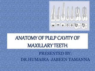
  • 1. ANATOMY OF PULP CAVITY OF MAXILLARY TEETH PRESENTED BY: DR.HUMAIRA JABEEN TAMANNA
  • 4. PULP Pulp is a soft tissue of mesenchymal origin residing within the pulp cavity.
  • 5. PULP CAVITY ā€¢ The entire internal space or central cavity enclosed by dentine except at the apical foramen. ā€¢ It consists of the following entities. ļƒ˜Pulp chamber ļƒ˜Root canals
  • 6. PULP CHAMBER PARTS: ļƒ˜ Roof ļƒ˜ Pulp horns A pulp horn is an accentuation of the pulp chamber directly under a cusp or a developmental lobe. ļƒ˜ Floor ļƒ˜ Canal orifice
  • 7. ROOT CANAL Portion of the pulp cavity from the canal orifice to the apical foramen Consists of three sections: ā€¢ Coronal ā€¢ Middle ā€¢ Apical
  • 8. LATERAL AND ACCESSORY CANALS ā€¢ A lateral canal is a canal that is located at approximately right angles to main root canal.
  • 9. ā€¢ According to Green , the incidence of accessory foramina ranged from 10% in maxillary central incisor and mandibular cuspids to 47% .
  • 10. ROOT CANAL ANATOMY ā€¢ The number of canals in a tooth correlates to the number of roots in a tooth. ā€¢ The space inside the root canals is filled with a highly vascularized and highly innervated loose connective tissue , called the dental pulp. ā€¢ The pulp tissue is in communication with the periodontium and the rest of the body through the apical foramen. ā€¢ Usually a root canal has curvature or constriction before terminating at apex.
  • 11. APICAL ROOT ANATOMY It is based on following anatomic and histological landmarks in the apical part of the root canal. APICAL CONSTRICTION (MINOR DIAMETER) ā€¢ It has an apical part of root canal having the narrowest diameter short of the apical foramina. ā€¢ It may or may not coincide with CDJ.
  • 12. APICAL FORAMEN (MAJOR DIAMETER) ā€¢ It is main apical opening on the surface of root canal through which blood vessels enter the canal. ā€¢ Its diameter is almost double the apical constriction giving it a funnel shape appearance.
  • 13. CEMENTODENTINAL JUNCTION ā€¢ Cementodentinal junction is the point in the canal where cementum meets dentin. ā€¢ The position of CDJ varies but usually it lies 0.5- 3.0mm short of the anatomic apex.
  • 14. APICAL DELTA It is a triangular area of root surrounded by main canal ,accessory canals and periradicular tissue.
  • 15. SIGNIFICANCE OF APICAL THIRD ā€¢ The root canal treatment of apical part of root is difficult sometimes because of presence of accessory and lateral canals, pulp stones , varying amounts of irregular secondary dentin and area of resorption. ā€¢ Most of the curvature occur in apical third. ā€¢ Obturation should end at apical constriction. ā€¢ Apical 3mm of root is generally resected during endodontic surgery in order to eliminate canal aberrations.
  • 16. ISTHMUS It is defined as narrow passage or anatomic part connecting two larger structures. It is commonly found in teeth with multiple canals. ā€¢ Isthumus is a narrow , ribbon shaped communication between two root canals which can be complete or incomplete. ā€¢ It contains pulp or pulpally derived tissue and acts as store house of bacteria so the isthmus should be well prepared and filled if seen on resected surface.
  • 17.
  • 18. CLASSIFICATION: Type I: two or three canals with no visible communication(incomplete isthmus) Type II: two canals showing definite connection with two main canals. Type III: three canals showing definite connection with main canals. Type IV: it is similar to type II or type III with canals extending to isthmus area. Type V: it is true connection throughout the section of root.
  • 19. The pulp canal system is complex, and canals may branch, divide, and rejoin (apical foramen). Weine categorized the root canal systems into four basic types:
  • 20. ā€¢ Type I- Single canal from pulp chamber to apex. ā€¢ Type II- Two separate canals leaving the pulp chamber but merging short of the apex to form only one canal. ā€¢ Type III- Two separate canals leaving the chamber and exiting the root in separate apical foramina. ā€¢ Type IV- One canal leaving the pulp chamber but dividing short of the apex into two separate and distinct canals with separate apical foramina.
  • 21. Vertucci et al, identified eight pulp space configurations, which briefly can be described as follows : ā€¢ Type I : A single canal extends from the pulp chamber to the apex ā€¢ Type II: Two separate canals leave the pulp chamber and join short of the apex to form one canal
  • 22. ā€¢ Type III: One canal leaves the pulp chamber and divides into two in the root; the two then merge to exit as one canal ā€¢ Type IV: Two separate, distinct canals extend from the pulp chamber to the apex ā€¢ Type V: One canal leaves the pulp chamber and divides short of the apex into two separate, distinct canals with separate apical foramina
  • 23. ā€¢ Type VI: Two separate canals leave the pulp chamber, merge in the body of the root, and redivide short of the apex to exit as two distinct canals . ā€¢ Type VII: One canal leaves the pulp chamber, divides and then rejoins in the body of the root, and finally redivides into two distinct canals short of the apex. ā€¢ Type VIII: Three separate, distinct canals extend from the pulp chamber to the apex.
  • 24. METHODS OF DETERMINING PULP ANATOMY 1. CLINICAL METHOD ā€¢ Anatomy studies ā€¢ Radiographs ā€¢ Exploration ā€¢ High resolution computed tomography ā€¢ Visualization endogram ā€¢ Fiber optic endoscope ā€¢ Magnetic resonance imaging
  • 25. 2. IN VITRO METHODS ā€¢ Sectioning of teeth ā€¢ Use of dyes ā€¢ Clearing of teeth ā€¢ Contrasting media ā€¢ Scanning electron microscopic analysis
  • 26. CLINICAL METHODS ANATOMIC STUDIES The knowledge of anatomy gained from various studies and books is commonly used method. RADIOGRAPHS They are also useful in assessing the root canal anatomy . Since , it is a two dimensional picture of three dimensional object , one has to analyze the radiograph carefully.
  • 27. HIGH RESOLUTION COMPUTED TOMOGRAPHY It shows three-dimensionl picture of root canal system using computer image processing. FIBER OPTIC ENDOSCOPE It is used to visualize canal anatomy. VISUALIZATION ENDOGRAM In this technique , an irrigant is used which helps in visualization of the canals on radiograph. This solution is called Ruddleā€™s solution. After injecting this solution into canal system , radiograph is taken to visualize the canal anatomy.
  • 28. MAGNETIC RESAONANCE IMAGING It produces data on computer which helps in knowing canal morphology. EXPLORATION On reaching pulpal floor one finds the grooves and anatomic dark lines which connect the canal orifices , this is called dentinal map.
  • 29. IN VITRO METHODS SECTIONING In this, teeth are sectioned longitudinally for visualization of root canal system. USE OF DYES Methylene blue or fluorescein sodium dyes(commonly used ) help in location pulp tissue present in pulp chamber because dyes stain any vital tissue present in pulp chamber or root canals.
  • 30. CLEARING OF ROOTS In this roots are initially decalcified using either 5 percent nitric acid or 10 percent hydrochloric acid and then dehydrated using different concentrations of alcohols and immersed in different clearing agents like methyl salicylate or xylene. By this treatment , tooth becomes transparent , then a dyes is injected and anatomy is visualized .
  • 31. HYPAQUE/CONTRASTING MEDIA It is iodine containing media which is injected into root canal space and visualized on radiograph. SCANNING ELECTRON MICROSCOPIC (SEM) ANALYSIS It also helps in evaluating root canal anatomy
  • 32. FACTORS AFFECTING INTERNAL ANATOMY Internal anatomy of teeth ,reflects the tooth form ,yet various enviromental factors whether physiological or pathological affect its shape and size because of pulpal and dentinal reaction to them. ā€¢ AGE ā€¢ IRRITANTS ā€¢ CALCIFICATIONS ā€¢ RESORPTION
  • 33. AGE With advancing age , there is continued dentin formation causing regression in shape and size of pulp cavity. Clinically it may pose problems in locating the pulp chamber and canals. IRRITANTS Various irritants like caries, periodontal disease , attrition, abrasion, erosion, cavity preparation and other operative procedures may stimulate dentin formation at the base of tubules resulting in change in shape of pulp cavity.
  • 34. CALCIFICATIONS pulp stones or diffuse calcifications are usually present in chamber and the radicular pulp. These alter the internal anatomy of teeth and may make the process of canal location difficult. RESORPTION Chronic inflammation or for unknown cause internal resorption may result in change of shape of pulp cavity making the treatment of such teeth challenging.
  • 35. VARIATION IN DEVELOPMENT ā€¢ GEMINATION-It arises from an attempt at division of a single tooth germ by an invagination resulting in incomplete formation of two teeth. ā€¢ FUSION it results in union two normally seperated tooth germ. Fused teeth may show separated tooth germ. Fused teeth may show separate or fused pulp space.
  • 36. ā€¢ CONCRESCENCE-In this fusion occurs after the root formation has completed. Teeth are joined by cementum only. ā€¢ TAURODONTISM In this ,body of tooth is enlarged at the expense of roots .Pulp chamber of this tooth is extremely large with a greater apico-occlusal height . Pulp lacks the normal costriction at cervical level of tooth.
  • 37. ā€¢ TALONā€™S CUSP It resembles eagleā€™s talon. ā€¢ In this , anamolous structure projects lingually from the cingulum area of maxillary or mandibular incisor . ā€¢ DILACERATION-It is an extraordinary curving of the roots of the teeth.Etiology of dilaceration is usually related to trauma during the root development in which movement of the crown and a part of root may result in sharp angulation after tooth completes development.
  • 38. ā€¢ DENTINOGENESIS IMPERFECTA It results in defective formation of dentin. It shows partial or total precocious obliteration of pulp chamber and root canals because of continued formation of dentin . ā€¢ DENTIN DYSPLASIA It is characterized by formation of normal enamel ,atypical dentin and abnormal pulpal morphology. In this root canals are obliterated so need special care while instrumentation.
  • 39. ā€¢ DENS IN DENTE OR DENS INVAGINATUS This condition represents an exaggeration of the lingual pit.Tooth with dens invaginatus has tendency for plaque accumulation which predisposes it to early decay and thus pulpitis. ā€¢ DENS EVAGINATUS In this condition an anomalous tubercle or cusp is located on the occlusal surface .Because of occlusal abrasion, this tubercle wears off fast causing early exposure of accessory pulp horn that extends into the tubercle .
  • 40.
  • 41. VARIATION IN SHAPE OF PULP CAVITY ā€¢ GRADUAL CURVE-It is most common condition in which root canal gradually curves from orifice to the apical foramen. ā€¢ APICAL CURVE-In this root canal is generally straight but at apex it shows curve. ā€¢ C-SHAPED CANAL This type of canal is usually found in mandibular molars.It is named so because of its morphology. Pulp chamber in C-shaped molar is single ribbon shaped with 180degree arc or more.
  • 42. ā€¢ BAYONET-SHAPED CANAL It is commonly seen in premolars. ā€¢ SICKLE-SHAPED CANALS In this, canal is sickle shaped. Cross section of this canal shows ribbon shape.
  • 43. VARIATION IN PULP CAVITY DUE TO PATHOLOGY ā€¢ PULP STONES AND CALCIFICATION Pulp stones are nodular calcified masses present in either coronal and radicular pulp or both these. They are present in at least 50 percent of teeth. Presence of pulp stones may alter the internal anatomy of the pulp cavity,making the access cavity of the tooth difficult.
  • 44. ā€¢ INTERNAL RESORPTION It is unusual form of tooth resorption that begins centrally within the tooth, initiated in most cases by a peculiar inflammation of the pulp . It is characterized by oval shaped enlargement of root canal space. ā€¢ EXTERNAL RESORPTION It is initiated in the periodontium and it effects the external or lateral surface of the root.
  • 45. VARIATION IN APICAL THIRD ā€¢ DIFFERENT LOCATIONS OF APICAL FORAMEN Apical foramen may exist on mesial ,distal , buccal or lingual surface of the root. ā€¢ ACCESSORY AND LATERAL CANALS They are the lateral branches of the main canal that form a communication between the pulp and periodontium . They can be seen anywhere from furcation to apex but tend to be more common apical third and posterior teeth. ā€¢ OPEN APEX It also referred as blunderbuss canal . ā€¢ In vital teeth with open apex, treatment should be apex-o- genesis and in nonvital teeth, it is apexification.
  • 46. VARIATION IN SIZE OF ROOT ā€¢ MACRODONTIA In this condition , pulp space and teeth are enlarged throughout the dentition . Commonly seen in giggantism. ā€¢ MICRODONTIA In this condition , pulpspace and teeth appear smaller in size . It is commonly seen in cases of dwarfism.
  • 47. ANATOMY OF PULP CAVITY OF MAXILLARY TEETH
  • 48. MAXILLARY CENTRAL INCISOR ā€¢ Average length of the tooth: 23.3mm ā€¢ Root curvature (most common to least common): straight(75%), labial, distal ā€¢ Single canal-99.4% ā€¢ Two canals-0.6%
  • 49.
  • 50. PULP CHAMBER: ā€¢ The pulp chamber of the maxillary central incisor is located in the center of the crown equidistant from the dentinal walls ā€¢ The pulp chamber usually follows the contours of the crown and has three pulp horns. ā€¢ It is broad mesiodistally than buccolingually, with its broadest part incisally
  • 51. ROOT CANAL ā€¢ Central incisor has one root with one canal. ā€¢ Coronally the root canal is wider buccopalatally. ā€¢ Lateral canals may be present (24% ) usually in the apical third. ā€¢ Most of the time canal is found to be straight
  • 52. ANOMALIES ā€¢ TALONā€™S CUSP- due to overdevelopment of the dental tubercle on the lingual side. A small cusp like projection with a pulp horn is seen on the lingual side of the teeth. ā€¢ Dens invaginatus ā€¢ Fusion ā€¢ Gemination
  • 53. CLINICAL CONSIDERATION: ā€¢ Placing the access cavity too far palatally makes straight line access difficult. ā€¢ Lateral canals are always found in apical third. ā€¢ Labial perforation is most commonly seen during access cavity preparation. ā€¢ Most of the canal is straight ,but 15 to 20 percent of roots show palatal curve .
  • 54. Maxillary lateral incisor ā€¢ Average length of the tooth: 21.8mm ā€¢ Average age of calcification: 11years ā€¢ Root curvature: distal(53%), straight(30%) ā€¢ Single canal 93.4% ā€¢ Two canals-6.6%
  • 55.
  • 56. PULP CHAMBER ā€¢ The shape of the pulp chamber of the maxillary lateral incisor is similar to that of the maxillary central incisor but smaller. ā€¢ It has two pulp horns, corresponding to the developmental mammelons. ā€¢ It is broad mesiodistally, with its broadest part incisally. ā€¢ The incisal outline of the pulp chamber tends to be more rounded.
  • 57. ROOT CANAL ā€¢ Root canal has finer diameter than that of central incisor though the shape is similar to that. ā€¢ Labiopalatally the canal is wider. ā€¢ Canal is ovoid labiopalatally in cervical third, ovoid in the middle third and round in apical third. ā€¢ Apical region of the canal is usually curved in a palatal direction.
  • 58. CLINICAL CONSIDERATION: ā€¢ cervical constriction need to be removed during coronal preparation to produce smooth progression from pulp chamber to root canal. ā€¢ Lateral canals are most common than maxillary central incisors. ā€¢ Labial perforation is most common error during access cavity preparation.
  • 59. ANOMALIES ā€¢ Peg lateral (gardnerā€™s syndrome) ā€¢ Fusion (with central incisor) ā€¢ Gemination ā€¢ Dens Invaginatus:
  • 60. MAXILLARY CANINE ā€¢ Average length of the tooth: 26.5mm ā€¢ Average age of calcification: 13-15 years ā€¢ Root curvature: straight(39%),distal (32%) ā€¢ Single canal-96.5% ā€¢ Two canals-3.5%
  • 61.
  • 62. PULP CHAMBER ā€¢ The pulp chambers of the maxillary cuspids are the largest of any single-rooted teeth. ā€¢ Labiopalatally, the chamber is triangular in shape, with the apex pointed incisally. ā€¢ Mesiodistally it is narrow. ā€¢ In cross section, the chamber is ovoid in shape, with the greater diameter labiopalatally. ā€¢ One pulp horn is present, corresponding to one cusp. ā€¢ The external access outline form is oval or slot shaped because no mesial or distal pulps horns are present
  • 63. ROOT CANAL ā€¢ There is single root canal which is wider labiopalatally than in mesiodistal aspect. ā€¢ Cross section at cervical and middle third show its oval shape, at apex it becomes circular. ā€¢ Canal is usually straight but may show a distal apical curvature.
  • 64. ANOMALIES ā€¢ Dilaceration ā€¢ Dens invaginatus ā€¢ 2 canals 2 root ā€¢ Facial talon cusp has been reported
  • 65. CLINICAL CONSIDERATION ā€¢ Cervical constriction needs to be shaped during corornal flaring to produce uniformly tapered preparations. ā€¢ Surgical access sometimes become difficult because of long length of the tooth. ā€¢ lateral canals are present in apical third. ā€¢ While preparing care must be taken to avoid blockage.
  • 66. MAXILLARY FIRST PREMOLAR ā€¢ Average length: 21.8mm ā€¢ No. of roots: 2roots(54.6%), 1 root(43%), 3roots(2.4%) ā€¢ Root curvature: Buccal- lingual(36%), straight(28%),bayonet curve(8%) Palatal- straight(45%), buccal(28%) ā€¢ Single root- straight(38%), distal(37%), buccal(15%) ā€¢ Average time of calcification: 12-13years
  • 67.
  • 68. PULP CHAMBER ā€¢ The pulp chamber is wider buccopalatally with two pulp horns,one under each cusp. ā€¢ The buccal pulp horn is more prominent than the palatal. ā€¢ The floor of the pulp chamber is convex, usually with two canal orifices, Palatal orifice is larger and is kidney shaped. ā€¢ In cross section, the pulp chamber is wide and ovoid in a buccopalatal dimension. ā€¢ Roof of the pulp chamber is coronal to the cervical line.
  • 69. ROOT CANAL ā€¢ Most commonly two roots and two root canals (buccal & palatal), three canals can also occur rarely(palatal, mesiobuccal & distobuccal) ā€¢ The palatal canal is generally the larger of the 2 canals. ā€¢ Buccal canal is directly under the buccal cusp and palatal canal under the palatal cusp. ā€¢ The root canals are usually straight and divergent.
  • 70. CLINICAL CONSIDERATION: ā€¢ To locate both the canals good quality radiograph should be taken so as to avoid superimposition of the canals. ā€¢ Palatal canal is usually larger than the buccal canal. ā€¢ Avoid over flaring of the coronal part of the buccal root to avoid perforation of palatal groove.
  • 71. ANOMALIES ā€¢ DENS EVAGINATUS: ā€¢ Gemination (rare) ā€¢ Taurodontism (rare) ā€¢ PRESENCE OF THREE CANALS ā€¢ one canal -21.3% ā€¢ Two canals-75.8% ā€¢ Three canals-1.4%
  • 73. PULP CHAMBER ā€¢ It is wider buccopalatally than the maxillary first premolar. It also has two pulp horns and is narrow mesiodistally. ā€¢ The buccal pulp horn is larger. ā€¢ In cross section, the pulp chamber has a narrow, ovoid shape.
  • 74. ROOT CANAL ā€¢ In cross section, the canals in the cervical third are ovoid and narrow In the middle third, when 1 canal is present it is ovoid, and when 2 canals are present they are round; in the apical third, the cross section is round regardless of whether 1 or 2 canals are present. ā€¢ Lateral canals are present in 59.5% of cases; 1.6% occur in the furcation area. ā€¢ canal is wider buccopalatally forming ribbon like shape.
  • 75. CLINICAL CONSIDERATION: ā€¢ Narrow ribbon like canal is always difficult to clean and obturate effectively. ā€¢ If one canal is present ,orifice is indistinct ,but if two canals are present ,two orifices is seen.
  • 76. ANOMALIES ā€¢ Presence of three canals. one canal-50.3% two canals-46.5% three canals-1.2% ā€¢ Dens invaginatus ā€¢ Deep distal root concavity
  • 77. MAXILLARY FIRST MOLAR ā€¢ Average length: 19.9mm ā€¢ Root curvature: MB root-distal(78%),straight(21%) DB root- straight(54%), mesial(19%), distal(17%) Palatal root- buccal(55%), straight(40%) ā€¢ Three canals-41.1% Four canals-56.5% Five canals-2.4% Incidence of MB2 canal is 61.9% ā€¢ Average age of calcification:9-10 years
  • 78.
  • 79. ā€¢ The pulp chamber of the maxillary first molar is the largest in the dental arch, with four pulp horns. ā€¢ The arrangement of the four pulp horns gives the pulp roof a rhomboidal shape in cross section. ā€¢ The four walls forming the roof converge toward the floor where the lingual wall almost disappears; the floor of the pulp chamber thus has a triangular form in cross section.
  • 80. ā€¢ The palatal orifice is the largest, round or oval in shape and easily accessible for exploration. ā€¢ The mesiobuccal orifice is under the mesiobuccal cusp, long buccopalatally and may have a depression at the palatal end in which the orifice of a fourth canal may be present. The location of the MB2 canal varies greatly ā€¢ The distobuccal orifice is located slightly distal and palatal to the mesiobuccal orifice .
  • 81. ROOT CANAL ā€¢ Maxillary first molar has generally three roots with three canals. ā€¢ Two canals in mesiobuccal root are closely interconnected and sometimes merge into one canal. ā€¢ The palatal root is the longest and has the largest diameter. It contains mostly only one canal, though two or three root canals can also occur. From its orifice the palatal canal is flat, ribbon like, and wider in a mesiodistal direction.
  • 82. ā€¢ The distobuccal root is conical and may have one or two canals. From its orifice, the canal(s) first is oval and then becomes round as it approaches the apical third of the root. Lateral canals are present in 36% of cases . ā€¢ Single mesiobuccal canal is oval and wider buccolingually two or three canals are more circular. The mesiobuccal root has lateral canals in 1 % of cases
  • 83. ANOMALIES ā€¢ Pulp stones may be present in the pulp chamber ā€¢ Rarely a second canal may be present in the palatal root(1%). ā€¢ Maxillary molar with a single root and single root canal. ā€¢ C-shaped canal
  • 84. CLINICAL CONSIDERATION: ā€¢ Buccal curvature of palatal canal (56%)may not be visible on radiographs,leading to procedural errors. ā€¢ Mesiobuccal canal shows curvature sometimes which is not visible radiographically. ā€¢ Perforation of palatal root is commonly caused by assuming canal to be straight .
  • 85. MAXILLARY SECOND MOLAR ā€¢ Average length:20.2mm ā€¢ 3 roots present(54%) fused (46%) ā€¢ Root curvature: MB- distal(54%),straight(22%) MB2 Canal incidence--(37%) DB- straight(54%),mesial(17%) Palatal root- straight(63%), buccal(37%) ā€¢ Average age of calcification: 14-16yrs
  • 86.
  • 87. PULP CHAMBER ā€¢ It is similar to that of the maxillary first molar, except it is narrow mesiodistally. ā€¢ Roof of pulp chamber is more rhomboidal in cross-section and floor is an obtuse triangle. ā€¢ When four canals are present, the access cavity preparation of the maxillary second molar has a rhomboid shape and is a smaller version of the access cavity for the maxillary first molar. If only three canals are present, the access cavity is a rounded triangle with the base to the buccal side.
  • 88. ROOT CANAL ā€¢ Similar to first molar except that in maxillary second molar roots tend to be less divergent and may be fused. ā€¢ Fewer lateral canals are present in roots and furcation area than in first molar.
  • 89. ANOMALIES: ā€¢ Root fusion ā€¢ Single root single canal ā€¢ Incidence of pulp stone ā€¢ 2 palatal canal in double palatal root
  • 90. CLINICAL CONSIDERATION: ā€¢ Similar to maxillary first molar. ā€¢ Maxillary second molar lies closer to the maxillary sinus than first molar.
  • 91. MAXILLARY THIRD MOLAR ā€¢ Average tooth length -17mm PULP CHAMBER AND ROOT CANAL ā€¢ It is similar to second molar but displays great variations in shape , size and form of both pulp chamber as well as root canal. ā€¢ There may be presence of one, two ,three or more canals sometimes. ā€¢ The roots are often fused forming one large root. ā€¢ About 45.7%had fused roots ,2%had c-shaped canals and 2% had four separate roots.
  • 92. ā€¢ Average time of eruption-: 17-22Years ā€¢ Average age of calcification-: 18-25Years ā€¢ Average tooth length-: 17mm ā€¢ Average crown length-: 6.5mm ā€¢ Average root length-: 11mm ā€¢ M-D of crown-: 8.5mm ā€¢ Labio-lingual diameter of crown-: 10mm CLINICAL CONSIDERATION: Maxillary third molar is closely related to maxillary sinus.
  • 93. CONCLUSION ā€¢ A successful treatment outcome depends on complete debridement and disinfection of all canals . ā€¢ Through the knowledge of root canal anatomy it enables the clinician to produce endodontic treatments of high quality and considerable longitivity.