SlideShare a Scribd company logo
1 of 91
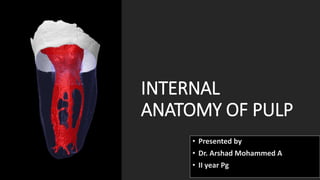
INTERNAL
ANATOMY OF PULP
• Presented by
• Dr. Arshad Mohammed A
• II year Pg
INTRODUCTION
The knowledge of
common root canal
morphology and
its
frequent
variations is a basic
requirement for
success during
root canal
procedures.
DEFINITION
CLASSIFICATION OF ROOT CANAL MORPHOLOGY
ISTHMUS
CLASSIFICATION OF C-SHAPED CANALS
METHODS OF DETERMINING PULP ANATOMY
FACTORS AFFECTING INTERNAL ANATOMY
ANATOMY OF PULP CAVITY OF MAXILLARY & MANDIBULAR TEETH
CLINICAL SIGNIFICANCE
CONCLUSION
PULP CAVITY
CLASSIFICATION
VERTUCCI’S CLASSIFIACTION
SERT AND BAYIRIL CLASSIFICATION
Gulabivala
supplemental
canal
configurations
Weine
classification
Walker classification
ISTHMUS
“A narrow passage or anatomic part connecting two
larger structures (root canals) “
-Identified using METHYLENE BLUE DYE
-Contains pulp or pulpally derived tissue and acts as
store house for bacteria
-Identification and treatment of isthumi are vital to success of
surgical procedure
-Investigators identified 5 types of isthumi that can be found on
a beveled root surface
CLASSIFICATION
OF ISTHUMI
Kim et al
1
Yeung Yi Hsu
2
Kim et al
Yeung Yi Hsu
CLASSIFICATION OF C-SHAPED CANALS
Melton’s classification
FAN’S CLASSIFICATION
METHODS OF DETERMINING PULP ANATOMY
1. Clinical methods:
• a. Anatomic studies
• b. Radiographs
• c. Exploration
• d. Visualization endogram
• e. Fiberoptic endoscope
• f. Magnetic resonance imaging (MRI)
• g. Dental operating microscope
• a. Clearing study
• b. Scanning electron microscope (SEM)
• c. Sectioning
• d. Microcomputed tomography
• e. Cone beam computed tomography (CBCT)
2. In vitro methods:
ANATOMIC STUDIES
The knowledge of anatomy gained from various studies and books
is commonly used method.
RADIOGRAPHS
They are also useful in assessing the root canal anatomy.
Since , it is a two dimensional picture of three dimensional object ,
one has to analyse the radiograph carefully.
HIGH RESOLUTION COMPUTED TOMOGRAPHY
It shows three- dimensionl picture of root canal system using computer
image processing.
FIBER OPTIC ENDOSCOPE
It is used to visualize canal anatomy. It is a new method of magnied intra
canal visualization using bro-optic endoscopes (orascope) or rod lens
endoscopes.
VISUALIZATION ENDOGRAM
In this technique , an irrigant is used which helps in visualization of the
canals on radiograph. This solution is called Ruddle’s solution. After
injecting this solution into canal system ,radiograph is taken to visualize
the canal anatomy.
MAGNETIC RESAONANCE IMAGING
It produces data on computer which helps in knowing canal
morphology.
EXPLORATION
On reaching pulpal floor one finds the grooves and anatomic dark
lines which connect the canal orifices , this is called dentinal map.
SECTIONING
In this, teeth are sectioned longitudinally for visualization of root
canal system.
CLEARING OF ROOTS
In this roots are initially decalcified using either 5 percent nitric
acid or 10 percent hydrochloric acid and then dehydrated using
different concentrations of alcohols and immersed in different
clearing agents like methyl salicylate or xylene.
By this treatment , tooth becomes transparent , then a dyes is
injected and anatomy is visualized.
HYPAQUE/CONTRASTING MEDIA
It is iodine containing media which is injected into root canal
space and visualized on radiograph.
SCANNING ELECTRON MICROSCOPIC (SEM) ANALYSIS
It also helps in evaluating root canal anatomy
FACTORS AFFECTING INTERNAL ANATOMY
Internal anatomy of teeth ,reflects the tooth form ,yet various
environmental factors whether physiological or pathological affect its
shape and size because of pulpal and dentinal reaction to them.
• AGE
• IRRITANTS
• CALCIFICATIONS
• RESORPTION
AGE
With advancing age , there is continued dentin formation causing
regression in shape and size of pulp cavity. Clinically it may pose
problems in locating the pulp chamber and canals.
IRRITANTS
Various irritants like caries, periodontal disease , attrition, abrasion,
erosion, cavity preparation and other operative procedures may
stimulate dentin formation at the base of tubules resulting in change
in shape of pulp cavity.
CALCIFICATIONS
pulp stones or diffuse calcifications are usually present in chamber and
the radicular pulp. These alter the internal anatomy of teeth and may
make the process of canal location difficult.
RESORPTION
Chronic inflammation or for unknown cause internal resorption may
result in change of shape of pulp cavity making the treatment of such
teeth challenging.
VARIATIONS IN INTERNAL ANATOMY
Variation in shape
 Gradual curve
 Apical curve
 C-shaped
 Bayonet shaped
 Sickle shaped
Variations because of pathology
 Pulp stones
 Internal resorption
 External resorption
Variations in apical third
 Different position of apical foramen
 Open apex
 Lateral and accessory canals
Variations in development
 Gemination
 Fusion
 Concrescence
 Taurodontism
 Talon’s cusp
 Dilaceration
 Extra root canal
 Dens invaginatus
 Dens evaginatus
Variations in size of root
• MACRODONTIA
In this condition , pulp space and teeth are enlarged throughout
the dentition .
• MICRODONTIA
In this condition , pulp space and teeth appear smaller in size .
ANATOMY OF PULP CAVITY OF MAXILLARY
TEETH
MAXILLARY CENTRAL
INCISOR
PULP CHAMBER:
• The pulp chamber of the maxillary central incisor is located in
the centre of the crown equidistant from the dentinal walls
• The pulp chamber usually follows the contours of the crown and
has three pulp horns.
• It is broad mesiodistally than buccolingually, with its broadest part
incisally
ROOT CANAL
• Central incisor has one root with one canal.
• Coronally the root canal is wider buccopalatally.
• Lateral canals may be present (24% ) usually in the apical third.
• Most of the time canal is found to be straight
Maxillary lateral
incisor
PULP CHAMBER
• The shape of the pulp chamber of the maxillary lateral incisor
is similar to that of the maxillary central incisor but smaller.
• It has two pulp horns, corresponding to the developmental
mammelons.
• It is broad mesiodistally, with its broadest part incisally.
• The incisal outline of the pulp chamber tends to be more
rounded.
ROOT CANAL
• Root canal has finer diameter than that of central incisor
though the shape is similar to that.
• Labiopalatally the canal is wider.
• Canal is ovoid labiopalatally in cervical third, ovoid in the
middle third and round in apical third.
• Apical region of the canal is usually curved
in a palatal direction.
MAXILLARY CANINE
PULP CHAMBER
• The pulp chambers of the maxillary cuspids are the largest of any
single-rooted teeth.
• Labiopalatally, the chamber is triangular in shape, with the apex
pointed incisally.
• Mesiodistally it is narrow.
• In cross section, the chamber is ovoid in shape, with the greater
diameter labiopalatally.
• One pulp horn is present, corresponding to one cusp.
• The external access outline form is oval or slot shaped because no
mesial or distal pulps horns are present
ROOT CANAL
• There is single root canal which is wider labiopalatally
than in mesiodistal aspect.
• Cross section at cervical and middle third show its oval
shape, at apex it becomes circular.
• Canal is usually straight but may show a distal apical
curvature.
MAXILLARY FIRST
PREMOLAR
PULP CHAMBER
• The pulp chamber is wider buccopalatally with two pulp horns, one
under each cusp.
• The buccal pulp horn is more prominent than the palatal.
• The floor of the pulp chamber is convex, usually with two canal
orifices, Palatal orifice is larger and is kidney shaped.
• In cross section, the pulp chamber is wide and ovoid in a
buccopalatal dimension.
• Roof of the pulp chamber is coronal to the cervical line.
ROOT CANAL
• Most commonly two roots and two root canals (buccal &
palatal), three canals can also occur rarely(palatal, mesiobuccal &
distobuccal)
• The palatal canal is generally the larger of the 2 canals.
• Buccal canal is directly under the buccal cusp and palatal canal
under the palatal cusp.
• The root canals are usually straight and divergent.
MAXILLARY SECOND
PREMOLAR
PULP CHAMBER
• It is wider buccopalatally than the maxillary first premolar. It
also has two pulp horns and is narrow mesiodistally.
• The buccal pulp horn is larger.
• In cross section, the pulp chamber has a narrow, ovoid shape.
ROOT CANAL
• In cross section, the canals in the cervical third are ovoid and
narrow In the middle third, when 1 canal is present it is
ovoid,and when 2 canals are present they are round; in the
apical third, the cross section is round regardless of whether 1
or 2 canals are present.
• Lateral canals are present in 59.5% of cases; 1.6% occur in the
furcation area.
• Canal is wider buccopalatally forming ribbon like shape.
MAXILLARY FIRST
MOLAR
PULP CHAMBER
• largest in the dental arch, with four pulp horns.
• The arrangement of the four pulp horns gives the pulp roof a
rhomboidal shape in cross section.
ROOT CANAL
• Maxillary first molar has generally three roots with three canals.
• Two canals in mesiobuccal root are closely interconnected and
sometimes merge into one canal.
• The palatal root is the longest and has the largest diameter. It
contains mostly only one canal, though two or three root
canals can also occur. From its orifice the palatal canal is flat,
ribbon like, and wider in a mesiodistal direction.
• The distobuccal root is conical and may have one or two canals.
MAXILLARY SECOND
MOLAR
PULP CHAMBER
• It is similar to that of the maxillary first molar, except it is
narrow mesiodistally.
• Roof of pulp chamber is more rhomboidal in cross-section and
floor is an obtuse triangle.
ROOT CANAL
• Similar to first molar except that in maxillary second molar
roots tend to be less divergent and may be fused.
• Fewer lateral canals are present in roots and furcation area
than in first molar.
MAXILLARY IIIrd
MOLAR
PULP CHAMBER
• Anatomically resembles the second molar.
• The pulp chamber can be similar to that of the maxillary second
molar with three canal orifices, but it may also have an odd-shaped
chamber with four or five root canal orifices or a conical chamber
with only one root canal.
ROOT CANAL
• The maxillary third molar may have three well- developed roots that
are closely grouped.
• It may also have fused roots, one conical root, or four or more
independent roots.
• The roots may be straight, curved, or dilacerated, and may be fully
or partially developed.
• Root canals vary from one to four or even five in number, depending
on the number of roots.
• One may find a “C-shaped” pulp chamber with a “C-shaped” root
canal.
ANATOMY OF PULP CAVITY OF MANDIBULAR
TEETH
MANDIBULAR INCISOR
PULP CHAMBER
• Smallest tooth in the arch.
• The pulp chamber is small and flat mesiodistally
ROOT CANAL
• One root.
• Which is flat and narrow mesiodistally but wide labiolingually.
• The root is straight in 60% of cases. The canal configuration varies from.
• One canal exiting in one apical foramen, as in 70% of these teeth.
• One canal bifurcating into two canals, coming together, and exiting into one
apical foramen (22%)
MANDIBULAR LATERAL
INCISOR
PULP CHAMBER
• The configuration of the pulp chamber of the mandibular lateral
incisor is similar to that of the mandibular central incisor, but the
lateral incisor has larger dimensions.
ROOT CANAL
• Larger than that of the mandibular central incisor,
• Distal curve of the lateral incisors is sharper.
MANDIBULAR CANINE
PULP CHAMBER
• Resembles the maxillary canine, but it is smaller in all dimensions.
• The pulp chamber is narrow mesiodistally.
ROOT CANAL
• Although the tooth usually has a single root, it may have two (2.3%).
• Most of these teeth have a straight root (68%), but some have distal
curvatures (20%).
• The mandibular canine usually has one canal exiting in one apical
foramen (78%).
MANDIBULAR Ist
PREMOLAR
PULP CHAMBER
• The mesiodistal width of the pulp chamber is narrow.
• Buccolingually, the pulp chamber is wide, with a prominent buccal
pulp horn that extends under a well-developed buccal cusp.
ROOT CANAL
• Usually has a short, conical root.
• This root may divide in the apical third into two or three roots.
• The root is usually straight (48%), but some roots curve distally (35%).
• One canal and one foramen are present in 70% of cases.
• One canal bifurcates into two canals and exits in two foramina in 24% of
cases.
• Two canals exit in two foramina in 1.5% of cases.
• One canal bifurcates into two canals uniting into one canal in the apical
third and then exiting in one foramen in 4% of cases.
• Three canals exit in three foramina in 0.5% of cases.
MANDIBULAR IInd
PREMOLAR
PULP CHAMBER
Similar to the mandibular first premolar, except that the lingual horn is
more prominent under a well-developed lingual cusp.
ROOT CANAL
• Usually has a single root, but on rare occasions two to three roots are
present.
• The root of the mandibular second premolar may curve distally (40%),
although in 39% of cases it is straight.
MANDIBULAR Ist
MOLAR
PULP CHAMBER
• The roof of the pulp chamber of the mandibular first molar is often
rectangular in shape.
• The mesial wall is straight, the distal wall round, and the buccal and
lingual walls converge to meet the mesial and distal walls and form
a rhomboidal floor. The roof of the pulp chamber has four pulp
horns: mesiobuccal, mesiolingual, distobuccal, and distolingual.
ROOT CANAL
• Usually, two well-differentiated roots are present in the mandibular first
molar, one mesial and one distal.
• Both roots are wide and flat buccolingually, with a depression in the
middle of the root buccolingually.
MANDIBULAR IInd
MOLAR
PULP CHAMBER
• Smaller than that of the mandibular first molar, and the root canal
orifices are smaller and closer together.
ROOT CANAL
• The majority of the mandibular second molars have two roots (71%),
but teeth with one root (27%) and teeth with three roots (2%) are also
seen.
MANDIBULAR IIIrd
MOLAR
PULP CHAMBER
• Anatomically resembles the pulp chamber of the mandibular first and
second molars.
• It is large and possesses many anomalous configurations such as C-
shaped root canal orifices.
ROOT CANAL
• The mandibular third molar usually has two roots and two canals, but
occasionally one root and one canal or three roots and three canals.
The root canals are generally large and short.
Conclusion
A thorough knowledge of the root canal anatomy, careful
interpretation of radiographs, and adequate access to exploration of
the tooth’s interiors are the prerequisites for a successful endodontic
treatment.
With recent advances, studying the pulp morphology has become
much more ecient. The clinician must be aware of the latest
developments so that he/she can render quality treatment to the
patient.
REFERENCES
Grossman’s ENDODONTIC PRACTICE-13th Edition
Cohen’s PATHAWAYS of the PULP-11th Edition
Determination of the internal anatomy of a permanent dentition: A review
{International Journal of Contemporary Dental and Medical Reviews (2015)}
Google

More Related Content

What's hot

Root canal preparation techniques _ endodontic treatment
Root canal preparation techniques _ endodontic treatment Root canal preparation techniques _ endodontic treatment
Root canal preparation techniques _ endodontic treatment Israa Awadh
 
Traumatic injuries of teeth
Traumatic injuries of teethTraumatic injuries of teeth
Traumatic injuries of teethKUMARAVEL SM
 
Access cavity preparation
Access cavity preparationAccess cavity preparation
Access cavity preparationIAU Dent
 
Obturation technique
Obturation technique Obturation technique
Obturation technique Deepashri Tekam
 
Biomechanical preparation in endodontics
Biomechanical preparation in endodonticsBiomechanical preparation in endodontics
Biomechanical preparation in endodonticsKarishma Ashok
 
Protaper endodontic system
Protaper endodontic systemProtaper endodontic system
Protaper endodontic systemmuthanna Al-Jubory
 
Anatomy of the pulp space and access cavity
Anatomy of the pulp space and access cavityAnatomy of the pulp space and access cavity
Anatomy of the pulp space and access cavityZubia Arshad
 
Internal anatomy of tooth
Internal  anatomy of  toothInternal  anatomy of  tooth
Internal anatomy of toothSNEHA RATNANI
 
Obturation techniques
Obturation techniquesObturation techniques
Obturation techniquesMaulee Sheth
 
Shade selection
Shade selectionShade selection
Shade selectionNakul Patidar
 
Bevels and flares in dental restoration
Bevels and flares in dental restorationBevels and flares in dental restoration
Bevels and flares in dental restorationDr. Mayank Nahta
 
5.full metal crown
5.full metal crown5.full metal crown
5.full metal crownLama K Banna
 
Endodontic Diagnosis: Pulp Vitality Tests
Endodontic Diagnosis: Pulp Vitality TestsEndodontic Diagnosis: Pulp Vitality Tests
Endodontic Diagnosis: Pulp Vitality TestsIraqi Dental Academy
 
Endodontic Diagnosis and Treatment Planning
Endodontic Diagnosis and Treatment PlanningEndodontic Diagnosis and Treatment Planning
Endodontic Diagnosis and Treatment PlanningIraqi Dental Academy
 
Everything you need to know about endo files
Everything you need to know about endo filesEverything you need to know about endo files
Everything you need to know about endo filesVitalticks Pvt Ltd
 
Removal of broken endodontic instruments
Removal of broken endodontic instrumentsRemoval of broken endodontic instruments
Removal of broken endodontic instrumentsPalaniselvi Kamaraj
 

What's hot (20)

Canal curvatures and determination method final
Canal curvatures and determination method finalCanal curvatures and determination method final
Canal curvatures and determination method final
 
Root canal preparation techniques _ endodontic treatment
Root canal preparation techniques _ endodontic treatment Root canal preparation techniques _ endodontic treatment
Root canal preparation techniques _ endodontic treatment
 
Endodontic diagnosis
Endodontic diagnosisEndodontic diagnosis
Endodontic diagnosis
 
Traumatic injuries of teeth
Traumatic injuries of teethTraumatic injuries of teeth
Traumatic injuries of teeth
 
working length
working lengthworking length
working length
 
Anatomy of root apex
Anatomy of root apexAnatomy of root apex
Anatomy of root apex
 
Access cavity preparation
Access cavity preparationAccess cavity preparation
Access cavity preparation
 
Obturation technique
Obturation technique Obturation technique
Obturation technique
 
Biomechanical preparation in endodontics
Biomechanical preparation in endodonticsBiomechanical preparation in endodontics
Biomechanical preparation in endodontics
 
Protaper endodontic system
Protaper endodontic systemProtaper endodontic system
Protaper endodontic system
 
Anatomy of the pulp space and access cavity
Anatomy of the pulp space and access cavityAnatomy of the pulp space and access cavity
Anatomy of the pulp space and access cavity
 
Internal anatomy of tooth
Internal  anatomy of  toothInternal  anatomy of  tooth
Internal anatomy of tooth
 
Obturation techniques
Obturation techniquesObturation techniques
Obturation techniques
 
Shade selection
Shade selectionShade selection
Shade selection
 
Bevels and flares in dental restoration
Bevels and flares in dental restorationBevels and flares in dental restoration
Bevels and flares in dental restoration
 
5.full metal crown
5.full metal crown5.full metal crown
5.full metal crown
 
Endodontic Diagnosis: Pulp Vitality Tests
Endodontic Diagnosis: Pulp Vitality TestsEndodontic Diagnosis: Pulp Vitality Tests
Endodontic Diagnosis: Pulp Vitality Tests
 
Endodontic Diagnosis and Treatment Planning
Endodontic Diagnosis and Treatment PlanningEndodontic Diagnosis and Treatment Planning
Endodontic Diagnosis and Treatment Planning
 
Everything you need to know about endo files
Everything you need to know about endo filesEverything you need to know about endo files
Everything you need to know about endo files
 
Removal of broken endodontic instruments
Removal of broken endodontic instrumentsRemoval of broken endodontic instruments
Removal of broken endodontic instruments
 

Similar to Internal Anatomy of Pulp cavity

Internal anatomy of each tooth
Internal anatomy of each toothInternal anatomy of each tooth
Internal anatomy of each toothEdward Kaliisa
 
Anatomy of pulp cavity of maxillary teeth (2)
Anatomy of pulp cavity of maxillary teeth (2)Anatomy of pulp cavity of maxillary teeth (2)
Anatomy of pulp cavity of maxillary teeth (2)Humaira Tamanna
 
Anatomy of pulp canal and its access opening
Anatomy of pulp canal and its access openingAnatomy of pulp canal and its access opening
Anatomy of pulp canal and its access openingPrattoo
 
Root canal anatomy
Root canal anatomyRoot canal anatomy
Root canal anatomyLama K Banna
 
Morphology and internal anatomy of root canal system
Morphology and internal anatomy of root canal systemMorphology and internal anatomy of root canal system
Morphology and internal anatomy of root canal systemAkansha Tilokani
 
PULP SPACE (Dr. SONA)
PULP SPACE (Dr. SONA)PULP SPACE (Dr. SONA)
PULP SPACE (Dr. SONA)MINDS MAHE
 
rootcanalanatomy-190808050207.pptx
rootcanalanatomy-190808050207.pptxrootcanalanatomy-190808050207.pptx
rootcanalanatomy-190808050207.pptxSiddharthSingh639
 
Anatomy of dental pulp.pdf
Anatomy of dental pulp.pdfAnatomy of dental pulp.pdf
Anatomy of dental pulp.pdfSajedaElgbaili
 
CLASSIFICATION (1).pdf
CLASSIFICATION (1).pdfCLASSIFICATION (1).pdf
CLASSIFICATION (1).pdfAltilbaniHadil
 
Internal anatomy of anterior tooth
Internal anatomy of anterior toothInternal anatomy of anterior tooth
Internal anatomy of anterior toothJigyasha Timsina
 
Apex and its significance in endodontics
Apex and its significance in endodonticsApex and its significance in endodontics
Apex and its significance in endodonticsRockyJohn6
 
Myth of Easy Root Canals
Myth of Easy Root CanalsMyth of Easy Root Canals
Myth of Easy Root CanalsDr. Rajat Sachdeva
 
access opening of molar teeth-shivangi.pptx
access opening of molar teeth-shivangi.pptxaccess opening of molar teeth-shivangi.pptx
access opening of molar teeth-shivangi.pptxssuser502d85
 
Locating root canal orifice in molar RCT
Locating root canal orifice in molar RCTLocating root canal orifice in molar RCT
Locating root canal orifice in molar RCTPrasanth Balan
 
Access cavity ppt
Access cavity pptAccess cavity ppt
Access cavity pptnehasingh1671
 

Similar to Internal Anatomy of Pulp cavity (20)

Internal anatomy of each tooth
Internal anatomy of each toothInternal anatomy of each tooth
Internal anatomy of each tooth
 
Anatomy of pulp cavity of maxillary teeth (2)
Anatomy of pulp cavity of maxillary teeth (2)Anatomy of pulp cavity of maxillary teeth (2)
Anatomy of pulp cavity of maxillary teeth (2)
 
Anatomy of pulp canal and its access opening
Anatomy of pulp canal and its access openingAnatomy of pulp canal and its access opening
Anatomy of pulp canal and its access opening
 
Root canal anatomy
Root canal anatomyRoot canal anatomy
Root canal anatomy
 
Morphology and internal anatomy of root canal system
Morphology and internal anatomy of root canal systemMorphology and internal anatomy of root canal system
Morphology and internal anatomy of root canal system
 
PULP SPACE (Dr. SONA)
PULP SPACE (Dr. SONA)PULP SPACE (Dr. SONA)
PULP SPACE (Dr. SONA)
 
rootcanalanatomy-190808050207.pptx
rootcanalanatomy-190808050207.pptxrootcanalanatomy-190808050207.pptx
rootcanalanatomy-190808050207.pptx
 
Access opening of molar teeth
Access opening of molar teethAccess opening of molar teeth
Access opening of molar teeth
 
Anatomy of dental pulp.pdf
Anatomy of dental pulp.pdfAnatomy of dental pulp.pdf
Anatomy of dental pulp.pdf
 
Root canal anatomy
Root canal anatomyRoot canal anatomy
Root canal anatomy
 
CLASSIFICATION (1).pdf
CLASSIFICATION (1).pdfCLASSIFICATION (1).pdf
CLASSIFICATION (1).pdf
 
Internal anatomy of anterior tooth
Internal anatomy of anterior toothInternal anatomy of anterior tooth
Internal anatomy of anterior tooth
 
ACPrep.pdf
ACPrep.pdfACPrep.pdf
ACPrep.pdf
 
Apex and its significance in endodontics
Apex and its significance in endodonticsApex and its significance in endodontics
Apex and its significance in endodontics
 
Myth of Easy Root Canals
Myth of Easy Root CanalsMyth of Easy Root Canals
Myth of Easy Root Canals
 
Pulp cavities
Pulp cavitiesPulp cavities
Pulp cavities
 
access opening of molar teeth-shivangi.pptx
access opening of molar teeth-shivangi.pptxaccess opening of molar teeth-shivangi.pptx
access opening of molar teeth-shivangi.pptx
 
Locating root canal orifice in molar RCT
Locating root canal orifice in molar RCTLocating root canal orifice in molar RCT
Locating root canal orifice in molar RCT
 
Neurophysiology of pulp
Neurophysiology of pulpNeurophysiology of pulp
Neurophysiology of pulp
 
Access cavity ppt
Access cavity pptAccess cavity ppt
Access cavity ppt
 

Recently uploaded

Employee wellbeing at the workplace.pptx
Employee wellbeing at the workplace.pptxEmployee wellbeing at the workplace.pptx
Employee wellbeing at the workplace.pptxNirmalaLoungPoorunde1
 
MARGINALIZATION (Different learners in Marginalized Group
MARGINALIZATION (Different learners in Marginalized GroupMARGINALIZATION (Different learners in Marginalized Group
MARGINALIZATION (Different learners in Marginalized GroupJonathanParaisoCruz
 
Meghan Sutherland In Media Res Media Component
Meghan Sutherland In Media Res Media ComponentMeghan Sutherland In Media Res Media Component
Meghan Sutherland In Media Res Media ComponentInMediaRes1
 
Introduction to AI in Higher Education_draft.pptx
Introduction to AI in Higher Education_draft.pptxIntroduction to AI in Higher Education_draft.pptx
Introduction to AI in Higher Education_draft.pptxpboyjonauth
 
ECONOMIC CONTEXT - PAPER 1 Q3: NEWSPAPERS.pptx
ECONOMIC CONTEXT - PAPER 1 Q3: NEWSPAPERS.pptxECONOMIC CONTEXT - PAPER 1 Q3: NEWSPAPERS.pptx
ECONOMIC CONTEXT - PAPER 1 Q3: NEWSPAPERS.pptxiammrhaywood
 
Types of Journalistic Writing Grade 8.pptx
Types of Journalistic Writing Grade 8.pptxTypes of Journalistic Writing Grade 8.pptx
Types of Journalistic Writing Grade 8.pptxEyham Joco
 
MICROBIOLOGY biochemical test detailed.pptx
MICROBIOLOGY biochemical test detailed.pptxMICROBIOLOGY biochemical test detailed.pptx
MICROBIOLOGY biochemical test detailed.pptxabhijeetpadhi001
 
Introduction to ArtificiaI Intelligence in Higher Education
Introduction to ArtificiaI Intelligence in Higher EducationIntroduction to ArtificiaI Intelligence in Higher Education
Introduction to ArtificiaI Intelligence in Higher Educationpboyjonauth
 
Historical philosophical, theoretical, and legal foundations of special and i...
Historical philosophical, theoretical, and legal foundations of special and i...Historical philosophical, theoretical, and legal foundations of special and i...
Historical philosophical, theoretical, and legal foundations of special and i...jaredbarbolino94
 
Proudly South Africa powerpoint Thorisha.pptx
Proudly South Africa powerpoint Thorisha.pptxProudly South Africa powerpoint Thorisha.pptx
Proudly South Africa powerpoint Thorisha.pptxthorishapillay1
 
Alper Gobel In Media Res Media Component
Alper Gobel In Media Res Media ComponentAlper Gobel In Media Res Media Component
Alper Gobel In Media Res Media ComponentInMediaRes1
 
call girls in Kamla Market (DELHI) 🔝 >༒9953330565🔝 genuine Escort Service 🔝✔️✔️
call girls in Kamla Market (DELHI) 🔝 >༒9953330565🔝 genuine Escort Service 🔝✔️✔️call girls in Kamla Market (DELHI) 🔝 >༒9953330565🔝 genuine Escort Service 🔝✔️✔️
call girls in Kamla Market (DELHI) 🔝 >༒9953330565🔝 genuine Escort Service 🔝✔️✔️9953056974 Low Rate Call Girls In Saket, Delhi NCR
 
Difference Between Search & Browse Methods in Odoo 17
Difference Between Search & Browse Methods in Odoo 17Difference Between Search & Browse Methods in Odoo 17
Difference Between Search & Browse Methods in Odoo 17Celine George
 
Full Stack Web Development Course for Beginners
Full Stack Web Development Course  for BeginnersFull Stack Web Development Course  for Beginners
Full Stack Web Development Course for BeginnersSabitha Banu
 
POINT- BIOCHEMISTRY SEM 2 ENZYMES UNIT 5.pptx
POINT- BIOCHEMISTRY SEM 2 ENZYMES UNIT 5.pptxPOINT- BIOCHEMISTRY SEM 2 ENZYMES UNIT 5.pptx
POINT- BIOCHEMISTRY SEM 2 ENZYMES UNIT 5.pptxSayali Powar
 
How to Make a Pirate ship Primary Education.pptx
How to Make a Pirate ship Primary Education.pptxHow to Make a Pirate ship Primary Education.pptx
How to Make a Pirate ship Primary Education.pptxmanuelaromero2013
 
Solving Puzzles Benefits Everyone (English).pptx
Solving Puzzles Benefits Everyone (English).pptxSolving Puzzles Benefits Everyone (English).pptx
Solving Puzzles Benefits Everyone (English).pptxOH TEIK BIN
 
Presiding Officer Training module 2024 lok sabha elections
Presiding Officer Training module 2024 lok sabha electionsPresiding Officer Training module 2024 lok sabha elections
Presiding Officer Training module 2024 lok sabha electionsanshu789521
 
ECONOMIC CONTEXT - LONG FORM TV DRAMA - PPT
ECONOMIC CONTEXT - LONG FORM TV DRAMA - PPTECONOMIC CONTEXT - LONG FORM TV DRAMA - PPT
ECONOMIC CONTEXT - LONG FORM TV DRAMA - PPTiammrhaywood
 

Recently uploaded (20)

Employee wellbeing at the workplace.pptx
Employee wellbeing at the workplace.pptxEmployee wellbeing at the workplace.pptx
Employee wellbeing at the workplace.pptx
 
MARGINALIZATION (Different learners in Marginalized Group
MARGINALIZATION (Different learners in Marginalized GroupMARGINALIZATION (Different learners in Marginalized Group
MARGINALIZATION (Different learners in Marginalized Group
 
Meghan Sutherland In Media Res Media Component
Meghan Sutherland In Media Res Media ComponentMeghan Sutherland In Media Res Media Component
Meghan Sutherland In Media Res Media Component
 
Introduction to AI in Higher Education_draft.pptx
Introduction to AI in Higher Education_draft.pptxIntroduction to AI in Higher Education_draft.pptx
Introduction to AI in Higher Education_draft.pptx
 
ECONOMIC CONTEXT - PAPER 1 Q3: NEWSPAPERS.pptx
ECONOMIC CONTEXT - PAPER 1 Q3: NEWSPAPERS.pptxECONOMIC CONTEXT - PAPER 1 Q3: NEWSPAPERS.pptx
ECONOMIC CONTEXT - PAPER 1 Q3: NEWSPAPERS.pptx
 
Types of Journalistic Writing Grade 8.pptx
Types of Journalistic Writing Grade 8.pptxTypes of Journalistic Writing Grade 8.pptx
Types of Journalistic Writing Grade 8.pptx
 
MICROBIOLOGY biochemical test detailed.pptx
MICROBIOLOGY biochemical test detailed.pptxMICROBIOLOGY biochemical test detailed.pptx
MICROBIOLOGY biochemical test detailed.pptx
 
Introduction to ArtificiaI Intelligence in Higher Education
Introduction to ArtificiaI Intelligence in Higher EducationIntroduction to ArtificiaI Intelligence in Higher Education
Introduction to ArtificiaI Intelligence in Higher Education
 
Historical philosophical, theoretical, and legal foundations of special and i...
Historical philosophical, theoretical, and legal foundations of special and i...Historical philosophical, theoretical, and legal foundations of special and i...
Historical philosophical, theoretical, and legal foundations of special and i...
 
Proudly South Africa powerpoint Thorisha.pptx
Proudly South Africa powerpoint Thorisha.pptxProudly South Africa powerpoint Thorisha.pptx
Proudly South Africa powerpoint Thorisha.pptx
 
Alper Gobel In Media Res Media Component
Alper Gobel In Media Res Media ComponentAlper Gobel In Media Res Media Component
Alper Gobel In Media Res Media Component
 
call girls in Kamla Market (DELHI) 🔝 >༒9953330565🔝 genuine Escort Service 🔝✔️✔️
call girls in Kamla Market (DELHI) 🔝 >༒9953330565🔝 genuine Escort Service 🔝✔️✔️call girls in Kamla Market (DELHI) 🔝 >༒9953330565🔝 genuine Escort Service 🔝✔️✔️
call girls in Kamla Market (DELHI) 🔝 >༒9953330565🔝 genuine Escort Service 🔝✔️✔️
 
Difference Between Search & Browse Methods in Odoo 17
Difference Between Search & Browse Methods in Odoo 17Difference Between Search & Browse Methods in Odoo 17
Difference Between Search & Browse Methods in Odoo 17
 
Full Stack Web Development Course for Beginners
Full Stack Web Development Course  for BeginnersFull Stack Web Development Course  for Beginners
Full Stack Web Development Course for Beginners
 
POINT- BIOCHEMISTRY SEM 2 ENZYMES UNIT 5.pptx
POINT- BIOCHEMISTRY SEM 2 ENZYMES UNIT 5.pptxPOINT- BIOCHEMISTRY SEM 2 ENZYMES UNIT 5.pptx
POINT- BIOCHEMISTRY SEM 2 ENZYMES UNIT 5.pptx
 
9953330565 Low Rate Call Girls In Rohini Delhi NCR
9953330565 Low Rate Call Girls In Rohini  Delhi NCR9953330565 Low Rate Call Girls In Rohini  Delhi NCR
9953330565 Low Rate Call Girls In Rohini Delhi NCR
 
How to Make a Pirate ship Primary Education.pptx
How to Make a Pirate ship Primary Education.pptxHow to Make a Pirate ship Primary Education.pptx
How to Make a Pirate ship Primary Education.pptx
 
Solving Puzzles Benefits Everyone (English).pptx
Solving Puzzles Benefits Everyone (English).pptxSolving Puzzles Benefits Everyone (English).pptx
Solving Puzzles Benefits Everyone (English).pptx
 
Presiding Officer Training module 2024 lok sabha elections
Presiding Officer Training module 2024 lok sabha electionsPresiding Officer Training module 2024 lok sabha elections
Presiding Officer Training module 2024 lok sabha elections
 
ECONOMIC CONTEXT - LONG FORM TV DRAMA - PPT
ECONOMIC CONTEXT - LONG FORM TV DRAMA - PPTECONOMIC CONTEXT - LONG FORM TV DRAMA - PPT
ECONOMIC CONTEXT - LONG FORM TV DRAMA - PPT
 

Internal Anatomy of Pulp cavity

  • 1. INTERNAL ANATOMY OF PULP • Presented by • Dr. Arshad Mohammed A • II year Pg
  • 2. INTRODUCTION The knowledge of common root canal morphology and its frequent variations is a basic requirement for success during root canal procedures.
  • 3. DEFINITION CLASSIFICATION OF ROOT CANAL MORPHOLOGY ISTHMUS CLASSIFICATION OF C-SHAPED CANALS METHODS OF DETERMINING PULP ANATOMY FACTORS AFFECTING INTERNAL ANATOMY ANATOMY OF PULP CAVITY OF MAXILLARY & MANDIBULAR TEETH CLINICAL SIGNIFICANCE CONCLUSION
  • 5.
  • 8. SERT AND BAYIRIL CLASSIFICATION
  • 13. “A narrow passage or anatomic part connecting two larger structures (root canals) “ -Identified using METHYLENE BLUE DYE -Contains pulp or pulpally derived tissue and acts as store house for bacteria -Identification and treatment of isthumi are vital to success of surgical procedure -Investigators identified 5 types of isthumi that can be found on a beveled root surface
  • 14. CLASSIFICATION OF ISTHUMI Kim et al 1 Yeung Yi Hsu 2
  • 20. METHODS OF DETERMINING PULP ANATOMY
  • 21. 1. Clinical methods: • a. Anatomic studies • b. Radiographs • c. Exploration • d. Visualization endogram • e. Fiberoptic endoscope • f. Magnetic resonance imaging (MRI) • g. Dental operating microscope
  • 22. • a. Clearing study • b. Scanning electron microscope (SEM) • c. Sectioning • d. Microcomputed tomography • e. Cone beam computed tomography (CBCT) 2. In vitro methods:
  • 23. ANATOMIC STUDIES The knowledge of anatomy gained from various studies and books is commonly used method. RADIOGRAPHS They are also useful in assessing the root canal anatomy. Since , it is a two dimensional picture of three dimensional object , one has to analyse the radiograph carefully.
  • 24. HIGH RESOLUTION COMPUTED TOMOGRAPHY It shows three- dimensionl picture of root canal system using computer image processing. FIBER OPTIC ENDOSCOPE It is used to visualize canal anatomy. It is a new method of magnied intra canal visualization using bro-optic endoscopes (orascope) or rod lens endoscopes. VISUALIZATION ENDOGRAM In this technique , an irrigant is used which helps in visualization of the canals on radiograph. This solution is called Ruddle’s solution. After injecting this solution into canal system ,radiograph is taken to visualize the canal anatomy.
  • 25.
  • 26. MAGNETIC RESAONANCE IMAGING It produces data on computer which helps in knowing canal morphology. EXPLORATION On reaching pulpal floor one finds the grooves and anatomic dark lines which connect the canal orifices , this is called dentinal map.
  • 27. SECTIONING In this, teeth are sectioned longitudinally for visualization of root canal system.
  • 28. CLEARING OF ROOTS In this roots are initially decalcified using either 5 percent nitric acid or 10 percent hydrochloric acid and then dehydrated using different concentrations of alcohols and immersed in different clearing agents like methyl salicylate or xylene. By this treatment , tooth becomes transparent , then a dyes is injected and anatomy is visualized.
  • 29. HYPAQUE/CONTRASTING MEDIA It is iodine containing media which is injected into root canal space and visualized on radiograph. SCANNING ELECTRON MICROSCOPIC (SEM) ANALYSIS It also helps in evaluating root canal anatomy
  • 31. Internal anatomy of teeth ,reflects the tooth form ,yet various environmental factors whether physiological or pathological affect its shape and size because of pulpal and dentinal reaction to them. • AGE • IRRITANTS • CALCIFICATIONS • RESORPTION
  • 32. AGE With advancing age , there is continued dentin formation causing regression in shape and size of pulp cavity. Clinically it may pose problems in locating the pulp chamber and canals. IRRITANTS Various irritants like caries, periodontal disease , attrition, abrasion, erosion, cavity preparation and other operative procedures may stimulate dentin formation at the base of tubules resulting in change in shape of pulp cavity.
  • 33. CALCIFICATIONS pulp stones or diffuse calcifications are usually present in chamber and the radicular pulp. These alter the internal anatomy of teeth and may make the process of canal location difficult. RESORPTION Chronic inflammation or for unknown cause internal resorption may result in change of shape of pulp cavity making the treatment of such teeth challenging.
  • 35. Variation in shape  Gradual curve  Apical curve  C-shaped  Bayonet shaped  Sickle shaped
  • 36. Variations because of pathology  Pulp stones  Internal resorption  External resorption
  • 37. Variations in apical third  Different position of apical foramen  Open apex  Lateral and accessory canals
  • 38. Variations in development  Gemination  Fusion  Concrescence  Taurodontism  Talon’s cusp  Dilaceration  Extra root canal  Dens invaginatus  Dens evaginatus
  • 39. Variations in size of root • MACRODONTIA In this condition , pulp space and teeth are enlarged throughout the dentition . • MICRODONTIA In this condition , pulp space and teeth appear smaller in size .
  • 40. ANATOMY OF PULP CAVITY OF MAXILLARY TEETH
  • 42. PULP CHAMBER: • The pulp chamber of the maxillary central incisor is located in the centre of the crown equidistant from the dentinal walls • The pulp chamber usually follows the contours of the crown and has three pulp horns. • It is broad mesiodistally than buccolingually, with its broadest part incisally
  • 43. ROOT CANAL • Central incisor has one root with one canal. • Coronally the root canal is wider buccopalatally. • Lateral canals may be present (24% ) usually in the apical third. • Most of the time canal is found to be straight
  • 45. PULP CHAMBER • The shape of the pulp chamber of the maxillary lateral incisor is similar to that of the maxillary central incisor but smaller. • It has two pulp horns, corresponding to the developmental mammelons. • It is broad mesiodistally, with its broadest part incisally. • The incisal outline of the pulp chamber tends to be more rounded.
  • 46. ROOT CANAL • Root canal has finer diameter than that of central incisor though the shape is similar to that. • Labiopalatally the canal is wider. • Canal is ovoid labiopalatally in cervical third, ovoid in the middle third and round in apical third. • Apical region of the canal is usually curved in a palatal direction.
  • 48. PULP CHAMBER • The pulp chambers of the maxillary cuspids are the largest of any single-rooted teeth. • Labiopalatally, the chamber is triangular in shape, with the apex pointed incisally. • Mesiodistally it is narrow. • In cross section, the chamber is ovoid in shape, with the greater diameter labiopalatally. • One pulp horn is present, corresponding to one cusp. • The external access outline form is oval or slot shaped because no mesial or distal pulps horns are present
  • 49. ROOT CANAL • There is single root canal which is wider labiopalatally than in mesiodistal aspect. • Cross section at cervical and middle third show its oval shape, at apex it becomes circular. • Canal is usually straight but may show a distal apical curvature.
  • 51. PULP CHAMBER • The pulp chamber is wider buccopalatally with two pulp horns, one under each cusp. • The buccal pulp horn is more prominent than the palatal. • The floor of the pulp chamber is convex, usually with two canal orifices, Palatal orifice is larger and is kidney shaped. • In cross section, the pulp chamber is wide and ovoid in a buccopalatal dimension. • Roof of the pulp chamber is coronal to the cervical line.
  • 52. ROOT CANAL • Most commonly two roots and two root canals (buccal & palatal), three canals can also occur rarely(palatal, mesiobuccal & distobuccal) • The palatal canal is generally the larger of the 2 canals. • Buccal canal is directly under the buccal cusp and palatal canal under the palatal cusp. • The root canals are usually straight and divergent.
  • 54. PULP CHAMBER • It is wider buccopalatally than the maxillary first premolar. It also has two pulp horns and is narrow mesiodistally. • The buccal pulp horn is larger. • In cross section, the pulp chamber has a narrow, ovoid shape.
  • 55. ROOT CANAL • In cross section, the canals in the cervical third are ovoid and narrow In the middle third, when 1 canal is present it is ovoid,and when 2 canals are present they are round; in the apical third, the cross section is round regardless of whether 1 or 2 canals are present. • Lateral canals are present in 59.5% of cases; 1.6% occur in the furcation area. • Canal is wider buccopalatally forming ribbon like shape.
  • 57. PULP CHAMBER • largest in the dental arch, with four pulp horns. • The arrangement of the four pulp horns gives the pulp roof a rhomboidal shape in cross section.
  • 58. ROOT CANAL • Maxillary first molar has generally three roots with three canals. • Two canals in mesiobuccal root are closely interconnected and sometimes merge into one canal. • The palatal root is the longest and has the largest diameter. It contains mostly only one canal, though two or three root canals can also occur. From its orifice the palatal canal is flat, ribbon like, and wider in a mesiodistal direction. • The distobuccal root is conical and may have one or two canals.
  • 60. PULP CHAMBER • It is similar to that of the maxillary first molar, except it is narrow mesiodistally. • Roof of pulp chamber is more rhomboidal in cross-section and floor is an obtuse triangle.
  • 61. ROOT CANAL • Similar to first molar except that in maxillary second molar roots tend to be less divergent and may be fused. • Fewer lateral canals are present in roots and furcation area than in first molar.
  • 63. PULP CHAMBER • Anatomically resembles the second molar. • The pulp chamber can be similar to that of the maxillary second molar with three canal orifices, but it may also have an odd-shaped chamber with four or five root canal orifices or a conical chamber with only one root canal.
  • 64. ROOT CANAL • The maxillary third molar may have three well- developed roots that are closely grouped. • It may also have fused roots, one conical root, or four or more independent roots. • The roots may be straight, curved, or dilacerated, and may be fully or partially developed. • Root canals vary from one to four or even five in number, depending on the number of roots. • One may find a “C-shaped” pulp chamber with a “C-shaped” root canal.
  • 65. ANATOMY OF PULP CAVITY OF MANDIBULAR TEETH
  • 67. PULP CHAMBER • Smallest tooth in the arch. • The pulp chamber is small and flat mesiodistally
  • 68. ROOT CANAL • One root. • Which is flat and narrow mesiodistally but wide labiolingually. • The root is straight in 60% of cases. The canal configuration varies from. • One canal exiting in one apical foramen, as in 70% of these teeth. • One canal bifurcating into two canals, coming together, and exiting into one apical foramen (22%)
  • 70. PULP CHAMBER • The configuration of the pulp chamber of the mandibular lateral incisor is similar to that of the mandibular central incisor, but the lateral incisor has larger dimensions.
  • 71. ROOT CANAL • Larger than that of the mandibular central incisor, • Distal curve of the lateral incisors is sharper.
  • 73. PULP CHAMBER • Resembles the maxillary canine, but it is smaller in all dimensions. • The pulp chamber is narrow mesiodistally.
  • 74. ROOT CANAL • Although the tooth usually has a single root, it may have two (2.3%). • Most of these teeth have a straight root (68%), but some have distal curvatures (20%). • The mandibular canine usually has one canal exiting in one apical foramen (78%).
  • 76. PULP CHAMBER • The mesiodistal width of the pulp chamber is narrow. • Buccolingually, the pulp chamber is wide, with a prominent buccal pulp horn that extends under a well-developed buccal cusp.
  • 77. ROOT CANAL • Usually has a short, conical root. • This root may divide in the apical third into two or three roots. • The root is usually straight (48%), but some roots curve distally (35%). • One canal and one foramen are present in 70% of cases. • One canal bifurcates into two canals and exits in two foramina in 24% of cases. • Two canals exit in two foramina in 1.5% of cases. • One canal bifurcates into two canals uniting into one canal in the apical third and then exiting in one foramen in 4% of cases. • Three canals exit in three foramina in 0.5% of cases.
  • 79. PULP CHAMBER Similar to the mandibular first premolar, except that the lingual horn is more prominent under a well-developed lingual cusp.
  • 80. ROOT CANAL • Usually has a single root, but on rare occasions two to three roots are present. • The root of the mandibular second premolar may curve distally (40%), although in 39% of cases it is straight.
  • 82. PULP CHAMBER • The roof of the pulp chamber of the mandibular first molar is often rectangular in shape. • The mesial wall is straight, the distal wall round, and the buccal and lingual walls converge to meet the mesial and distal walls and form a rhomboidal floor. The roof of the pulp chamber has four pulp horns: mesiobuccal, mesiolingual, distobuccal, and distolingual.
  • 83. ROOT CANAL • Usually, two well-differentiated roots are present in the mandibular first molar, one mesial and one distal. • Both roots are wide and flat buccolingually, with a depression in the middle of the root buccolingually.
  • 85. PULP CHAMBER • Smaller than that of the mandibular first molar, and the root canal orifices are smaller and closer together.
  • 86. ROOT CANAL • The majority of the mandibular second molars have two roots (71%), but teeth with one root (27%) and teeth with three roots (2%) are also seen.
  • 88. PULP CHAMBER • Anatomically resembles the pulp chamber of the mandibular first and second molars. • It is large and possesses many anomalous configurations such as C- shaped root canal orifices.
  • 89. ROOT CANAL • The mandibular third molar usually has two roots and two canals, but occasionally one root and one canal or three roots and three canals. The root canals are generally large and short.
  • 90. Conclusion A thorough knowledge of the root canal anatomy, careful interpretation of radiographs, and adequate access to exploration of the tooth’s interiors are the prerequisites for a successful endodontic treatment. With recent advances, studying the pulp morphology has become much more ecient. The clinician must be aware of the latest developments so that he/she can render quality treatment to the patient.
  • 91. REFERENCES Grossman’s ENDODONTIC PRACTICE-13th Edition Cohen’s PATHAWAYS of the PULP-11th Edition Determination of the internal anatomy of a permanent dentition: A review {International Journal of Contemporary Dental and Medical Reviews (2015)} Google